Characterization and Identification of Organic
Selenium-enriched Bacteria Isolated from Rumen Fluid and Hot Spring Water
A. M. Dalia
1,2, T. C. Loh
1, A. Q. Sazili
1,3, M.F. Jahromi
3, and A. A. Samsudin
1*
1
Department of Animal Science, Faculty of Agriculture, Universiti Putra Malaysia, 43400, Serdang, Selangor, Malaysia
2
Department of Animal Nutrition, Faculty of Animal Production, University of Khartoum, Khartoum, Sudan
3
Institute of Tropical Agriculture, Universiti Putra Malaysia, 43400, Serdang, Selangor, Malaysia
Received: December 13, 2017 / Accepted: December 18, 2017
Introduction
Selenium (Se) is a micronutrient of vital environmen- tal importance, it is essential for animals and humans, with a relatively narrow gap between toxic and essential values [1, 2]. Both oxyanions of Se, selenite (SeO3
2−) and selenate (SeO4
2−), are water soluble and acutely toxic, especially in high concentrations [3]. Selenite can dam- age the cellular antioxidant system, affect cellular respi-
ration, and block DNA repair [4, 5]. However, selenium in its elemental form is insoluble with less toxicity and less availability [6]. The preferred form of Se is its organic form (selenoproteins), which is common in plants like garlic, onion, Brazil nuts, and Se-enriched yeast. This form is considered to be an efficient Se source with nutritional bioavailability, which can be absorbed and accumulated in animals and humans more easily than inorganic selenium [7]. Recently, different studies have suggested that finding a proper source of selenium supplementation is important, especially in Se-deficient regions; therefore, organic selenium might be a potential alternative source of Se.
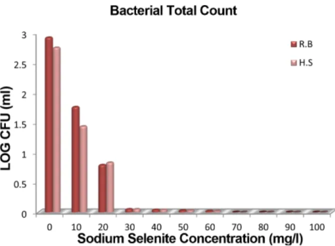
In the present study, the isolation of selenium (Se)-enriched bacteria from rumen fluid and hot spring water was carried out. Rumen fluid samples were taken from cannulated goats fed a basal diet and the water samples were collected from Selayang hot spring, Selangor- Malaysia. A total number of 140 Se-toler- ant isolates were obtained aerobically using an Se-enriched medium and spread plate technique. All the isolates were initially screened for the ability to transform the Se-containing medium to a red-orange cul- ture using a spectrophotometer. Twenty isolates of dark red-orange medium were selected for a screening of the highest Se-containing protein accumulating strains using the dialysis technique and icp.ms to mea- sure the Se content. Four isolates, identified as Enterobacter cloacae (ADS1, ADS7, and ADS11), and Klebsiella pneumoniae (ADS2) from rumen fluid origin, as well as, one isolate from hot spring water (Stenotro- phomonas maltophilia (ADS18)), were associated with the highest biomass organic Se-containing protein when grown in a medium enriched with 10 µg/ml sodium selenite. In addition, around 50 µg/100 µg of the absorbed inorganic Se was accumulated as an organic form. Organic Se-containing protein in all the selected strains showed antioxidant properties in the range of 0.306 to 0.353 Trolox equivalent antioxidant capacity (TEAC) mg/ml. Therefore, these strains may offer a potential source of organic Se due to their Se-tolerant nature and higher biomass organic to inorganic Se ratio.
Keywords: Accumulation, antioxidant, bacteria, isolation, organic selenium
*Corresponding author
Tel: +0389474878, Fax: +0389432954 E-mail: [email protected]
© 2017, The Korean Society for Microbiology and Biotechnology
Organic Se can be produced biologically through selenate or selenite microbial reduction. Selenium resistance microorganisms can challenge selenite and selenate when grown in an Se-enriched medium; this resistance action is achieved through two different pro- cesses: reduction to red elemental Se form [8], or meta- bolic conversion to organic Se, such as selenocysteine and selenomethionine [9]. Bacterial selenite reduction results in a red-orange culture in liquid media due to the accu- mulation of intracellular deposits of red elemental Se [10]. Recently, a number of microorganisms, such as Lactobacillus spp., Bifidobacterium spp., and Enterococ- cus, have been reported to take up and accumulate Se in their cells and can be used as Se-enriched probiotics [11 −13]. The bacterial strain, Lactobacillus retire Lb2 BMDSM 16143, can uptake inorganic Se from the medium and metabolize it into an organic Se form and incorporate it into proteins as SeCys; however, this is associated with a bacterial biomass reduction [14]. The same result has been found for L. bulgaricus; the bio- mass is (p < 0.05) affected when the selenite concentra- tion is greater than 0.46 mM [15]. Therefore, the poor selenite tolerance of lactic acids bacteria limits the appli- cation of Se-enriched Lactobacillus spp. in the food industry. In contrast, Gram-negative bacteria isolated from soil and metalloid water shows (p < 0.05) selenite resistance associated with less Se effect on their biomass [16]. However, although most of these Gram-negative strains are resistant to very high concentrations of the toxic Se and reduce it to a less toxic elemental Se, this process may be associated with organic Se accumulation as an intermediate step. Therefore, organic Se produced by un-probiotic bacteria can be extracted and used to deliver dietary levels of Se to livestock through feed sup- plementation.
Evidence has been accumulated that most of the microbial selenite reduction was in aerobic conditions however, some studies reported the ability of anaerobic conditions in selenite reduction [17]. Previous studies clearly demonstrated that natural ecosystem such as hot spring water contained a variety of microorganisms which they useful in metals bioremediation process [18].
As well as, rumen microorganisms which contain facul- tative anaerobic bacteria were capable of reduction inor- ganic Se and incorporating it into the microbial protein [19]. Therefore, once no more bacterial strains have been
characterized and obtained in laboratory culture as an organic Se source, it is interesting to identify some strains as organic Se-enriched bacteria since it is able to absorb medium selenite and accumulate it as selenopro- teins. Thus, the objective of this study is to isolate, enu- merate, and to characterize several bacterial strains from rumen fluid and hot spring water that resist and reduce medium selenite and accumulate it as high bio- mass organic selenium.
Materials and Methods
Chemicals
All the chemicals and microbiological media used in this study were of analytical grade. Nutrient agar and nutrient broth were purchased from Merck (Darmstadt, Germany), sodium selenite, Na
2SeO
3, ≥99%, were pur- chased from Sigma-Aldrich.
Sample collection
Rumen fluid samples were taken from cannulated goats fed a basal diet (Field 2, Department of Animal Science, Faculty of Agriculture, Universiti Putra Malay- sia), and the water samples were collected from Selayang hot spring located in the Gombak, Selangor, Malaysia (N.03
o15.542’ and E.101
o38.766’). Both sam- ples sources were collected in triplicates at different points from the sampling sites and were transported to the laboratory, in sterile capped bottles in a proper tem- perature and directly diluted serially for inoculation.
Culture media and bacterial growth
The selective medium of selenium-tolerant bacteria
was prepared using nutrient agar media enriched with
10, 20, 30, 40, 50, and 100 μg/ml of Sodium selenite, as
described by Shahverdi et al. [20]. A sodium selenite
stock solution (2.19 g/l corresponding to 1 g/l of Se) was
prepared and sterilized by filtration (single use syringe
filter, 0.20 mm, Sartorius Stedim Biotech). The collected
samples were serially diluted in sterile deionized water
and spread onto the nutrient agar plates. Inoculated
plates of rumen fluid and hot spring water samples were
incubated aerobically for 48 h at 39 ℃ and 30℃, respec-
tively. Each plate holding between 30 and 300 colonies
was selected to be counted as colony-forming units
(CFU) per ml of sample.
Isolation of selenium-enriched bacteria . A total of 140 red single colonies of different morphological appear- ance were selected and re-streaked on new nutrient agar media supplemented with 10 μ g/ml Na
2SeO
3to obtain a pure bacterial culture of the isolates. The pure agar cul- tures were sub-cultured by the transfer of a single colony to a nutrient broth medium enriched with the same Na
2SeO
3concentration. Among these cultures, 20 isolates were selected for this study according to their higher capability to reduce selenite to red elemental Se, which was chosen, based on the red color intensity using a spectrophotometer (624 nm). The pure cultures of the isolates were kept at -20 ℃ using 30% glycerol [21].
Screening of organic selenium accumulated strains. The screening was carried out by determina- tion of organic selenium according to the method described by [12]. Aliquots of fresh culture (24 h) con- taining 1 × 10
6of isolated bacterial cells were used to ensure that all the cultures were inoculated by the same amount of cells [22]. The culture was centrifuged at 3,220 × g for 15 min to harvest the bacterial pellets and then washed two times using deionized water to remove inorganic selenium which might adsorb to the bacterial cells. The selenium-enriched bacterial cells were lyo- philized at -20ºC for further use.
To determine the organic selenium, all measurements of the samples were made in triplicate; one gram of bac- terial cells from each strain was dialyzed using dialysis sacks of flat width 25 mm, 12,000 Da, (Sigma-Aldrich).
The dialysis process was performed against deionized water, which was changed every 12 h for a total of 96 hours to separate inorganic Se from its organic form [12].
The content in the dialysis tube was lyophilized and then used to determine the Se concentration using a Perkin Elmer ICP.MS by the same protocol as for the determi- nation of the total selenium concentration [23]. The accumulating rate of Se in the bacterial biomass was cal- culated according to the following equation:
Accumulation rate (%) =
Determination of Selenium Concentration
Selenium was determined in the supernatant and pel-
let (cell) fractions according to Garbisu et al. [24]. The supernatant fraction was analyzed directly; the cell frac- tion was subjected to an acid digestion procedure (30%
H
2O
2in 16 M HNO
3, 100 ℃, overnight) followed by reduction of any selenite generated with 6 N HC1 (100 ℃, 1 h). The samples were analyzed by inductively coupled plasma mass spectrometry.
Characterization and identification of isolated bacteria The best five organic Se producing bacteria ADS1, ADS2, ADS7, ADS11, and ADS18 were identified using phenotypic characterization and genetic characterization:
Phenotypic characterization. All isolates were subjected to Gram staining according to standard micro- biological protocol. The colonies were distinguished through visual observation of the colony morphology.
Individual colonies were characterized by a specific bio- chemical test using commercially available biochemical Kits (Api-20E) API
®bioMérieuxs. API 20E data were compared to those in the bioMérieux’s database (bioMérieux’s 1990). All tests were performed in dupli- cate, and negative controls were obtained using a fresh medium. Their characteristics are summarized in Table 4.
Genetic characterization. Genomic DNA was extracted using PureLink
®Genomic DNA Kits (Invitro- gen). The DNA of each bacterial isolate was PCR ampli- fied directly with primer pair 27F/1492R (27F: 5’-AGA GTT TGA TCC TGG CTC AG-3’; 1492R: 5’-TAC CTT GTT ACG ACT T-3’). The reaction product was analyzed using agarose gel electrophoresis. The PCR products were sent to a private laboratory (First Base, Malaysia) for purification and sequencing. The sequences obtained were analyzed using the National Centre for Biotechnology International (NCBI) BLAST, which is available on the Internet at http://blast.ncbi.nlm.nih.gov/Blast.cgi. The sequences in the FASTA form were aligned in this soft- ware. BLAST was used to search for a similar sequence in the GenBank and compared to the query sequence.
Antioxidant capacity using ABTS
•+method
The ABTS
•+radical cation decolorization assay was determined according to Chan et al. [25]. ABTS was pro- duced by reacting 7 mM ABTS aqueous solution with
(organic Se content in bacteria × biomass in 10 ml medium)× 100 Se content in 10 ml medium