대한화상학회지 제 19 권 제 2 호
88
Journal of Korean Burn SocietyVol. 19, No. 2, 88-91, 2016
CASE REPORT
자리 옮김 근막 피부 피판을 이용한 머리덮개와 머리덮개뼈의 4도 접촉 화상의 수복: 증례 보고
안건형ㆍ주홍실ㆍ임수아ㆍ송진경ㆍ임성윤
한전의료재단 한일병원 성형외과Coverage of a 4
thDegree Contact Burn of Scalp and Calvarium Using a Fasciocutaneous Transposition Flap: A Case Report
Gun Hyung Ahn, M.D., Hong Sil Ju, M.D., Ph.D., Soo A Lim, M.D., Ph.D., Jin Kyung Song, M.D., Ph.D. and Seong Yoon Lim, M.D.
Department of Plastic and Reconstructive Surgery, Hanil General Hospital, Seoul, Korea
Scalp and calvarium defects are caused by trauma, burn, tumor resection, or congenital diseases. We experienced a few cas- es of severe electrical burn of scalp and calvarium, but fourth-degree contact burn of scalp and calvarium is a rare case. A 67 years old man was presented with a 25% total body surface area contact burn. A 20 cm×15 cm thick eschar on the patient’s scalp was observed. Among various techniques for scalp reconstruction, we planned fasciocutaneous transposition flap with split thickness skin graft for coverage of large defect. Considering aesthetically satisfactory outcome, we designed a fas- ciocutaneous transposition flap including the hair-bearing areas. We additionally used skin graft for uncovered surrounding areas. There were no flap necrosis, graft loss, or any other surgical complications after the surgical flap and skin graft. At 6-month follow-up, the operation site was stable. The patient satisfied with functional and aesthetical outcomes, so we report this case. (J Korean Burn Soc 2016;19:88 -91)
Key Words: Burn, Scalp, Surgical flap, Skin graft
Received: 2016. 10. 16, Revised: 2016. 10. 23, Accepted: 2016. 10. 25 Corresponding author: Hong Sil Ju, Department of Plastic and Reconstructive Surgery, Hanil General Hospital, 308 Uicheon-ro, Dobong-gu, Seoul 01450, Korea
Tel: 82-2-901-3109, Fax: 82-2-901-3104 E-mail: [email protected]
INTRODUCTION
Defects in scalp and calvarium are caused by trauma, burn, tumor resection, or congenital diseases. Since the anatomical characteristics of scalp and calvarium as well as aesthetical results have to be considered in covering defect of these areas, it is challenging problem for plastic surgeons
1,2). Reconstruction techniques of these areas vary from using a simple primary closure or local flap to using a tissue expander, to performing a skin graft after pro- liferation of granulation tissue by perforating burr holes
in the necrotic calvarium, to a distant or free flap
3). The choice of reconstruction technique is made considering the size, location, depth and etiology of the defect area;
the quality of surrounding tissue; previous or active in-
fection; the exposure status of vital structures such as
bone, dura mater, or brain parenchyma; and the age and
general condition of a patient; aesthetic considerations
such as hair growth pattern, hair line and aesthetic sub-
unit
4). We have treated a few burn patients who had re-
ceived damage to the scalp and calvarium due to a
high-voltage electrical burn. A fourth-degree contact burn
on the scalp and calvarium is extremely rare. Considering
aesthetically satisfactory outcome, we planned a fas-
ciocutaneous transposition flap including the hair-bearing
areas located in center. Additionally we used skin graft
for uncovered surrounding areas. Although only one
stage surgery was done, we took functional and aesthet-
ical successful outcomes, so we report this case with liter-
Gun Hyung Ahn, et al:Coverage of 4th Degree Burn of Scalp and Calvarium
89
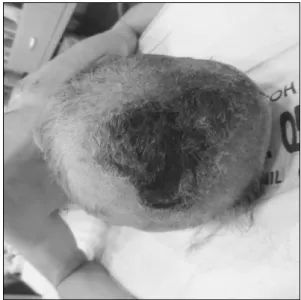
Fig. 1. A 20 cm×15 cm thick black eschar on the scalp.
Fig. 3. Fasciocutaneous transposition flap and split-thickness skin graft.
Fig. 2. The calvarium was invaded by 4th degree contact burn.
ature review.
CASE REPORT
A 67 years old male patient, as loss of consciousness in sauna, was presented with a 25% total body surface area of contact burn in the scalp, back, hip and both legs.
A 20 cm×15 cm thick black eschar on the patient's scalp due to contact burn had already formed at admission (Fig. 1). He had been suffering from diabetes, chronic ob- structive pulmonary disease. Therefore, the risk and like- lihood of side effects were predicted to be high owing to a long operation time and the limitation in the donor area in case of performing the free tissue transfer procedure.
We planned one stage surgery using a fasciocutaneous transposition flap. Considering aesthetic result, hair-bear- ing flap was located in center, and donor site was covered with split thickness skin graft. After escharectomy, we knew necrosis invaded to some parts of the calvarium.
Necrotic calvarium was removed using a burr until fresh calvarium appeared (Fig. 2). Using doppler imaging, the distance from the perforation point to the end of defect area was set as the length of the flap. Because it was diffi- cult to cover the large defect of the scalp and calvarium with one single flap, another small flap of the occipital area was also used. Excessive electric cauterization was avoided in elevating the flap to keep it viable, and metic- ulous dissection was done in surrounding blood vessels.
Afterwards, the transposition flap was elevated and fixed to the central defect area and then a split-thickness skin graft was performed in the surrounding area (Fig. 3). On postoperative 15 days, there were no infection, necrosis of the flap, loss of the grafted skin (Fig. 4). The procedure was successful without loss or partial necrosis of the flap.
Functionally and aesthetically satisfactory outcome was
observed at the 6-month follow-up (Fig. 5).
90
대한화상학회지 Vol. 19, No. 2, 2016Fig. 4. Five days after the operation, there were no com- plications such as infection, flap necrosis, or skin loss.
Fig. 5. At 6-month follow-up, a well-healed and stable wound coverage was observed.
DISCUSSION
Various techniques have been used in reconstruction of scalp and calvarium defects depending on the size and depth of the defect area. Secondary intention healing or simple primary closure is mostly used for small defects, and tissue expansion or skin graft after trepanation is used for relatively larger defects. If there is large defect invading calvarium, regional flap, distant flap or free tis- sue transfer is generally used
4,5).
Secondary intention healing by granulation, contraction and re-epithelization is simplest way but it leads to pro- longed wound healing time, scarring and alopecia
4,6). A simple primary closure only used if the defect is smaller than 3 cm in diameter
4). Similarly, local flap procedure has a limitation in covering a wide area of defect. The tissue expansion procedure is possible when a scalp de- fect is relatively large, but it is also leads to prolonged wound healing time and needs safe coverage of the defect during the time of expansion
6). If the calvarium is ex- posed while the tissue expander is inflated, life threat- ening severe inflammation can occur. Skin graft after trepanation; it has many disadvantages in cases such as skin ulcers, bone exposure, swelling, scarring, delayed wound healing, alopecia and requiring secondary proce- dure of soft tissue and calvarium in the future
3).
Since first described by Mclean in 1972, free tissue transfer have become most common technique used in large defect of scalp and calvarium by many plastic and reconstructive surgeons
4,6). Various free flap have been used such as omental free flap, radial forearm free flap, latissimus dorsi free flap, rectus free flap and so on
4,6). It has superior vascular supply for survival of the flap to the wound, compared to any other scalp reconstructive techniques. However, for the free flap procedure, the sur- geon must be skilled in microsurgical techniques. And pro- longed operation time will be lead to cardiopulmonary complications. Morbidity in the donor area can also be a problem. The current patient was determined to have a high risk for side effects in case of performing a free flap due to old age, systemic weakness, and underlying diseases.
It is generally important aesthetical outcomes as well
as functional outcomes for scalp and calvarium defect
patients. Especially, scalp alopecia secondary to re-
construction of scalp defect is a serious problem for pa-
tient because it lead to social phobia and severe psycho-
logical trauma
7). So we considered not only functional
outcomes but aesthetical outcomes. A transposition flap
and skin graft use were planned to cover the defect in
the primary procedures. In designing a fasciocutaneous
transposition flap procedure, the hairline and hair dis-
tribution pattern and whether the vascular pedicle enters
the base well should also be fully considered
6).
Gun Hyung Ahn, et al:Coverage of 4th Degree Burn of Scalp and Calvarium
91
The fasciocutaneous transposition flap used in this case to cover the wound in the calvarium due to the fourth-de- gree contact burn has several advantages. First, duration of the operation and postoperative hospital stay are re- duced because micro techniques are not needed. Second, the morbidity potential in the donor area is low, and the donor area defects can be minimized because only a linear scar remains. Third, the aesthetic appearance is sat- isfactory because the flap preserve hair bearing area.
Although skin graft site result in alopecia, hair bearing area of fasciocutaneous flap can cover the alopecia. During follow up period, the patient has been satisfied with his aesthetical outcomes. Here, we report that a functionally and aesthetically satisfactory outcome was obtained in the donor and recipient areas after the procedure.
REFERENCES
1) Kim MC, Ko YL, Shim HS. Reconstruction of scalp and
forehead defect with local transposition split skin flap and remnant full-thickness skin graft. J Plast Reconstr Aesthet Surg. 2013;66:1436-1438.
2) Tenna S, Brunetti B, Aveta A. Scalp reconstruction with superficial temporal artery island flap: clinical experience on 30 consecutive cases. J Plast Reconstr Aesthet Surg. 2013;66:
660-666.
3) Lee HS, Kim YS. Regeneration of necrosed skull after high-tension electrical injury. J Korean Soc Plast Reconstr Surg. 1995;22:1483-1489.
4) Seitz IA, Gottlieb LJ. Reconstruction of scalp and forehead defects. Clinics Plast Surg. 2009;36:355-377.
5) Lee ET, Jin US, Lee YH. Experimental and clinical study on the healing process of burned dura mater. J Korean Burn Soc.
2003;6:158-163.
6) Ducic Y. Reconstruction of the scalp. FRCSC, FACS. 2009;17:
177-187.
7) Guzey S, Alhan D, Sahin I. Our experiences on the reconstruction of lateral scalp burn alopecia with tissue expanders. J Burns. 2015;41:631-637.