Clinical Features and Prognosis of Invasive Pulmonary
Aspergillosis in Korean Children with Hematologic/Oncologic Diseases
Invasive pulmonary aspergillosis (IPA) is the most frequent form of invasive fungal diseases in immunocompromised patients. However, there are only a few studies on IPA in immunocompromised children in Korea. This study was designed to characterize IPA in Korean children with hematologic/oncologic diseases. Medical records of children with hematologic/oncologic diseases receiving antifungal therapy were reviewed. The enrolled children were divided into the IPA group (proven and probable IPA) and non-IPA group, and the clinical characteristics and prognosis were compared between the two groups.
During the study period, 265 courses of antifungal therapy were administered to 166 children. Among them, two (0.8%) episodes of proven IPA, 35 (13.2%) of probable IPA, and 52 (19.6%) of possible IPA were diagnosed. More children in the IPA group suffered from neutropenia lasting for more than two weeks (51.4% vs. 21.9%, P < 0.001) and showed halo signs on the chest computed tomography (78.4% vs. 40.7%, P < 0.001) than in the non-IPA group. No other clinical factors showed significant differences between the two groups. Amphotericin B deoxycholate was administered as a first line antifungal agent in 33 (89.2%) IPA group episodes, and eventually voriconazole was administered in 27 (73.0%) episodes. Ten (27.0%) children in the IPA group died within 12 weeks of antifungal therapy. In conclusion, early use of chest computed tomography to identify halo signs in immunocompromised children who are expected to have prolonged neutropenia can be helpful for early diagnosis of IPA and improving prognosis of children with IPA.
Keywords: Invasive Pulmonary Aspergillosis; Immunocompromised Host; Child Seung Beom Han,1,2 Seong koo Kim,1,3
E Young Bae,1,2 Jae Wook Lee,1,3 Jong-Seo Yoon,1,2 Nack-Gyun Chung,1,3 Bin Cho,1,3 Dae Chul Jeong,1
Jin Han Kang,1,2 Hack-Ki Kim,1,3 Dong-Gun Lee,2,3,4 Hyun Sil Lee,5 and Soo Ah Im5
1Department of Pediatrics, 2Vaccine Bio Research Institute, 3Catholic Blood and Marrow Transplantation Center, 4Division of Infectious Diseases, Department of Internal Medicine, and
5Department of Radiology, College of Medicine, The Catholic University of Korea, Seoul, Korea Received: 24 December 2014
Accepted: 27 April 2015 Address for Correspondence:
Jong-Seo Yoon, MD
Department of Pediatrics, Seoul St. Mary’s Hospital, College of Medicine, The Catholic University of Korea, 222 Banpo-daero, Seocho-gu, Seoul 137-701, Korea
Tel: +82.2-2258-6190, Fax: +82.2-537-4544 E-mail: [email protected]
http://dx.doi.org/10.3346/jkms.2015.30.8.1121 • J Korean Med Sci 2015; 30: 1121-1128
INTRODUCTION
Aspergillus spp. became the most common pathogen causing invasive fungal disease (IFD) in immunocompromised patients after the introduction of fluconazole prophylaxis; therefore, in- vasive aspergillosis (IA) is the most common type of IFD in these patients (1-3). Immunocompromised patients with IA have a high mortality of 40%-50% (4-6), and mortality up to 70% has been reported in pediatric cancer patients with IA (7). In spite of the introduction of newer antifungal agents, such as voricon- azole and echinocandins, which have an improved effect on IA, the mortality of IA was recently reported as 25%-35% (8-11). A relatively higher mortality of 40%-50% due to IA is still cited in situations where amphotericin B deoxycholate was adminis- tered as the first line antifungal agent, as in Korea where avail- ability of first line antifungal agents was limited to amphotericin B deoxycholate or itraconazole under government regulations of the national health insurance system (12-14).
Given the high mortality of IA and improved prognosis with an early diagnosis and antifungal treatment (15, 16), empirical or pre-emptive antifungal therapy with antifungal agents hav- ing an anti-mold effect has been recommended in immuno- compromised patients with risk factors for IA (17, 18). On the other hand, universal empirical therapy in patients at risk for IA may expose those patients without a true fungal infection to unnecessary antifungal agents, and this may result in unwanted adverse effects and increased medical costs. Actually, only 8%- 20% of patients with hematologic/oncologic diseases who re- ceived empirical antifungal therapy were reported to have true IFD (19-21).
In Korea where empirical antifungal therapy has been stan- dard practice for children with hematologic/oncologic diseas- es, epidemiological data on IFD or IA in those children is scarce (12). The aim of this retrospective study was to investigate the clinical features and prognosis of invasive pulmonary aspergil- losis (IPA) in Korean children with hematologic/oncologic dis- Pediatrics
eases. This study was conducted with consideration of the fact that IA is the most frequent of IFD in immunocompromised pa- tients and most cases of IA are IPA (3, 11, 12, 15, 22, 23).
MATERIALS AND METHODS Subjects and study design
Medical records of children (< 20 yr of age) with hematologic/
oncologic diseases, who were admitted to Seoul St. Mary’s Hos- pital, Seoul, Korea between April 2009 and March 2013 were retrospectively reviewed. Among them, children who received antifungal therapy with agents having an anti-mold effect for more than 48 hr were enrolled. The enrolled children were di- agnosed with possible, probable, or proven IPA based on the revised definition of IFD in immunocompromised patients rec- ommended by the European Organization for Research and Treatment of Cancer/Invasive Fungal Infections Cooperative Group and the National Institute of Allergy and Infectious Dis- eases Mycoses Study Group (EORTC-MSG) Consensus Group in 2008 (24). Children with IA not involving the lung were ex- cluded. Children diagnosed with proven and probable IPA were included in the IPA group, whereas children diagnosed with possible IPA and those who did not fulfill the diagnostic criteria for three categories of IPA were included in the non-IPA group.
In the event that one child experienced several episodes of IPA, IPA diagnosed after the cessation of antifungal therapy for a previous episode of IPA was considered a separate episode of IPA and included in the present study. Clinical features of the IPA group were investigated, and clinical factors related to the development of IPA were evaluated by comparing clinical fea- tures of the IPA and non-IPA groups. The evaluated clinical fea- tures included sex, age, the type of underlying hematologic/on- cologic disease and its remission status, the type of therapy ad- ministered prior to antifungal therapy, the duration of fever and neutropenia, previous histories of IPA and hematopoietic cell transplantation (HCT), and the presence of concurrent infec- tions. In addition, mortality at 6 and 12 weeks after antifungal therapy was compared between the IPA and non-IPA groups.
Institutional strategies for antifungal therapy
In our hospital, antifungal prophylaxis and empirical antifungal therapy have been given based on the recommendations of the committee for “Guidelines for the Empirical Therapy of Neutro- penic Fever Patients based on Literature in Korea” (25). Anti- fungal prophylaxis with oral fluconazole (3 to 5 mg/kg/day) was given from the initiation of anti-cancer chemotherapy until re- covery from neutropenia. In children who received HCT, intra- venous micafungin (1 mg/kg/day) prophylaxis was given from the start of pre-HCT conditioning therapy to engraftment, and then oral fluconazole was given until the discontinuation of im- mune suppressants. Empirical antifungal therapy was adminis-
tered when fever lasted for 3 to 5 days in children who were ex- pected to have neutropenia for longer than seven days or who received immune suppressants after allogeneic HCT. If chest computed tomography (CT) findings consistent with fungal pneumonia or positive serum galactomannan index (GMI) were observed, pre-emptive antifungal therapy was given re- gardless of the duration of fever. Serum GMI was tested once or twice a week during neutropenia in children receiving anti-can- cer chemotherapy and from pre-HCT conditioning therapy un- til discharge from hospital in children receiving HCT. Chest CT was performed in children with abnormal findings on chest x- ray or positive serum GMI.
Definition
The diagnosis of IPA was based on the definition of IFD recom- mended by the EORTC-MSG Consensus Group, which was ap- plicable to patients with host factors of compromised immunity (24). Proven IPA was defined as when Aspergillus spp. were ob- served with accompanying tissue damage or Aspergillus spp.
were identified in culture on sterile materials (24). Probable IPA was defin ed as when clinical features and mycological evidence for IPA were observed in patients with host factors, and possible IPA was defined as the presence of clinical features of IPA with- out mycological evidence (24). One of the chest CT findings among well-circumscribed lesions with or without halo sign (consolidation and surrounding ground glass opacity), air-cres- cent sign and cavity fulfills clinical criteria for IPA, and myco- logical criteria consisted of the presence of mold or identifica- tion of Aspergillus spp. from culture of sputum, bronchoalveolar lavage (BAL) fluid or bronchial brush and positive GMI from serum, plasma or BAL fluid (24). Neutropenia was defined as a neutrophil count < 500/μL (24), and chest CT was reviewed by two independent radiologists (HYL and SAI). The GMI was mea- sured using the PLATELIATM ASPERGILLUS EIA kit (Bio-Rad, Marnes-la-Cocquette, France) in accordance with the manu- facturer’s recommendations. Serum GMI > 0.5 and BAL fluid GMI > 1.0 were defined as positive based on the 4th European Conference on Infections in Leukemia (ECIL-4) guidelines (17).
Positive GMI results were confirmed by a repeat measurement of GMI in the same sample. Because it was difficult to clearly define whether immunocompromised patients with IPA died of IPA or other complicated causes (26), overall mortality at 6 and 12 weeks after antifungal therapy were evaluated.
Statistical analysis
Categorical variables were compared using the chi-square test and continuous variables were compared using student’s t-test or the Mann-Whitney test based on the distribution of the data.
Fischer’s exact test was used to compare the incidence of IPA during the study period. Mortality at 12 weeks after antifungal therapy was compared between the IPA and non-IPA groups
using a log-rank test. Statistical analysis was performed using SPSS Statistics 17.0 (SPSS Inc., Chicago, IL, USA), and statistical significance was defined as a two-tailed P value < 0.05.
Ethics statement
This study was approved by the institutional review board of the Seoul St. Mary’s Hospital with an exemption of acquiring inform- ed consent (Approval No.: KC14RISI0791).
RESULTS
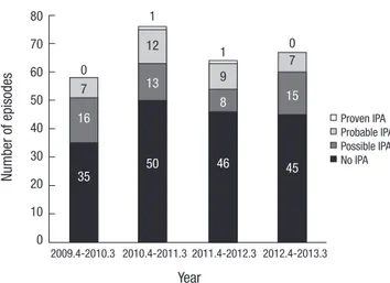
A total of 268 courses of antifungal therapy with anti-mold agents were administered to 168 children. Among them, one episode of probable cerebral IA and two cases of possible fungal sinus- itis without involvement of the lung were excluded. Among the remaining 265 courses of antifungal therapy, 52 (19.6%) episodes of possible IPA, 35 (13.2%) episodes of probable IPA, and two (0.8%) episodes of proven IPA were diagnosed. Accordingly, 37 (14.0%) and 228 (86.0%) episodes were included in the IPA and non-IPA groups, respectively. Candidemia was diagnosed in five (1.9%) episodes in the non-IPA group, and IFD caused by other fungi was not reported. The yearly distribution of IPA was not significantly different during the study period (Fig. 1, P = 0.603).
Characteristics of children with invasive pulmonary aspergillosis
The mean age of children in the IPA group was 11.4 ± 5.3 yr, and 23 (62.2%) episodes of IPA were diagnosed in boys (Table 1).
Acute myeloid leukemia (AML) and acute lymphoblastic leu- kemia (ALL) were the most common underlying diseases, and 23 (74.2%) episodes were diagnosed in non-complete remission (CR) of the underlying hematologic malignancies. In accordance with these results, re-induction or salvage chemotherapy was
the most common therapy preceding antifungal therapy (35.1%), and allogeneic HCT was the next most common (16.2%).
Prolonged neutropenia lasting for longer than 10 days was most frequent among the host factors for defining IFD recom- mended by the EORTC-MSG Consensus Group (Table 2). A halo sign was the most common finding on chest CT, and serum GMI was positive in 30 (81.1%) episodes. BAL fluid analyses were per- formed in 11 episodes, and BAL fluid GMI was positive in six (54.5%) episodes. Both serum and BAL fluid GMIs were posi- tive in one (2.7%) episode. In the five episodes of BAL cases, BAL fluid GMI were negative, despite of positive serum GMIs. Accor- ding to GMI results by specimen, BAL fluid(+)/serum(-) group and BAL fluid(-)/serum(+) group showed no significant differ- ences in host factors or in clinical presentation. Bronchoscopy was performed earlier after starting antifungal therapy in the BAL fluid(+)/serum(-) group than in the BAL fluid(-)/serum(+) group, although statistically insignificant (median 3 days, inter- quartile range [IQR]: 1-12 vs. median 6 days, IQR: 1-11 days, P = 0.173). Aspergillus spp. were identified in BAL fluid cultures in two episodes. Two children diagnosed with proven IPA under- went a pulmonary lobectomy, and the Aspergillus spp. were iden- tified in their excised lung tissue.
Antifungal prophylaxis was performed in 26 (70.3%) episodes, and fluconazole (5 to 10 mg/kg/day) was administered most frequently (Table 3). Amphotericin B deoxycholate was admin- istered as the first line antifungal agent in 33 (89.2%) episodes.
The first line antifungal agent was changed to a second line drug in 35 (94.6%) of 37 episodes a median of 4 days (IQR: 2-7) later.
Among 33 episodes in which amphotericin B deoxycholate was administered as the first line antifungal agent, amphotericin B deoxycholate was changed to a second line antifungal agent in 32 (97.0%) episodes. Oral voriconazole was given as a second line agent on discharge from the hospital in three (9.4%) episo- des, and intravenous voriconazole was given with a diagnosis of probable IPA in four (12.5%) episodes. In six (18.8%) episodes, second line agents were administered due to sustained fever lasting for more than five days in spite of first line antifungal ther- apy. In the remaining 22 (68.8%) episodes, second line antifun- gal agents were given due to adverse effects of first line agents.
Infusion-related adverse effects of amphotericin B deoxycho- late were reported in 18 (56.3%) episodes, including 14 cases of chills, seven cases of high fever, five cases of nausea and vomit- ing, and one case each of dyspnea, skin rash and severe irrita- bility. Hypokalemia (serum potassium level < 3.0 mEq/L) de- veloped in 15 (46.9%) episodes during amphotericin B deoxy- cholate therapy in spite of potassium supplementation. Increas- ed serum creatinine greater than two times the baseline was observed in three (9.4%) episodes. Finally, antifungal therapy was completed with voriconazole in 27 (73.0%) episodes.
Mortality at 6 and 12 weeks after antifungal therapy was 16.2%
and 27.0%, respectively (Table 3). Between 10 children who died
Number of episodes
Year
2009.4-2010.3 2010.4-2011.3 2011.4-2012.3 2012.4-2013.3 80
70 60 50 40 30 20 10 0
Proven IPA Probable IPA Possible IPA No IPA 0
7
12
9
7
16
13
8 15
35 50 46 45
0 1
1
Fig. 1. Yearly distribution of the incidence of invasive pulmonary aspergillosis (P = 0.603).
within 12 weeks after antifungal therapy and 27 children who survived longer than 12 weeks after antifungal therapy, there were no significant differences in the distribution of host factors and clinical and mycological criteria (data not shown). Voricon- azole was administered to 77.8% of the children who survived and 60.0% of the children who did not survive (P = 0.407). Vori- conazole therapy was begun a median of 7 days (IQR: 4-13) af- ter antifungal therapy in the children who survived and a medi- an of 14 days (IQR: 6-25) after antifungal therapy in the children who did not survive (P = 0.204).
Comparison between the IPA and non-IPA groups
Clinical features between the IPA and non-IPA groups were compared in order to identify any significant factors related to
the development of IPA in children who received antifungal therapy (Table 1). There were no significant differences in sex, age, underlying disease and its remission status, previous histo- ries of IPA and HCT, and the type therapy administered prior to antifungal therapy between the two groups. However, signifi- cantly more children in the IPA group experienced neutropenia lasting for longer than 2 weeks before antifungal therapy (P <
0.001), and accordingly the proportion of children with a total duration of neutropenia longer than 4 weeks was significantly higher in the IPA group compared to the non-IPA group (P <
0.001). Although fever duration was significantly longer in the IPA group than in the non-IPA group, the difference was only one day.
There was no difference in the distribution of host factors for Table 1. Characteristics of children receiving antifungal therapy
Factors Non-IPA group (n = 228) IPA group (n = 37) P value
Gender Male Female
139 (61.0) 89 (39.0)
23 (62.2) 14 (37.8)
0.890
Age (yr), mean ± SD 10.0 ± 5.4 11.4 ± 5.3 0.155
Underlying disease Acute myeloid leukemia Acute lymphoblastic leukemia Juvenile myelomonocytic leukemia Chronic myeloid leukemia Other leukemias Lymphoma Solid tumor Severe aplastic anemia
Hemophagocytic lymphohistiocytosis Myelodysplastic syndrome Other anemias
94 (41.2) 73 (32.0) 5 (2.2) 1 (0.4) 6 (2.6) 5 (2.2) 20 (8.8) 18 (7.9) 4 (1.8) 1 (0.4) 1 (0.4)
15 (40.5) 15 (40.5) 0 (0.0) 0 (0.0) 1 (2.7) 0 (0.0) 0 (0.0) 6 (16.2) 0 (0.0) 0 (0.0) 0 (0.0)
0.519
Status of underlying disease*
Complete remission state Uncontrolled state
82 (39.4) 126 (60.6)
8 (25.8) 23 (74.2)
0.144
Administered therapy prior to antifungal therapy No chemotherapy
Remission induction chemotherapy Re-induction or salvage chemotherapy Consolidation chemotherapy Maintenance chemotherapy Palliative chemotherapy Allogeneic HCT Autologous HCT
25 (11.0) 22 (9.6) 76 (33.3) 43 (18.9) 5 (2.2) 12 (5.3) 36 (15.8)
9 (3.9)
5 (13.5) 4 (10.8) 13 (35.1) 4 (10.8) 1 (2.7) 4 (10.8) 6 (16.2) 0 (0.0)
0.715
Previous history Post-HCT state
Previous IPA 94 (41.2)
19 (8.3) 19 (51.4)
4 (10.8)
0.248 0.541 Fever duration (days), median (IQR)
Before treatment
Total duration 3 (1-5)
7 (4-13) 2 (1-4)
7 (4-17) 0.017
0.641 Neutropenia duration
Before treatment ≤ 2 weeks > 2 weeks Total duration ≤ 4 weeks > 4 weeks
178 (78.1) 50 (21.9) 175 (76.8) 53 (23.2)
18 (48.6) 19 (51.4) 16 (43.2) 21 (56.8)
< 0.001
< 0.001
Concurrent other infections 60 (26.3) 10 (27.0) 0.927
*Complete remission state was determined in 239 episodes (208 in the non-IPA group and 31 in the IPA group). IPA, invasive pulmonary aspergillosis; SD, standard deviation;
HCT, hematopoietic cell transplantation; IQR, interquartile range.
defining IFD recommended by the EORTC-MSG Consensus Group between the two groups (Table 2). Chest CT was perform- ed in 118 episodes in the non-IPA group, and halo signs were
observed significantly more frequently in the IPA group than in the non-IPA group (P < 0.001). BAL fluid analyses were perform- ed in 28 episodes (11 in the IPA group and 17 in the non-IPA group) between April 2009 and March 2011. In the 17 episodes in the non-IPA group, there were no cases of positive BAL fluid GMI and only one (5.9%) episode of positive BAL fluid culture for Aspergillus spp.
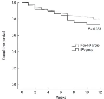
Antifungal prophylaxis was administered more frequently in the IPA group; however, the difference was not significant (Ta- ble 3). The type of first line antifungal agent was not significant- ly different between the two groups. However, second line and later administered antifungal agents were significantly different between the two groups - voriconazole and liposomal ampho- tericin B were most commonly administered in the IPA and non-IPA groups, respectively. Mortality at 6 and 12 weeks after antifungal therapy was higher in the IPA group compared to the non-IPA group, however, the differences were not statistically significant (Fig. 2, P = 0.353).
DISCUSSION
In the present study, clinical features and prognosis of IPA diag- nosed in children with hematologic/oncologic diseases were investigated, and clinical factors associated with the develop- ment of IPA in children who received antifungal therapy were evaluated. Prolonged neutropenia, corticosteroids or immune suppressant therapy, hematologic malignancy as a underlying disease, especially AML, relapsed leukemia, receiving an allo- geneic HCT, graft-versus-host disease and cytomegalovirus in- fection after allogeneic HCT, and other respiratory viral infec- tion after allogeneic HCT were reported as risk factors for IA in Table 2. The application of definitions of invasive aspergillogsis recommended by the
European Organization for Research and Treatment of Cancer/Invasive Fungal Infec- tions Cooperative Group and the National Institute of Allergy and Infectious Diseases Mycoses Study Group Consensus Group
Factors Non-IPA group
(n = 228)
IPA group
(n = 37) P value Host factors
Prolonged neutropenia Allogeneic HCT Prolonged steroid Immune suppressant
176/228 (77.2) 55/228 (24.1) 4/228 (1.8) 2/228 (0.9)
33/37 (89.2) 13/37 (35.1) 2/37 (5.4) 2/37 (5.4)
0.097 0.155 0.166 0.095 Clinical criteria
Halo sign Consolidation Air-crescent Cavity
48/118 (40.7) 14/118 (11.9) 2/118 (1.7) 0/118 (0.0)
29/37 (78.4) 8/37 (21.6) 0/37 (0.0) 2/37 (5.4)
< 0.001 0.138 1.000 0.056 Mycological criteria
Serum GMI positivity BAL GMI positivity BAL fluid Culture positivity
27/228 (11.8) 0/17 (0.0) 1/17 (5.9)
30/37 (81.1) 6/11 (54.5) 2/11 (18.2)
< 0.001 0.001 0.543 Data represent the number of corresponding children/the total number of included children (%). IPA, invasive pulmonary aspergillosis; HCT, hematopoietic cell transplan- tation; GMI, galactomannan index; BAL, bronchoalveolar lavage.
Table 3. Antifungal therapy and clinical outcomes in children receiving antifungal therapy
Factors Non-IPA group
(n = 228) IPA group
(n = 37) P value Anti-fungal prophylaxis
No prophylaxis Fluconazole Itraconazole Micafungin
95 (41.7) 101 (44.3) 5 (2.2) 27 (11.8)
11 (29.7) 21 (56.8) 2 (5.4) 3 (8.1)
0.279
Anti-fungal therapy 1st line therapy
Amphotericin B deoxycholate Liposomal amphotericin B Caspofungin
Itraconazole Voriconazole
202 (88.6) 12 (5.3)
2 (0.9) 9 (3.9) 3 (1.3)
33 (89.2) 2 (5.4) 0 (0.0) 2 (5.4) 0 (0.0)
0.913
2nd line therapy
Amphotericin B deoxycholate Liposomal amphotericin B Caspofungin
Itraconazole Voriconazole
166 (72.8) 2 (1.2) 133 (80.1)
24 (14.5) 3 (1.8) 4 (2.4)
35 (94.6) 0 (0.0) 16 (45.7)
6 (17.1) 1 (2.9) 12 (34.3)
< 0.001
Final therapy
Amphotericin B deoxycholate Liposomal amphotericin B Caspofungin
Itraconazole Voriconazole
44 (19.3) 119 (52.2) 33 (14.5) 10 (4.4) 22 (9.6)
2 (5.4) 3 (8.1) 4 (10.8) 1 (2.7) 27 (73.0)
< 0.001
Total duration of therapy (days),
median (IQR) 12 (7-21) 73 (22-175) < 0.001 Outcome
6 weeks mortality
12 weeks mortality 30 (13.2)
46 (20.2) 6 (16.2)
10 (27.0) 0.615 0.344 IPA, invasive pulmonary aspergillosis; IQR, interquartile range.
Fig. 2. Comparison of cumulative survival between the IPA and non-IPA groups.
Cumulative survival
Weeks
0 2 4 6 8 10 12 1.0
0.8
0.6
0.4
0.2
0.0
P = 0.353
Non-IPA group IPA group
patients with hematologic/oncologic diseases (5, 6, 8, 27-30). In accordance with previous reports, AML was the most frequent underlying disease, and more than half of the children with he- matologic malignancies were non-CR status in the present study.
IPA was diagnosed in 14.0% of children who received antifun- gal therapy in the present study, and the remaining children, over 80.0%, may have experienced adverse effects of the adminis- tered antifungal agents, increased medical costs, and prolonged duration of hospitalization due to unnecessary antifungal ther- apy. We tried to identify clinical factors predicting the develop- ment of IPA in those children to reduce unnecessary antifungal therapy in future patients, and only the duration of neutropenia was significantly different between the IPA and non-IPA groups.
Because prophylactic fluconazole has been administered to chil- dren at risk for IFD in our hospital, we decided to begin empiri- cal or pre-emptive antifungal therapy based on the risk for IA.
As a result, antifungal therapy was initiated based on similar clin- ical criteria for children both in the IPA and non-IPA groups.
Other hospitals where antifungal prophylaxis is performed should address such situations in a fashion similar to our hospital. There- fore, the initiation of antifungal therapy should be based not only on clinical features, but also on results of imaging and mycolo- gical tests, such as chest CT and serum and BAL fluid GMIs. In the present study, the incidence of halo sign on chest CT was significantly higher in the IPA group compared to the non-IPA group. Although there was no significant difference in outcome between the IPA and non-IPA groups, chest CT was helpful in diagnosing IPA.
Serum GMI is valuable in diagnosing IPA in children and adults (31-33), and the ECIL-4 guidelines recommend a serum GMI
> 0.5 as positive (17). Considering 16.2% of the IPA group in the present study showed negative serum GMI results but positive BAL fluid GMI or culture results, BAL fluid analyses should be encouraged in patients in whom IPA is suspected in spite of neg- ative serum GMI results. In addition, bronchoscopy should be performed in the early phase of IPA to acquire reliable BAL fluid GMI results. The ECIL-4 guidelines recommend a BAL fluid GMI
> 1.0 as positive (17). However, there have been only a few stud- ies on the application of BAL fluid GMI for diagnosing IPA in children (34, 35), and further studies to determine an appropri- ate BAL fluid GMI cut-off value for diagnosing IPA should be performed in children.
The 27.0% mortality in the IPA group was lower than recently reported 45% mortality in Korean adult and pediatric patients with IPA (12, 13), although 89.2% of the IPA group received am- photericin B deoxycholate as a first line antifungal agent in the present study. The difference in mortality was assumed to be a result of early changes in antifungal agents from amphotericin B deoxycholate to liposomal amphotericin B or voriconazole in our cases. In addition, about 80% of the IPA group showed halo signs on chest CT, which have been reported as characteristic
CT findings in the early phase of IPA as opposed to air-crescent, cavity, and simple consolidation (31, 36). Most patients in the IPA group may have been diagnosed with IPA in the early phase of disease in the present study, and an improved prognosis in IPA patients with halo signs compared to other CT findings has been reported (37).
The present study was performed when amphotericin B de- oxycholate or itraconazole were required to be administered as a first line antifungal agent due to government regulation of the national health insurance system in Korea. Now, in 2015, we are able to administer liposomal amphotericin B and caspofun- gin as first line antifungal agents in febrile neutropenic children based on a modified government policy in 2014. Because first line antifungal agents were adjusted to second line agents due to their adverse effects in more than half of IPA episodes in the present study, more safe and effective antifungal therapy is ex- pected in the future in Korea. Voriconazole treatment was asso- ciated with improved survival and fewer side effects compared to amphotericin B deoxycholate treatment of IPA (9), and em- pirical antifungal therapy with voriconazole was reported to be more economical than empirical therapy with amphotericin B deoxycholate (38, 39). In the present study, almost all IFD cases were IPA and no cases of mucormycosis were reported. In ad- dition, in the IPA group of the present study, children who died received voriconazole therapy later than children who survived, although this difference was not statistically significant. Consid- ering these results, voriconazole as well as liposomal ampho- tericin B and caspofungin could be considered as a first line an- tifungal agent in immunocompromised Korean children at risk for IPA to further improve prognosis in those children.
This study has several limitations including its retrospective nature. Cases of IFDs other than IPA were not thoroughly re- viewed in the whole population of children with hematologic/
oncologic diseases in our hospital. Risk factors for mortality and poor prognosis in children with IPA could not be evaluated due to the small number of children diagnosed with IPA. The enroll- ed children were heterogeneous - children with various types of underlying diseases were included and children receiving au- tologous or allogeneic HCT and not receiving HCT were includ- ed together. To overcome these limitations, a multicenter study including many children should be performed in the future.
In conclusion, 14.0% of children who received empirical an- tifungal therapy were diagnosed with IPA. Although amphoter- icin B deoxycholate was administered as a first line antifungal agent in most cases, mortality at 12 weeks after antifungal ther- apy was lower than previously reported in Korea. This improve- ment in prognosis of IPA was assumed to be arisen from early diagnosis of IPA. Therefore, early use of chest CT to identify halo signs in immunocompromised children who are expected to have prolonged neutropenia can be helpful for early diagnosis of IPA and improving prognosis of children with IPA. In addition,
the prognosis of IPA in immunocompromised children is ex- pected to be improved with the introduction of liposomal am- photericin B and caspofungin as first line antifungal agents in the future in Korea.
DISCLOSURE
There is no conflict of interest for all authors.
AUTHOR CONTRIBUTION
Study design: Han SB, Lee DG, Cho B, Kang JH. Data collection and analysis: Han SB, Kim SK, Bae EY, Lee JW, Yoon JS, Chung NG, Jeong DC. Review radiological findings: Lee HS, Im SA. Writ- ing: Han SB, Bae EY, Lee DG, Kang JH. Review and revision: Cho B, Kim HK.
ORCID
Seung Beom Han http://orcid.org/0000-0002-1299-2137 Seong koo Kim http://orcid.org/0000-0002-1860-3067 E Young Bae http://orcid.org/0000-0003-2777-210X Jae Wook Lee http://orcid.org/0000-0002-0735-0287 Jong-Seo Yoon http://orcid.org/0000-0002-5782-6175 Nack-Gyun Chung http://orcid.org/0000-0003-4855-390X Bin Cho http://orcid.org/0000-0001-6675-695X
Dae Chul Jeong http://orcid.org/0000-0003-0934-817X Jin Han Kang http://orcid.org/0000-0003-1610-6742 Hack-Ki Kim http:// orcid.org/0000-0001-7298-4596 Dong-Gun Lee http://orcid.org/0000-0003-4655-0641 Hyun Sil Lee http://orcid.org/0000-0001-8695-9113 Soo Ah Im http://orcid.org/0000-0002-1264-3690
REFERENCES
1. Marr KA, Seidel K, Slavin MA, Bowden RA, Schoch HG, Flowers ME, Corey L, Boeckh M. Prolonged fluconazole prophylaxis is associated with persistent protection against candidiasis-related death in allogeneic mar- row transplant recipients: long-term follow-up of a randomized, place- bo-controlled trial. Blood 2000; 96: 2055-61.
2. Kaya Z, Gursel T, Kocak U, Aral YZ, Kalkanci A, Albayrak M. Invasive fungal infections in pediatric leukemia patients receiving fluconazole prophylaxis. Pediatr Blood Cancer 2009; 52: 470-5.
3. Mor M, Gilad G, Kornreich L, Fisher S, Yaniv I, Levy I. Invasive fungal infections in pediatric oncology. Pediatr Blood Cancer 2011; 56: 1092-7.
4. Lin SJ, Schranz J, Teutsch SM. Aspergillosis case-fatality rate: systematic review of the literature. Clin Infect Dis 2001; 32: 358-66.
5. Patterson TF, Kirkpatrick WR, White M, Hiemenz JW, Wingard JR, Du- pont B, Rinaldi MG, Stevens DA, Graybill JR. Invasive aspergillosis. Dis- ease spectrum, treatment practices, and outcomes. I3 Aspergillus Study Group. Medicine (Baltimore) 2000; 79: 250-60.
6. Groll AH, Kurz M, Schneider W, Witt V, Schmidt H, Schneider M, Schw-
abe D. Five-year-survey of invasive aspergillosis in a paediatric cancer centre. Epidemiology, management and long-term survival. Mycoses 1999; 42: 431-42.
7. Abbasi S, Shenep JL, Hughes WT, Flynn PM. Aspergillosis in children with cancer: A 34-year experience. Clin Infect Dis 1999; 29: 1210-9.
8. Babor F, Schuster F, Mackenzie C, Meisel R, Schaper J, Sabir H, Sieper- mann M, Wessalowski R, Janßen G, Borkhardt A, et al. Invasive asper- gillosis in pediatric oncology patients: a rare event with poor prognosis- -case analysis to plan better targeted prophylactic or therapeutic mea- surement. Klin Padiatr 2012; 224: 160-5.
9. Herbrecht R, Denning DW, Patterson TF, Bennett JE, Greene RE, Oest- mann JW, Kern WV, Marr KA, Ribaud P, Lortholary O, et al.; Invasive Fungal Infections Group of the European Organisation for Research and Treatment of Cancer and the Global Aspergillus Study Group. Voricon- azole versus amphotericin B for primary therapy of invasive aspergillo- sis. N Engl J Med 2002; 347: 408-15.
10. Neofytos D, Horn D, Anaissie E, Steinbach W, Olyaei A, Fishman J, Pfall- er M, Chang C, Webster K, Marr K. Epidemiology and outcome of inva- sive fungal infection in adult hematopoietic stem cell transplant recipi- ents: analysis of Multicenter Prospective Antifungal Therapy (PATH) Al- liance registry. Clin Infect Dis 2009; 48: 265-73.
11. Steinbach WJ, Marr KA, Anaissie EJ, Azie N, Quan SP, Meier-Kriesche HU, Apewokin S, Horn DL. Clinical epidemiology of 960 patients with invasive aspergillosis from the PATH Alliance registry. J Infect 2012; 65:
453-64.
12. Choi SH, Kang ES, Eo H, Yoo SY, Kim JH, Yoo KH, Sung KW, Koo HH, Kim YJ. Aspergillus galactomannan antigen assay and invasive aspergil- losis in pediatric cancer patients and hematopoietic stem cell transplant recipients. Pediatr Blood Cancer 2013; 60: 316-22.
13. Kim SH, Moon SM, Han SH, Chung JW, Moon SY, Lee MS, Choo EJ, Choi YH, Kim SW, Bae IG, et al. Epidemiology and clinical outcomes of inva- sive pulmonary aspergillosis: a nationwide multicenter study in Korea.
Infect Chemother 2012; 44: 282-8.
14. Kwon JC, Kim SH, Park SH, Choi SM, Lee DG, Choi JH, Yoo JH, Kim YJ, Lee S, Kim HJ, et al. Prognosis of invasive pulmonary aspergillosis in pa- tients with hematologic diseases in Korea. Tuberc Respir Dis (Seoul) 2012;
72: 284-92.
15. Nivoix Y, Velten M, Letscher-Bru V, Moghaddam A, Natarajan-Amé S, Fohrer C, Lioure B, Bilger K, Lutun P, Marcellin L, et al. Factors associ- ated with overall and attributable mortality in invasive aspergillosis. Clin Infect Dis 2008; 47: 1176-84.
16. Caillot D, Casasnovas O, Bernard A, Couaillier JF, Durand C, Cuisenier B, Solary E, Piard F, Petrella T, Bonnin A, et al. Improved management of invasive pulmonary aspergillosis in neutropenic patients using early thoracic computed tomographic scan and surgery. J Clin Oncol 1997; 15:
139-47.
17. Groll AH, Castagnola E, Cesaro S, Dalle JH, Engelhard D, Hope W, Roi- lides E, Styczynski J, Warris A, Lehrnbecher T; Fourth European Con- ference on Infections in Leukaemia; Infectious Diseases Working Party of the European Group for Blood Marrow Transplantation (EBMT-ID- WP); Infectious Diseases Group of the European Organisation for Re- search and Treatment of Cancer (EORTC-IDG); International Immu- nocompromised Host Society (ICHS); European Leukaemia Net (ELN).
Fourth European Conference on Infections in Leukaemia (ECIL-4): guide- lines for diagnosis, prevention, and treatment of invasive fungal diseases
in paediatric patients with cancer or allogeneic haemopoietic stem-cell transplantation. Lancet Oncol 2014; 15: e327-40.
18. Freifeld AG, Bow EJ, Sepkowitz KA, Boeckh MJ, Ito JI, Mullen CA, Raad, II, Rolston KV, Young JA, Wingard JR, Infectious Diseases Society of Amer- ica. Clinical practice guideline for the use of antimicrobial agents in neu- tropenic patients with cancer: 2010 update by the infectious diseases so- ciety of america. Clin Infect Dis 2011; 52: e56-93.
19. Caselli D, Cesaro S, Ziino O, Ragusa P, Pontillo A, Pegoraro A, Santoro N, Zanazzo G, Poggi V, Giacchino M, et al. A prospective, randomized study of empirical antifungal therapy for the treatment of chemotherapy-in- duced febrile neutropenia in children. Br J Haematol 2012; 158: 249-55.
20. Cesaro S, Pagano L, Caira M, Carraro F, Luciani M, Russo D, Colombini A, Morello W, Viale P, Rossi G, et al.; Hema-e-chart Group. A prospec- tive, multicentre survey on antifungal therapy in neutropenic paediatric haematology patients. Mycoses 2013; 56: 21-5.
21. des Champs-Bro B, Leroy-Cotteau A, Mazingue F, Pasquier F, François N, Corm S, Lemaitre L, Poulain D, Yakoub-Agha I, Alfandari S, et al. In- vasive fungal infections: epidemiology and analysis of antifungal pre- scriptions in onco-haematology. J Clin Pharm Ther 2011; 36: 152-60.
22. Crassard N, Hadden H, Piens MA, Pondarré C, Hadden R, Galambrun C, Pracros JP, Souillet G, Basset T, Berthier JC, et al. Invasive aspergillo- sis in a paediatric haematology department: a 15-year review. Mycoses 2008; 51: 109-16.
23. Burgos A, Zaoutis TE, Dvorak CC, Hoffman JA, Knapp KM, Nania JJ, Prasad P, Steinbach WJ. Pediatric invasive aspergillosis: a multicenter retrospective analysis of 139 contemporary cases. Pediatrics 2008; 121:
e1286-94.
24. De Pauw B, Walsh TJ, Donnelly JP, Stevens DA, Edwards JE, Calandra T, Pappas PG, Maertens J, Lortholary O, Kauffman CA, et al.; European Organization for Research and Treatment of Cancer/Invasive Fungal Infections Cooperative Group; National Institute of Allergy and Infec- tious Diseases Mycoses Study Group (EORTC/MSG) Consensus Group.
Revised definitions of invasive fungal disease from the European Orga- nization for Research and Treatment of Cancer/Invasive Fungal Infec- tions Cooperative Group and the National Institute of Allergy and Infec- tious Diseases Mycoses Study Group (EORTC/MSG) Consensus Group.
Clin Infect Dis 2008; 46: 1813-21.
25. Lee DG, Kim SH, Kim SY, Kim CJ, Min CK, Park WB, Park YJ, Song YG, Jang JS, Jang JH, et al. Evidence-based guidelines for empirical therapy of neutropenic fever in Korea. Infect Chemother 2011; 43: 258-321.
26. Segal BH, Herbrecht R, Stevens DA, Ostrosky-Zeichner L, Sobel J, Vis- coli C, Walsh TJ, Maertens J, Patterson TF, Perfect JR, et al. Defining re- sponses to therapy and study outcomes in clinical trials of invasive fun- gal diseases: Mycoses Study Group and European Organization for Re- search and Treatment of Cancer consensus criteria. Clin Infect Dis 2008;
47: 674-83.
27. Marr KA, Carter RA, Boeckh M, Martin P, Corey L. Invasive aspergillosis in allogeneic stem cell transplant recipients: changes in epidemiology and risk factors. Blood 2002; 100: 4358-66.
28. Zaoutis TE, Heydon K, Chu JH, Walsh TJ, Steinbach WJ. Epidemiology, outcomes, and costs of invasive aspergillosis in immunocompromised children in the United States, 2000. Pediatrics 2006; 117: e711-6.
29. Mikulska M, Raiola AM, Bruno B, Furfaro E, Van Lint MT, Bregante S, Ibatici A, Del Bono V, Bacigalupo A, Viscoli C. Risk factors for invasive aspergillosis and related mortality in recipients of allogeneic SCT from alternative donors: an analysis of 306 patients. Bone Marrow Transplant 2009; 44: 361-70.
30. Steinbach WJ. Invasive aspergillosis in pediatric patients. Curr Med Res Opin 2010; 26: 1779-87.
31. Caillot D, Couaillier JF, Bernard A, Casasnovas O, Denning DW, Man- none L, Lopez J, Couillault G, Piard F, Vagner O, et al. Increasing volume and changing characteristics of invasive pulmonary aspergillosis on se- quential thoracic computed tomography scans in patients with neutro- penia. J Clin Oncol 2001; 19: 253-9.
32. Hayden R, Pounds S, Knapp K, Petraitiene R, Schaufele RL, Sein T, Walsh TJ. Galactomannan antigenemia in pediatric oncology patients with in- vasive aspergillosis. Pediatr Infect Dis J 2008; 27: 815-9.
33. Kontoyiannis DP, Marr KA, Park BJ, Alexander BD, Anaissie EJ, Walsh TJ, Ito J, Andes DR, Baddley JW, Brown JM, et al. Prospective surveillance for invasive fungal infections in hematopoietic stem cell transplant re- cipients, 2001-2006: overview of the Transplant-Associated Infection Sur- veillance Network (TRANSNET) Database. Clin Infect Dis 2010; 50: 1091- 100.
34. Desai R, Ross LA, Hoffman JA. The role of bronchoalveolar lavage ga- lactomannan in the diagnosis of pediatric invasive aspergillosis. Pediatr Infect Dis J 2009; 28: 283-6.
35. Hsu LY, Ding Y, Phua J, Koh LP, Chan DS, Khoo KL, Tambyah PA. Ga- lactomannan testing of bronchoalveolar lavage fluid is useful for diag- nosis of invasive pulmonary aspergillosis in hematology patients. BMC Infect Dis 2010; 10: 44.
36. Becker MJ, de Marie S, Fens MH, Verbrugh HA, Bakker-Woudenberg IA. Effect of amphotericin B treatment on kinetics of cytokines and pa- rameters of fungal load in neutropenic rats with invasive pulmonary as- pergillosis. J Antimicrob Chemother 2003; 52: 428-34.
37. Greene RE, Schlamm HT, Oestmann JW, Stark P, Durand C, Lortholary O, Wingard JR, Herbrecht R, Ribaud P, Patterson TF, et al. Imaging find- ings in acute invasive pulmonary aspergillosis: clinical significance of the halo sign. Clin Infect Dis 2007; 44: 373-9.
38. Bowden R, Chandrasekar P, White MH, Li X, Pietrelli L, Gurwith M, van Burik JA, Laverdiere M, Safrin S, Wingard JR. A double-blind, random- ized, controlled trial of amphotericin B colloidal dispersion versus am- photericin B for treatment of invasive aspergillosis in immunocompro- mised patients. Clin Infect Dis 2002; 35: 359-66.
39. Walsh TJ, Finberg RW, Arndt C, Hiemenz J, Schwartz C, Bodensteiner D, Pappas P, Seibel N, Greenberg RN, Dummer S, et al. Liposomal ampho- tericin B for empirical therapy in patients with persistent fever and neu- tropenia. National Institute of Allergy and Infectious Diseases Mycoses Study Group. N Engl J Med 1999; 340: 764-71.