ⓒ 2016 Korean Association of Physical Anthropologists
This is an Open Access article distributed under the terms of the Creative Commons Attribution Non-Commercial License(http://creativecommons.org/ licenses/by-nc/3.0) which permits unrestricted non-commercial use, distribution, and reproduction in any medium, provided the original work is properly cited.
ISSN 2287-626X (Online)·ISSN 1225-150X (Print)
http://dx.doi.org/10.11637/kjpa.2016.29.2.47 Original Article
Introduction
The central nervous system is composed of the brain and spinal cord. The spinal cord is in charge of transport- ing the afferent and efferent information between brain peripheral nervous system. The caudal end of the spinal cord is called conus medullaris. The anatomical under- standing about the tip level of the conus medullaris is es- sential for the lumbar puncture or other procedures in the
clinical field. Thus, many investigators evaluated the tip level of the conus medullaris. In the studies using magne- tic resonance imaging(MRI), the tip level of the conus medullaris ranged from vertebra T11 to vertebra L3, aver- age about L1 level[1-4]. Many reports and textbooks based on cadaver dissection studies described that the level of termination of the conus medullaris ranges between the T12 vertebra and L3 vertebra, average about L1-2 inter- vertebral disc level[5-10] or upper third of L2[11].
Despite of these previous efforts of researchers, no def- inite correlation was found between MRI and cadaver studies. There was a potential agreement or a recognized tendency for the tip of the conus medullaris. Termination level of the spinal cord by MRI studies is higher(to the head) than cadaver studies(Table 1). In addition to that,
The Tip Level of the Conus Medullaris by Magnetic Resonance Imaging and Cadaver Studies in Korean Adults
Soonwook Kwon
1, Tae Sik Kim
1, Hyung Soo Kim
2, Im Joo Rhyu
11Department of Anatomy, Korea University College of Medicine
2Department of Radiology, Wonkwang University College of Medicine (Received 19 May 2016, revised 21 June 2016, accepted 22 June 2016)
Abstract : The spinal cord is part of the central nervous system, and its caudal end is named as the conus medullaris. Many researchers have reported the tip level of the conus medullaris by magnetic resonance imaging studies; others by cadaver dissection. The tip level of magnetic resonance imaging studies seemed to be higher than that of cadaver studies.
We evaluated the tip level the conus medullaris with magnetic resonance imaging and cadaver dissection in Korean adult population. MR data were scanned with T1-weighted, mid-sagittal magnetic resonance imaging of 248 living persons(mean age, 42.3±16.0 years; range, 12-85 years) and cadaver data were collected by dissections of 118 cadavers(mean age, 56.0±14.9 years; range, 16-94 years). The mean level of conus tip was found to be at the middle third of 1st lumbar vertebra(range, lower third T12-lower third L2) from magnetic resonance imaging study and the upper third of 2nd lumbar vertebra(range, lower third T12-lower third L3) from cadaver dissection study. The tip level of conus medullaris from magnetic resonance imaging study was higher than that from cadaver dissection study(p<0.05).
Keywords : Spinal cord, Vertebra, MRI, Korean
The author(s) agree to abide by the good publication practice guideline for medical journals.
The author(s) declare that there are no conflicts of interest.
Correspondence to : Im Joo Rhyu(Department of Anatomy, Korea University College of Medicine, 73 Inchon-ro, Seongbuk-gu, Seoul 02841, Korea) E-mail : irhyu@korea.ac.kr
previous studies were reported mainly in western countries [1-3,5-11].
Therefore, we investigated the tip level of conus medul- laris both of adult cadavers and of living human using MRI in Korean population to provide anatomical basis of Koreans.
Materials and Methods
MRI study
In order to determine the tip level of the conus medul- laris in living human, the MRI data of 248 subjects(140 male, 108 female; mean age 42.3±16.0 years, range 12- 85 years) were used in this study. The age in male(37.7±
13.8 years) was significantly lower than that in female (48.3±16.8 years)(p<0.05). All subjects were Korean.
Although some of them had mild low back pain, visual inspection by a radiologist was performed resulting in no structural abnormality or deformity(e.g. scoliosis).
The MRI machines were 1.5T Magnetom vision(Sie- mans, Erlangen, Germany) and 1.0T Horizon LX(GE Medical system, Milwaukee, WI, U.S.A). The protocols used were T1(TR/TE =480/14msec), T2(40000/112), FOV(280×220mm), matrix number(512 ×256), slice thickness 4mm, interval 0.1mm and T1(TR/TE=500- 700/20-25msec), T2(3300-4300/90-120), FOV(260×260 mm) in Magnetom vision, matrix number(256 ×192), slice thickness 5mm, interval 0.3mm in Horizon LX, re- spectively.
The tip point of the conus medullaris was defined as the most distal point of the cord that could be visualized on
the sagittal plane, based on T1 image. A line perpendicular to the long axis of the cord was used to locate the conus level and define the relation with the adjacent vertebrae.
The vertebral body was divided into three equal portions (upper, middle, and lower one-thirds; U1/3, M1/3, L1/3, respectively) and the intervertebral disc was considered as a separate portion(Fig. 1A). The lowest vertebral body separated from the sacrum by a complete intervertebral disc was designated as fifth lumbar vertebra.
Cadaver dissection
Sixty-seven cadavers(49 male, 18 female; mean age 56.0±14.9 years, range 16-94 years) were dissected in cadaver dissection study. The difference of age between male(57.7±14.9 years) and female(51.4±14.3 years) Table 1. Comparison of the tip level of the conus medullaris in different populations.
Method Population Average tip level Authors(publication year)
MRI American L1(mean) Wilson and Prince(1989)
MRI British L1 lower third(mean) Saifuddin et al.(1998)
MRI Turkey T12-L1 intervertebral disc(mode) Demiryurek et al.(2002)
MRI Korean L1 lower third(median) Kim et al.(2003)
MRI Korean* L1 middle third(mean)*
Cadaver Thai L1-L2 intervertebral disc(mean) Boonpirak et al.(1994)
Cadaver White between upper border of L1 and L2(mode) McCotter(1916)
Cadaver American L1 lower third-L2 upper third(mode) Needle(1935)
Cadaver American L1-L2 intervertebral disc(?)(mean) Reimann and Anson(1944)
Cadaver Japanese L2 upper third(mean) Hara(1987)
Cadaver Korean* L2 upper third(mean)*
*, this study
Fig. 1. The vertebral body and intervertebral disc were divided into the equal portions in MRI image(A) and cadaver dissection (B). The arrow head indicates the tip level of the conus medullaris.
has no statistical significance(p=0.127).
In the prone position, superficial and deep back muscles were removed along the vertebral column. The laminec- tomy was performed using an electrical saw, and then the dura and arachnoid maters were removed. The tip point of the conus medullaris was determined at the transforming point to the filum terminale. The vertebral body was di- vided into three equal portions(upper, middle, and lower one-thirds; U1/3, M1/3, L1/3, respectively) and the inter- vertebral disc was defined as a separate portion(Fig. 1B).
One female cadaver had the conus medullaris fibrolipo- ma syndrome, and therefore, was excluded in this study.
Statistical analysis
Independent t-test and correlation analysis were perform- ed to reveal difference between groups with covariates of age and sex by SPSS version 21(SPSS Inc, Chicago, IL, USA). Different MRI data were considered as a result from the same scanner because of careful visual inspection and confirmation by a radiologist and anatomists. The p value of less than 0.05 was considered as statistical significance.
All data were expressed as mean±standard deviations.
Results
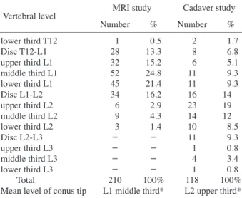
Table 2 showed that the mean level of the conus tip was the middle third of the L1 vertebra in MR study(range,
lower third T12-lower third L2). The termination level in male was significantly higher than that in female(p<0.05).
There was no significant correlation in the mean level of conus tip with age(r= -0.160, p=0.12).
In a cadaver dissection study, the mean level of the conus tip was upper third of L2 vertebra(range, lower third T12- lower third L3). No statistical difference of the termination level between in male and female was found(p=0.911).
There was no significant correlation in the mean level of conus tip with age(r= -0.028, p=0.821).
Discussion
In present study, we found that the mean tip of the conus medullaris in MRI was higher than that in cadaver dissec- tion(middle third L1 in MRI versus upper third L2 in ca- daver). Moreover, age and mean level of the tip of the conus had no correlation both in MR and cadaver studies.
Although we found no sex difference in cadaver dissec- tion, MRI revealed that male tip was higher than female.
Previous MR studies had similar results from our MR study with small variation(Table 1). The location of co- nus medullaris was revealed by MRI of 100 young adults (range, 21-40 years)[1]. The average location was at ver tebra L1 with range from T11-T12 to L2-L3. The T1- weighted, midline, sagittal MRI studies of 504 adult pa- tients were assessed to identify the tip of the conus medul- laris[2]. The mean conus position was the lower third of L1(range from the middle third of T12 to the upper third of L3). Demiryurek et al.[3] revealed that the level of the conus medullaris was most commonly located at the T12- L1 intervertebral disc level in 639 patients using MR im- aging. In Korea, Kim et al.(2003) reported that the conus medullaris was positioned at L1-lower(median), ranged from T12-upper to L3-upper, with MR imaging study [4]. Basically, methodological, racial, and age differences among studies should be considered, developmental dif- ference in spinal cord generation might not be important or revealed in MR imaging which made this study similar to previous MR studies. Sex difference in present MR study could be explained by different mean age between male and female. Because old female might have volume changes of reduction in their vertebral bodies due to aging process.
With respect to cadavers, Hara reported that the average Table 2. Distribution level of tip of the conus medullaris based on
MRI and cadaver studies.
Vertebral level MRI study Cadaver study
Number % Number %
lower third T12 1 0.5 2 1.7
Disc T12-L1 28 13.3 8 6.8
upper third L1 32 15.2 6 5.1
middle third L1 52 24.8 11 9.3
lower third L1 45 21.4 11 9.3
Disc L1-L2 34 16.2 16 14
upper third L2 6 2.9 23 19
middle third L2 9 4.3 14 12
lower third L2 3 1.4 10 8.5
Disc L2-L3 - - 11 9.3
upper third L3 - - 1 0.8
middle third L3 - - 4 3.4
lower third L3 - - 1 0.8
Total 210 100% 118 100%
Mean level of conus tip L1 middle third* L2 upper third*
*P<0.05
caudal level of cord termination located at the interverte- bral disc between L1 and L2 vertebrae or upper third of the L2 vertebra from the cadaver dissection study[11].
In 44(23.4%) among 188 adults, the caudal level was at upper third of the L2 vertebra. In 37.2% of Thai adults (129 cadavers), the spinal cord ended between L1 and L2 lumbar vertebrae[10]. These findings were not far from our cadaver data, which might be explained by gross con- sistency with little difference of cadaver studies among countries. The age of adults might be a factor affecting the extent of the spinal cord[11]. In our study, however, there was no significant correlation in the mean level of conus tip with age in cadaver dissection study. In many standard books, the tip level of the conus medullaris is at the intervertebral disc between the L1 and L2 lumbar ver- tebrae[8,9]. There were, however, no exact descriptions about method whether MRI or cadaver dissection in some books. Because MRI scanner was developed in the 1970s, their data in the books were assumed to be based on ca- daver dissection studies.
Although there are some differences of the tip level according to reports, no definite differences from race or population are confirmed. However, the tip level of the conus medullaris is not seen lower than the third lumbar vertebra in all reports. Therefore, the L3-L4 intervertebral disc space is acceptable for the standard lumbar puncture level in the clinical fields.
We could not understand and reveal exactly why the tip level of conus from MRI study was higher than that from cadaver dissection study. Barson reported that the level of the termination of the conus medullaris in cada- ver might be changed by the extension and flexion of the spinal skeleton[12]. Any changes such as the rigor mortis might be related to this difference. In addition to that, the fixative effect on vertebral muscle and intervertebral disc might shorten the vertebral column, might influence the tip level of conus medullaris. For more exact comparison in the further study, MRI and cadaver dissection studies
should have the same subject which might be helpful to reveal this methodological difference.
REFERENCES
1. Wilson DA, Prince JR. MR imaging determination of the location of the normal conus medullaris throughout child- hood. AJR 1989; 152:1029-32.
2. Saifuddin A, Burnett SJ, White J. The variation of position of the conus medullaris in an adult population. A magnetic resonance imaging study. Spine 1998; 23:1452-6.
3. Demiryurek D, Aydingoz U, Aksit MD, Yener N, Geyik PO.
MR imaging determination of the normal level of conus medullaris. Clin Imaging. 2002; 26:375-7.
4. Kim JT, Bahk JH, Sung J. Influence of age and sex on the position of the conus medullaris and Tuffier’s line in adults.
Anesthesiology. 2003; 99:1359-63.
5. McCotter RE. Regarding the length and extent of the human medulla spinalis. Anat Rec 1916; 26:559-64.
6. Needles JH. The caudal level of termination of the spinal cord in American whites and American Negroes. Anat Rec 1935; 63:417-24.
7. Reimann AF, Anson BJ. Vertebral level of termination of the spinal cord with report of a case of sacral cord. Anat Rec 1944; 83:127-38.
8. Williams PL, Warwick R.(eds) Spinal medulla or cord. In:
Gray’s Anatomy. 36th Ed. New York: Churchill Living- stone, 1980. pp. 864-896.
9. Carpenter MB, Sutin J.(eds) Spinal cord: gross anatomy and internal structure. In: Human neuroanatomy. 8th Ed. Balti- more: Lippincott William & Wilkins, 1983. pp. 232-264.
10. Boonpirak N, Apinhasmit W. Length and caudal level ter- mination of the spinal cord in Thai adults. Acta Anat(Basel) 1994; 149:74-8.
11. Hara K. Changes by age group in the caudal level of ter- mination of the spinal cord in Japanese adults. Acta Anat Nippon 1987; 62:329-33.
12. Barson AJ. The vertebral level of termination of the spinal cord during normal and abnormal development. J Anat 1970; 106:489-97.
해부와 자기공명영상으로 분석한 한국 성인의 척수원뿔 말단부 위치
권순욱
1, 김태식
1, 김형수
2, 유임주
11고려대학교 의과대학 해부학 교실, 2원광대학교 의과대학 영상의학과
간추림 : 척수는 중추신경계의 한 부분으로, 척수의 아래쪽 끝의 이름은 척수원뿔이다. 여러 연구자들이 척수원뿔의
끝 부분을 자기공명영상 연구를 통해 보고하였고, 다른 연구자들은 시신해부연구를 통해 보고하였다. 자기공명영상
연구의 결과가 시신연구 결과보다 더 높은 위치에 있는 것으로 보인다.
우리는 자기공명영상과 시신해부연구를 동시에 진행하여 척수원뿔의 높이를 알아내었다. 자기공명영상은 T1
강조영상이고 정중시상면의 248명을 대상으로 하였다(평균나이 42.3±16.0세, 범위 12~85세). 시신연구는 118 구의 시신을 해부하였다(평균나이 56.0±14.9세, 범위 16~94세).
척수원뿔의 평균 높이는 자기공명연구에서 L1 척추뼈의 중간 정도였다(범위, T12의 아래 1/3에서 L2의 아래 1/3까지). 그리고 시신연구에서는 L2 척추뼈 몸통의 위 1/3 높이에 위치하였다. 척수원뿔의 말단 끝이 자기공명 영상에서 시신연구보다 더 높은 위치로 나타났다(p<0.05).
찾아보기 낱말 : 척수, 척추, 자기공명영상, 한국인
교신저자 : 유임주(고려대학교 의과대학 해부학교실) 전자우편 : irhyu@korea.ac.kr