Long-term Prognosis of Anti-Neutrophil Cytoplasmic Antibody- Negative Renal Vasculitis: Cohort Study in Korea
Few studies have reported on the long-term prognosis of anti-neutrophil cytoplasmic antibody (ANCA)-negative renal vasculitis. Between April 2003 and December 2013, 48 patients were diagnosed with renal vasculitis. Their ANCA status was tested using indirect immunofluorescence and enzyme-linked immunosorbent assays. During a median (interquartile range) follow-up duration of 933.5 (257.5-2,079.0) days, 41.7% of patients progressed to end stage renal disease (ESRD) and 43.8% died from any cause. Of 48 patients, 6 and 42 were ANCA-negative and positive, respectively. The rate of ESRD within 3 months was higher in ANCA-negative patients than in ANCA-positive patients
(P = 0.038). In Kaplan-Meier survival analysis, ANCA-negative patients showed shorter renal survival than did ANCA-positive patients (log-rank P = 0.033). In univariate Cox- proportional hazard regression analysis, ANCA-negative patients showed increased risk of ESRD, with a hazard ratio 3.190 (95% confidence interval, 1.028-9.895, P = 0.045).
However, the effect of ANCA status on renal survival was not statistically significant in multivariate analysis. Finally, ANCA status did not significantly affect patient survival. In conclusion, long-term patient and renal survival of ANCA-negative renal vasculitis patients did not differ from those of ANCA-positive renal vasculitis patients. Therefore, different treatment strategy depending on ANCA status might be unnecessary.
Keywords: Anti-Neutrophil Cytoplasmic Antibody (ANCA); Kidney Failure, Chronic;
Vasculitis; Mortality; Sex; Proteinuria; Koreans; Prognosis; Survival; Cohort Studies Sung Woo Lee,1,2 Mi-Yeon Yu,2
Seon Ha Baek,2 Shin-Young Ahn,1,2 Sejoong Kim,2 Ki Young Na,2,3 Dong-Wan Chae,2,3 and Ho Jun Chin1,2,3
1Department of Immunology, Seoul National University Postgraduate School, Seoul, Korea;
2Department of Internal Medicine, Seoul National University Bundang Hospital, Seongnam, Korea;
3Department of Internal Medicine, Seoul National University College of Medicine, Seoul, Korea Received: 17 October 2015
Accepted: 23 December 2015 Address for Correspondence:
Ho Jun Chin, MD
Department of Internal Medicine, Seoul National University Bundang Hospital, 82 Gumi-ro 173-beon-gil, Bundang-gu, Seongnam 13620, Korea
E-mail: [email protected]
http://dx.doi.org/10.3346/jkms.2016.31.4.542 • J Korean Med Sci 2016; 31: 542-546
INTRODUCTION
Anti-neutrophil cytoplasmic antibody (ANCA) associated vas- culitis is a life-threatening disease characterized by inflamma- tion and necrosis of small to medium-sized vessels (1). Renal vasculitis, also known as pauci-immune necrotizing crescentic glomerulonephritis is a severe form of ANCA associated vascu- litis (2-4). ANCA has recently been proposed to be pathogno- monic in ANCA associated vasculitis due to neutrophil, mono- cyte and endothelial cell activation, which leads to endothelial damage (1). However, patients with vasculitis are not always positive for ANCA. Approximately, 10% of ANCA associated vasculitis patients are ANCA-negative (5-12). To our knowledg- es, no study has focused specifically on the treatment of ANCA- negative patients (5). Moreover, few studies have directly com- pared long-term patient and renal outcomes between ANCA- negative and positive renal vasculitis patients (6,7,9). Hence, the long-term prognosis of ANCA-negative renal vasculitis re- mains unknown (6,7,9). Despite the paucity of evidence, cur- rent guidelines recommend that ANCA-negative receive treat- ments similar to those of ANCA-positive patients (5).
Therefore, the present study compared the long-term prog- noses of ANCA-negative and positive renal vasculitis.
MATERIALS AND METHODS Patients
This retrospective cohort study included 48 patients diagnosed with renal vasculitis at Seoul National University Bundang Hos- pital, a tertiary care hospital, between April 2003 and December 2013. The requirement for informed consent was waived be- cause the study did not infringe the patients’ privacy or health status. Demographic, physiologic, therapeutic and laboratory data were gathered from the electronic medical records data- base. Manual data verification was performed after patient da- tasets were merged. The long-term outcomes included end stage renal disease (ESRD) and all-cause mortality. ESRD de- velopment and all-cause mortality were determined from the ESRD registry database of the Korean Society of Nephrology and the Korea Statistics Service database, respectively.
Measurement and definitions
All 48 patients were tested for ANCA by indirect immunofluo- rescence (IIF) assay, but only 66.7% (32/48) were tested by en- zyme-linked immunosorbent assay (ELISA) for myeloperoxi- dase (MPO) and/or proteinase 3 (PR3). ANCA testing was per- formed following the manufacturer’s instructions by the immu- Nephrology
nology department of Green Cross Laboratories (Yongin-si, Ko- rea). During the study period, the IIF test kit was changed from INOVA Lite ANCA (INOVA Diagnostics, San Diego, California, USA) to Kallestad ANCA IFA (Bio-Rad, Hercules, California, USA) in April 2010, and the results of the two kits were found to be comparable. Quanta Lite (INOVA Diagnostics) was used for the ELISA.
Renal vasculitis was defined as cases which fulfilled the fol- lowing two criteria: 1) evidence of glomerular crescent and/or necrosis but no other explainable pathologic diagnosis, such as IgA nephropathy or lupus nephritis and 2) no evidence of im- mune-complex deposition in immunofluorescence staining and electron microscopic examination. Hypertension was de- fined as systolic blood pressure (BP) ≥ 140 mmHg or diastolic BP ≥ 90 mmHg, use of antihypertensive drugs, or a physician’s diagnosis of hypertension. Diabetes was defined as random glucose ≥ 200 mg/dL, use of insulin and/or oral hypoglycemic agents, or a physician’s diagnosis of diabetes. The disease extent index was scored as described previously (13). The physiologic and laboratory data used for analysis were the worst values ob- served at index admission. Severe proteinuria and hematuria were defined as ≥ 3 positive on the dipstick protein test and
≥ 100 red blood cells per high power field in urine sediment microscopic examination, respectively. The estimated glomer- ular filtration rate (eGFR) was calculated using the equation from the chronic kidney disease epidemiology collaboration
(14).
For renal pathology, each glomerulus was evaluated for the presence of global sclerosis, segmental sclerosis, crescent, and necrosis. Glomerular lesions were reported as a proportion of the total number of glomeruli in each specimen. Glomerular ischemia was defined as the presence of ischemic wrinkling and/or collapse. Glomerular cellularity and size and tubule-in- terstitial lesions were assessed semi-quantitatively as normal to severe (15). Vascular abnormality was defined as the presence of arteriolar hyalinosis and arteriosclerosis.
Statistical analysis
Values were expressed as median (interquartile range, IQR) for continuous variables and No./total (%) for categorical variables.
Difference was analyzed by Mann-Whitney U-test for continu- ous variables and Fisher’s exact test for categorical variables.
The Kaplan-Meier method was used to estimate survival, and statistical significance was determined using the log-rank test.
Univariate and multivariate Cox-proportional hazard regres- sion analyses were performed for the factors related to renal and patient survival. Variables associated with clinical outcomes or ANCA status were entered in the multivariate analysis, along with age and sex. P < 0.05 was considered statistically signifi- cant. All analyses were performed using IBM SPSS for Windows, version 22 (IBM Corp., Armonk, NY, USA).
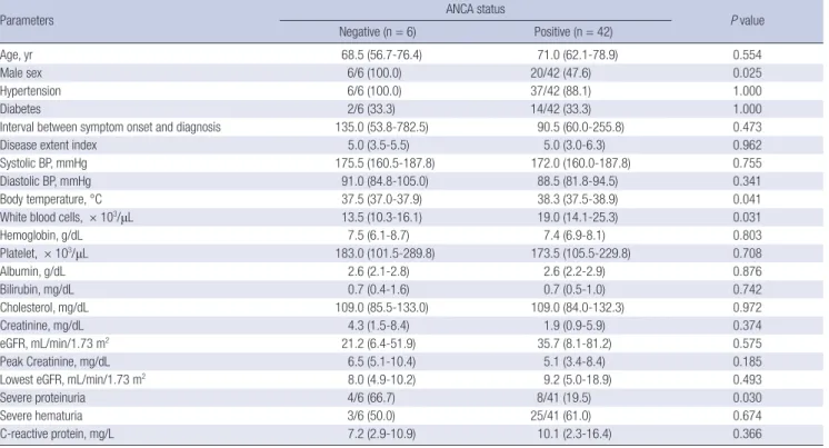
Table 1. Baseline characteristics according to anti-neutrophil cytoplasmic antibody status
Parameters ANCA status
P value
Negative (n = 6) Positive (n = 42)
Age, yr 68.5 (56.7-76.4) 71.0 (62.1-78.9) 0.554
Male sex 6/6 (100.0) 20/42 (47.6) 0.025
Hypertension 6/6 (100.0) 37/42 (88.1) 1.000
Diabetes 2/6 (33.3) 14/42 (33.3) 1.000
Interval between symptom onset and diagnosis 135.0 (53.8-782.5) 90.5 (60.0-255.8) 0.473
Disease extent index 5.0 (3.5-5.5) 5.0 (3.0-6.3) 0.962
Systolic BP, mmHg 175.5 (160.5-187.8) 172.0 (160.0-187.8) 0.755
Diastolic BP, mmHg 91.0 (84.8-105.0) 88.5 (81.8-94.5) 0.341
Body temperature, °C 37.5 (37.0-37.9) 38.3 (37.5-38.9) 0.041
White blood cells, × 103/μL 13.5 (10.3-16.1) 19.0 (14.1-25.3) 0.031
Hemoglobin, g/dL 7.5 (6.1-8.7) 7.4 (6.9-8.1) 0.803
Platelet, × 103/μL 183.0 (101.5-289.8) 173.5 (105.5-229.8) 0.708
Albumin, g/dL 2.6 (2.1-2.8) 2.6 (2.2-2.9) 0.876
Bilirubin, mg/dL 0.7 (0.4-1.6) 0.7 (0.5-1.0) 0.742
Cholesterol, mg/dL 109.0 (85.5-133.0) 109.0 (84.0-132.3) 0.972
Creatinine, mg/dL 4.3 (1.5-8.4) 1.9 (0.9-5.9) 0.374
eGFR, mL/min/1.73 m2 21.2 (6.4-51.9) 35.7 (8.1-81.2) 0.575
Peak Creatinine, mg/dL 6.5 (5.1-10.4) 5.1 (3.4-8.4) 0.185
Lowest eGFR, mL/min/1.73 m2 8.0 (4.9-10.2) 9.2 (5.0-18.9) 0.493
Severe proteinuria 4/6 (66.7) 8/41 (19.5) 0.030
Severe hematuria 3/6 (50.0) 25/41 (61.0) 0.674
C-reactive protein, mg/L 7.2 (2.9-10.9) 10.1 (2.3-16.4) 0.366
Values are expressed as median (interquartile range) for continuous variables and No./total (%) for categorical variables. Difference was analyzed by Mann-Whitney U-test for continuous variables and Fisher’s exact test for categorical variables. Severe proteinuria and hematuria were defined as dipstick urine protein ≥ 3+ and urine red cells ≥ 100/
high power field, respectively. ANCA, anti-neutrophil cytoplasmic antibody; BP, blood pressure; AKI, acute kidney injury; eGFR, estimated glomerular filtration rate.
Ethics statement
This study was approved by the Seoul National University Bun- dang Hospital institutional review board (IRB number: B-1410/
272-119). The requirement for informed consent was waived because the study did not infringe the patients’ privacy or health status.
RESULTS
Of 48 patients, the median (IQR) follow-up duration was 933.5 (257.5-2,079.0) days. The median (IQR) age was 71.0 (61.5-78.8) years, and nearly half of the patients were male (26/48, 54.2%).
During the follow-up period, 21 patients died from any cause (all-cause mortality 43.8%), and 20 progressed to ESRD (ESRD rate 41.7%). Among 48 patients with renal vasculitis, 6 (12.5%) were ANCA-negative and 42 (87.5%) were ANCA-positive.
We compared baseline characteristics according to ANCA status (Table 1). Unlike patients with ANCA, those without AN- CA were predominantly male. Furthermore, ANCA-negative patients had a lower body temperatures and white blood cell counts than ANCA-positive patients. Although the serum creat- inine level and eGFR were similar between ANCA-negative and positive patients, the rate of severe proteinuria was higher in ANCA-negative patients. We also compared pathologic find- ings depending on ANCA status, but there were no statistically significant differences between groups (Table 2). The treatment strategy did not differ between ANCA-negative and positive pa- tients (Table 3).
ANCA-negative patients had a higher rate of ESRD within 3 months than did ANCA-positive patients (Table 3). In Kaplan- Meier survival curves, the renal survival of ANCA-negative pa- tients was significantly shorter than that of ANCA-positive pa- tients: median (95% confidence interval [CI]) 15.0 (0.0-63.0) days vs. 2,941.0 (90.9-5,791.1) days (log-rank P = 0.033, Fig. 1A).
In univariate Cox proportional hazard regression analysis, AN- CA-negative patients showed significantly higher risk of ESRD
than did ANCA-positive patients with a hazard ratio of 3.190 (95% CI, 1.028-9.895, P = 0.045). We performed multivariate analysis to adjust for confounding effects among the variables.
Adjusting only for age did not affect the significance of ANCA status on renal survival. However, after adjusting for sex and se- vere proteinuria, the association between ANCA status and re- nal survival was not statistically significant (Table 4). Patient survival did not differ between groups (Table 3, Fig. 1B).
Table 2. Pathologic finding according to anti-neutrophil cytoplasmic antibody status
Findings ANCA status
P value Negative (n = 6) Positive (n = 42) Number of glomeruli/biopsy 32.0 (19.0-47.0) 28.0 (14.5-49.3) 0.827 Global sclerosis, % 9.8 (4.2-17.4) 13.9 (7.1-45.4) 0.242 Segmental sclerosis, % 0.0 (0.0-13.8) 0.0 (0.0-0.4) 0.512 Crescent, % 52.6 (23.2-88.4) 27.9 (15.0-60.1) 0.156
Necrosis, % 0.0 (0.0-12.5) 0.0 (0.0-15.1) 0.271
Other glomerular finding Increased cellularity Increased size Ischemia
3/6 (50.0) 2/6 (33.3) 0/6 (0.0)
12/42 (28.6) 9/42 (21.4) 4/48 (9.5)
0.360 0.609 1.000 Tubular atrophy
Normal Mild Moderate Severe
0/6 (0.0) 1/6 (16.7) 2/6 (33.3) 3/6 (50.0)
5/42 (14.0) 9/42 (21.4) 7/42 (16.7) 21/42 (50.0)
0.671 1.000 1.000 0.312 1.000 Interstitial inflammation
Normal Mild Moderate Severe
0/5 (0.0) 1/5 (20.0) 1/5 (20.0) 3/5 (60.0)
0/42 (0.0) 5/42 (11.9) 13/42 (31.0) 24/42 (57.1)
0.812 0.511 1.000 1.000 Interstitial fibrosis
Normal Mild Moderate Severe
0/5 (0.0) 1/5 (20.0) 1/5 (20.0) 3/5 (60.0)
10/42 (23.8) 8/42 (19.0) 6/42 (14.3) 18/42 (42.9)
0.663 0.569 1.000 0.571 0.644 Vascular abnormality 3/6 (50.0) 34/42 (81.0) 0.124 Values are median (interquartile range) for continuous variables and No./total (%) for categorical variables. Difference was analyzed by Mann-Whitney U-test for continu- ous variables and Fisher’s exact test for categorical variables. ANCA, anti-neutrophil cytoplasmic antibody.
Table 3. Therapeutic and clinical courses according to anti-neutrophil cytoplasmic antibody status
Status ANCA status
P value
Negative (n = 6) Positive (n = 42)
Plasmapheresis 0/6 (0.0) 8/42 (19.0) 0.571
Steroid use 6/6 (100.0) 39/42 (92.9) 1.000
Steroid dose, g 6.7 (3.4-13.8) 7.7 (3.1-10.9) 0.803
Steroid duration, day 119.0 (50.3-866.8) 185.5 (56.0-572.3) 0.803
Cyclophosphamide use
Oral cyclophosphamide use 6/6 (100.0)
4/6 (66.7) 32/42 (76.2)
29/42 (69.0) 0.320
1.000
Cyclophosphamide dose, g 1.1 (0.9-3.3) 1.2 (0.1-6.6) 0.851
Cyclophosphamide duration, day 23.5 (7.8-56.5) 30.0 (0.8-146.0) 0.802
ESRD 4/6 (66.7) 16/42 (38.1) 0.218
ESRD within 3 months 4/6 (66.7) 9/42 (21.4) 0.038
All-cause mortality 2/4 (33.3) 19/42 (45.2) 0.683
Values were median (interquartile range) for continuous variables and No./total (%) for categorical variables. The difference was analyzed by Mann-Whitney U-test in continuous variables and Fisher’s exact test in categorical variables. ANCA, anti-neutrophil cytoplasmic antibody; ESRD, end stage renal disease.
Table 4. Factors associated with renal survival in Cox-proportional hazard model
Factors Univariate Multivariate* Multivariate† Multivariate‡
HR (95% CI) P HR (95% CI) P HR (95% CI) P HR (95% CI) P
ANCA (- vs. +) 3.190 (1.028-9.895) 0.045 3.200 (1.030-9.938) 0.044 1.923 (0.492-7.522) 0.347 1.568 (0.347-7.081) 0.559 Age, yr§ 1.004 (0.966-1.044) 0.826 1.005 (0.966-1.046) 0.805 1.018 (0.976-1.063) 0.403 1.057 (0.995-1.122) 0.070
Sex (M vs. F) 1.317 (0.536-3.235) 0.549 - - 1.121 (0.406-3.098) 0.825 0.809 (0.228-2.872) 0.743
Severe PU (+ vs. -) 2.859 (1.145-7.138) 0.024 - - 2.833 (0.989-8.118) 0.053 3.314 (1.109-9.903) 0.032
Cr, mg/dL§ 1.213 (1.090-1.349) < 0.001 - - - - 1.328 (1.133-1.556) < 0.001
WBC, 103/μL§ 0.978 (0.922-1.037) 0.460 - - - - 1.015 (0.932-1.105) 0.732
BT, °C§ 0.894 (0.507-1.577) 0.700 - - - - 0.524 (0.165-1.669) 0.274
*Adjusted for age; †Adjusted for age, sex and severe PU; ‡Variables associated with either renal survival or ANCA status were included for the full adjustment; §Per 1 unit increase.
BT, body temperature; Cr, creatinine; PU, proteinuria; WBC, white blood cells; ANCA, anti-neutrophil cytoplasmic antibody; HR, hazard ratio; CI, confidence interval.
Fig. 1. Kaplan-Meier survival curves according to anti-neutrophil cytoplasmic antibody anti-neutrophil cytoplasmic antibody (ANCA) status. Renal and patient survival are shown in (A) and (B), respectively. The gray and black lines represent ANCA-negative and positive groups, respectively.
Renal survival (%)
Follow-up duration (year)
0 2 4 6 8 10 12 100
80
60
40
20
0
Log rank P = 0.033
ANCA positive ANCA negative
Patient survival (%)
Follow-up duration (year)
0 2 4 6 8 10 12 100
80
60
40
20
0
Log rank P = 0.692
ANCA positive ANCA negative
A B
DISCUSSION
Although ANCA is thought to play a pathogenic role in vasculi- tis (1), it is not always positive. According to Kidney Disease: Im- proving Global Outcome guideline, 10% of patients with signs and symptoms of vasculitis are persistently ANCA-negative;
these patients are treated similarly to ANCA-positive patients (5). To our knowledge, only three studies have directly compared the long-term prognosis of ANCA-negative and positive renal vasculitis (6,7,9); however, their findings were not consistent.
Weidner et al. (9) analyzed 80 patients with vasculitis, and show- ed excellent long-term outcome in ANCA negative patients: pa- tients for an average of 46.7 months; in this population, no death or ESRD developed in 6 ANCA-negative patients, but 17 deaths and 18 cases of ESRD were identified in 74 ANCA-positive pa- tients. In contrast, Chen et al. (6) reported poor renal, but simi- lar patient survival in 28 ANCA-negative vasculitis patients, com- pared to 57 ANCA-positive vasculitis patients. Meanwhile, Hung et al. (7) reported similar renal and patient survival between 15 ANCA-negative and 25 ANCA-positive patients. In our study, we found similar patient survival between ANCA-negative and positive renal vasculitis patients after 933.5 days’ follow-up, con- cordant with previous studies (6,7). However, the seemingly poor
renal survival in ANCA-negative renal vasculitis patients could be a result confounded by various factors.
According to our study results, ANCA-negative renal vasculi- tis patients showed shorter renal survival than ANCA-positive renal vasculitis patients. The rate of ESRD within 3 months was significantly higher in ANCA-negative renal vasculitis patients than in ANCA-positive renal vasculitis patients. The hazard of ESRD in ANCA-negative patients was 3.190 times higher than that in ANCA-positive patients. These findings are concordant with those of Chen et al. (6). However, the poor renal outcome in ANCA-negative renal vasculitis patients might result from the confounding effects of sex and proteinuria. In our study, the ANCA-negative patients were predominantly male and had higher rates of severe proteinuria, both of which are known risk factors for the progression of kidney disease (16,17). The fact that the significance of ANCA status on renal survival was not statistically significant after adjusting for sex and proteinuria supports our assumption. The similar clinical presentation and pathologic results between ANCA-negative and positive renal vasculitis patients in our study, which are different from previ- ous studies (6,7), also strengthened the suspicion that the seem- ingly poor renal outcome in ANCA-negative patients is not real.
This study has several limitations. First, because the incidence
of vasculitis is low (18,19), the number of study patients was small.
Because of the small sample size, the differences in clinical pre- sentation and pathologic results between ANCA-negative and positive patients could be under-represented for statistical sig- nificance. However, despite this weakness, renal survival be- tween ANCA-negative and positive patients was significantly different. Moreover, the similarity of clinical and pathologic find- ings between ANCA-negative and positive patients was paral- leled by similar patient and renal survival. Therefore, the prob- lem of small sample size could be acceptable in this respect.
Second, because of the retrospective study design, data such as crescent characteristics or the relapse pattern were unavailable.
Third, the ELISA findings to confirm ANCA and classify wheth- er the ANCA is for MPO or PR3 were not always available. In addition, complete ANCA data during the follow-up period was not available. Finally, the study patients were from a single cen- ter and single nation, which limits the generalizability of the findings.
In conclusion, long-term patient and renal survival of ANCA- negative renal vasculitis patients was not good or bad, compar- ed to that of ANCA positive renal vasculitis patients. The results of our study support current treatment recommendations for ANCA-negative vasculitis patients. However, future large stud- ies are necessary to confirm our results.
DISCLOSURE
The authors have no potential conflicts of interest to disclose.
AUTHOR CONTRIBUTION
Study design: Na KY, Chae DW, Chin HJ. Data production and collection: Lee SW, Na KY, Chin HJ. Data analysis and interpre- tation: Lee SW, Yu MY, Chae DW, Baek SH, Ahn SY, Kim S, Chin HJ. Writing: Lee SW. Review and revision of manuscript: Chin HJ. Approval of final manuscript: all authors.
ORCID
Sung Woo Lee http://orcid.org/0000-0002-4419-3938 Ho Jun Chin http://orcid.org/0000-0003-1185-2631 REFERENCES
1. Wilde B, van Paassen P, Witzke O, Tervaert JW. New pathophysiological insights and treatment of ANCA-associated vasculitis. Kidney Int 2011;
79: 599-612.
2. Tarzi RM, Cook HT, Pusey CD. Crescentic glomerulonephritis: new as- pects of pathogenesis. Semin Nephrol 2011; 31: 361-8.
3. Rutgers A, Sanders JS, Stegeman CA, Kallenberg CG. Pauci-immune nec- rotizing glomerulonephritis. Rheum Dis Clin North Am 2010; 36: 559-72.
4. Han JY, Yoon SA, Woo JY, Park IS, Kim SY, Chang YS, Bang BK. ‘Pauci-im- mune’ rapidly progressive glomerulonephritis associated with systemic vasculitis. J Korean Med Sci 1992; 7: 264-70.
5. Chapter 13: pauci-immune focal and segmental necrotizing glomerulo- nephritis. Kidney Int Suppl (2011) 2012; 2: 233-9.
6. Chen M, Yu F, Wang SX, Zou WZ, Zhao MH, Wang HY. Antineutrophil cy- toplasmic autoantibody-negative Pauci-immune crescentic glomerulo- nephritis. J Am Soc Nephrol 2007; 18: 599-605.
7. Hung PH, Chiu YL, Lin WC, Chiang WC, Chen YM, Lin SL, Wu KD, Tsai TJ. Poor renal outcome of antineutrophil cytoplasmic antibody negative Pauci-immune glomerulonephritis in Taiwanese. J Formos Med Assoc 2006; 105: 804-12.
8. Bajema IM, Hagen EC, Hermans J, Noël LH, Waldherr R, Ferrario F, Van Der Woude FJ, Bruijn JA. Kidney biopsy as a predictor for renal outcome in ANCA-associated necrotizing glomerulonephritis. Kidney Int 1999; 56:
1751-8.
9. Weidner S, Geuss S, Hafezi-Rachti S, Wonka A, Rupprecht HD. ANCA-as- sociated vasculitis with renal involvement: an outcome analysis. Nephrol Dial Transplant 2004; 19: 1403-11.
10. Hauer HA, Bajema IM, van Houwelingen HC, Ferrario F, Noël LH, Wald- herr R, Jayne DR, Rasmussen N, Bruijn JA, Hagen EC, et al. Renal histolo- gy in ANCA-associated vasculitis: differences between diagnostic and se- rologic subgroups. Kidney Int 2002; 61: 80-9.
11. Little MA, Nazar L, Farrington K. Outcome in glomerulonephritis due to systemic small vessel vasculitis: effect of functional status and non-vas- culitic co-morbidity. Nephrol Dial Transplant 2004; 19: 356-64.
12. Hedger N, Stevens J, Drey N, Walker S, Roderick P. Incidence and outcome of pauci-immune rapidly progressive glomerulonephritis in Wessex, UK:
a 10-year retrospective study. Nephrol Dial Transplant 2000; 15: 1593-9.
13. de Groot K, Gross WL, Herlyn K, Reinhold-Keller E. Development and validation of a disease extent index for Wegener’s granulomatosis. Clin Nephrol 2001; 55: 31-8.
14. Levey AS, Stevens LA, Schmid CH, Zhang YL, Castro AF 3rd, Feldman HI, Kusek JW, Eggers P, Van Lente F, Greene T, et al. A new equation to esti- mate glomerular filtration rate. Ann Intern Med 2009; 150: 604-12.
15. Oh SW, Kim S, Na KY, Chae DW, Kim S, Jin DC, Chin HJ. Clinical implica- tions of pathologic diagnosis and classification for diabetic nephropathy.
Diabetes Res Clin Pract 2012; 97: 418-24.
16. Möllsten A, Svensson M, Waernbaum I, Berhan Y, Schön S, Nyström L, Arnqvist HJ, Dahlquist G; Swedish Childhood Diabetes Study Group; Di- abetes Incidence Study in Sweden, et al. Cumulative risk, age at onset, and sex-specific differences for developing end-stage renal disease in young patients with type 1 diabetes: a nationwide population-based co- hort study. Diabetes 2010; 59: 1803-8.
17. Jafar TH, Stark PC, Schmid CH, Landa M, Maschio G, de Jong PE, de Ze- euw D, Shahinfar S, Toto R, Levey AS, et al. Progression of chronic kidney disease: the role of blood pressure control, proteinuria, and angiotensin- converting enzyme inhibition: a patient-level meta-analysis. Ann Intern Med 2003; 139: 244-52.
18. Gatenby PA. Anti-neutrophil cytoplasmic antibody-associated systemic vasculitis: nature or nurture? Intern Med J 2012; 42: 1066-7.
19. Sinico RA, Di Toma L, Radice A. Renal involvement in anti-neutrophil cy- toplasmic autoantibody associated vasculitis. Autoimmun Rev 2013; 12:
477-82.