INTRODUCTION
Among the general population, it has been assumed that an average of 150 to 250 g of lead is ingested through food each day, five to ten percent of which is absorbed (1, 2). The other sources for lead poisoning may be lead-soldered kettles, cans, and lead-glazed pottery, which release lead when acidic fluids are stored or cooked in them (2).
The lead in blood is equilibrated with that of lead in tis- sues, including many potential target organs such as the brain and kidney. The kinetic model of lead in the body is not defined well, but usually it consists of two pools: in the blood and in the skeleton, which may be divided further into labile and deep pools (3). The kidney is the main organ excre- ting lead from pool. Thus, lead burdens have a tendency to be accumulated in the patients with renal failure (4).
The skeleton acts as a reservoir of lead, and it may be mobi- lized by physiological and pathological states including preg- nancy, lactation, and osteoporosis (5, 6). In patients with end- stage renal disease (ESRD), hyperparathyroidism, abnormal vitamin D metabolism, and consequent osteoporosis have frequently been observed (7). Taken together with the lack of renal excretion and stimulated bone resorption, the blood level of lead seems to increase in ESRD and may aggravate uremic symptoms such as peripheral neuropathy and anemia.
The classic clinical manifestations of industrial lead poison- ing include colic, anemia, peripheral neuropathy, encephalopa- thy, renal impairment, hypertension, and reproductive dis- ability (8). More recently, recognition has become widespread that, in addition to its clinically evident toxicity, lead also causes a spectrum of adverse effects at levels of exposure insuf- ficient to produce obvious signs and symptoms. The premise underlying this recognition is that there exist asymptotic, subclinical counterparts. Thus, clinically obvious manifesta- tions of lead poisoning such as anemia, peripheral neuropa- thy, and renal failure lie at the upper end of the range of toxi- city, while such covert effects as impaired synthesis of heme, altered excretion of uric acid, and slowed nerve conduction are their subclinical correlate (9).
Diseases of the peripheral nervous system are among the most prevalent neurologic conditions in ESRD (10). Nonethe- less, differential diagnosis of peripheral nerve disease is chal- lenging because the catalogue of disorders that can produce neuropathies is extensive in ESRD. First of all, many patients with renal failure develop electrophysiologic evidence of peripheral neuropathy (11). The primary pathophysiology of uremic neuropathy remains unknown. According to one theory (12), some slowly dializable substances of intermedi- ate molecular weight alter the peripheral nerve in some man- ner, resulting in axonal dysfunction and eventually degener-
Yeng-Soo Kim, Jae-Ho Park, Joong-Rock Hong, Hyo-Wook Gil, Jong-Oh Yang, Eun-Young Lee, Sae-Yong Hong
Departments of Internal Medicine, Soonchunhyang University Cheonan Hospital, Cheonan, Korea
Address for correspondence Hyo-Wook Gil, M.D.
Department of Internal Medicine, Soonchunhyang University Cheonan Hospital, 23-20 Bongmyung-dong, Cheonan 330-100, Korea
Tel : +82.41-570-3671, Fax : +82.41-574-5762 E-mail : [email protected]
290
Influence of Blood Lead Concentration on the Nerve Conduction Velocity in Patients with End-Stage Renal Disease
Diseases of the peripheral nervous system are the most prevalent in patients with end-stage renal disease (ESRD). Although increased blood levels of lead in ESRD have been reported, the role of lead remains to be elucidated. The purpose of this study was to determine the connection of blood lead concentration with peripheral nerve conduction velocity. One hundred ninety-eight healthy subjects (control group) and 68 patients with ESRD undergoing hemodialysis (ESRD group) were enrolled.
Nerve conduction was measured within two hours after hemodialysis. Orthodromic sensory nerve action potentials and compound muscle action potentials were record- ed on the median, ulnar, and radial nerves. Hemoglobin-corrected blood lead was significantly higher in ESRD patients than in controls (9.1±±2.8 g/dL vs. 5.9±±2.3 g/dL, p<0.001). 32.4% of 68 ESRD patients with diabetes mellitus were significantly related to poorer motor and sensory nerve conduction velocity (p<0.001). However, blood lead was not a significant predictor of the nerve conduction velocity (p>0.05).
Our result suggested that even though the blood lead levels were high in ESRD, they were not associated with the decline of peripheral nerve function. Diabetes mellitus is a primary independent risk of neuropathy in ESRD patients.
Key Words : Lead; Kidney Failure, Chronic; End-stage Renal Disease; Neural Conduction; Diabetes Mellitus;
Peripheral Nervous System Diseases
Received : 28 July 2005 Accepted : 12 October 2005
ation. The other causes of the peripheral neuropathy in ESRD are diabetes mellitus, pharmaceutic neurotoxins, alcohol-nutri- tional neuropathy, and industrial and environmental neuro- toxins such as lead, mercury, arsenic, and thallium.
Although many investigators have described increased blood levels of lead in renal failure patients (13-16), the role of lead on the clinical characteristics remains to be elucidat- ed. The purpose of this study is to determine the blood con- centration of lead in patients with end-stage renal disease and to validate the relation with peripheral motor and sen- sory nerve conduction velocity as clinical correlates.
MATERIALS AND METHODS Subjects
One hundred ninety-eight healthy subjects (control group, 147 men, 51 women; mean age, 43.2±11.5 yr) and 68 patients with ESRD undergoing hemodialysis (ESRD group, 31 men, 37 women; mean age, 50.1±12.3 yr) were enrolled for the blood lead measurement. The control group com- prised healthy volunteers recruited during a regular physi- cal examination at Soonchunhyang University Health Pro- motion Center (Cheonan, Korea). Blood samples for lead con- centration were drawn from the antecubital vein in the morn- ing after overnight fasting.
The ESRD patients were undergoing regular hemodialysis eight to twelve hours weekly using a cellulose acetate hollow- fiber dialyser (surface area 1.2 m2; Gambro, Sweden, or Bax- ter, U.S.A.) at a hemodialysis unit of the Soonchunhyang Cheonan Hospital. In order to avoid any influence of carpal tunnel syndrome and/or ischemic neuropathy on the result of peripheral nerve conduction study, any patients who had vascular access on the right arm and had past histories of cere- brovascular disease, with or without hemiparesis, were ex- cluded from this study. Among 124 ESRD patients on hemo- dialysis, 68 cases were consistent with these terms.
This study was approved by the Institutional Review Board of Soonchunhyang Cheonan Hospital (Cheonan, Korea), and all human subjects provided written informed consent Measurement of nerve conduction velocity
Nerve conduction was measured within two hours after hemodialysis. The upper limb temperatures were maintained above 34℃and lower limb temperatures above 32℃, using hot packs when necessary. Orthodromic sensory nerve action potentials and compound muscle action potentials were recorded in response to supramaximal constant-voltage stim- ulation (duration=0.1 msec; sensory=20 mA, motor=25-35 mA). Stimuli were delivered from a bipolar stimulator; stim- ulating electrodes were 0.8 cm in diameter, and their centers separated by 1.8 cm. Motor and sensory responses were record-
ed with 0.8 cm disk electrodes. The amplifier band pass was 2-10.000 Hz for all nerve conduction study. A Dantec Key- point version 2.0 electrodiagnostic system (Dantec Com, Skovlunde, Denmark) was employed. Studies were performed by two physicians among the authors and were assisted by a technologist.
For excluding idiopathic carpal tunnel syndrome, which could also be caused by diabetes and uremia, we included both the motor conduction study in the wrist-elbow segment of the median and ulnar nerves and the motor and sensory conduction studies in the radial nerve. Results were com- pared with age-specific normative data obtained in the same laboratory.
Measurement of blood lead concentration
Blood samples for lead level, CBC, and general chemistry were drawn from a venous line at the beginning of hemodial- ysis. Blood lead level was analyzed in duplicate with a Zee- man background-corrected atomic absorption spectropho- tometer (Hitachi Z-8100, Japan) with NIOSH’s standard addition method (17) at Soonchunhyang University Insti- tute of Industrial Medicine, a certified reference laboratory for lead in Korea.
Statistical analysis
Data were presented as a mean (SD) for continuous vari- ables and frequency (%) for categorical variables. Pearson’s correlation was applied between outcomes and covariates.
Multiple linear regression was used to determine associa- tions between blood lead and nerve conduction tests, adjust- ing for potential confounders. For easy presentation of results, the final regression models were selected with the same set of confounding variables (i.e., age, sex, duration of hemodial- ysis, Kt/V, and protein catabolic rate). All models were evalu- ated for violation of the assumptions of linear regression. A value of p<0.05 was considered statistically significant, and all statistical analysis was performed using a Stata program (Stata Release 5, College Station, Texas, U.S.A.).
RESULTS
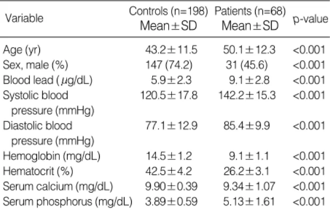
In the control group, mean systolic and diastolic blood pressure were 120.5±17.8 mmHg and 77.1±12.9 mmHg, respectively. Hemoglobin and hematocrit were 14.5±2.2 mg/dL and 42.5±4.2%. Serum calcium and phosphorus were 9.9±0.39 mg/dL and 3.89±0.59 mg/dL (Table 1).
In ESRD group, the median duration of hemodialysis was 42.5 months (range; 1 to 210 months). The causes of renal disease were diabetes mellitus (n=22), hypertension (n=20), polycystic kidney disease (n=1), glomerulonephritis (n=20), and unknown factors (n=7). Kt/V was 1.30±0.27. Normal-
ized protein catabolic rate (nPCR) was 1.2±0.2. Mean sys- tolic blood pressure was 142.2±15.5 mmHg, and mean diastolic blood pressure was 85.4±9.9 mmHg. Sixty two of 68 patients have had anti-hypertensive medications. Sev- enteen cases (25%) were current smokers and 8 patients (11.8%) were regular drinkers (Table 2). The mean serum albumin level was 3.7±0.4 g/dL. Hemoglobin was 9.1± 1.1 mg/dL and hematocrit was 26.2±3.1%. Serum calci- um and phosphorus were 9.34±1.07 mg/dL and 5.13± 1.61 mg/dL, respectively.
Blood lead levels were corrected by hemoglobin (15 mg/
dL) in both groups, and compared. After the correction, blood lead levels were significantly higher in the ESRD group than in control group (p<0.001). All other study variables were sig- nificantly different between the groups (p<0.001) (Table 1).
Summary of nerve conduction tests is displayed in Fig. 1. Blood lead was significantly correlated with duration of hemodial- ysis (p<0.05) (Table 3). However, other study variables were not related to blood lead. Also, motor or sensory nerve con- duction velocity did not show significant correlation with blood lead in ESRD patients (Table 4). The positive correla- tion was observed between nutritional marker, nPCR, and ulnar motor nerve conduction velocity. Also, the positive
Variable Controls (n=198)
Mean±SD
Patients (n=68) Mean±SD p-value
Age (yr) 43.2±11.5 50.1±12.3 <0.001
Sex, male (%) 147 (74.2) 31 (45.6) <0.001 Blood lead ( g/dL) 5.9±2.3 9.1±2.8 <0.001 Systolic blood 120.5±17.8 142.2±15.3 <0.001
pressure (mmHg)
Diastolic blood 77.1±12.9 85.4±9.9 <0.001 pressure (mmHg)
Hemoglobin (mg/dL) 14.5±1.2 9.1±1.1 <0.001 Hematocrit (%) 42.5±4.2 26.2±3.1 <0.001 Serum calcium (mg/dL) 9.90±0.39 9.34±1.07 <0.001 Serum phosphorus (mg/dL) 3.89±0.59 5.13±1.61 <0.001 Table 1.Comparison of general characteristics between con- trols and patients with end-stage renal failure
Variable Mean±SD Range
Duration of dialysis (yr) 3.6±3.4 0-12.0 Antihypertensive medication (%) 55 (90.9)
Diabetes mellitus (%) 22 (32.4)
Albumin (g/dL) 3.7±0.4 2.0-4.4
Normalized protein catabolic rate 1.03±0.38 0.43-3.1
Kt/V 1.30±0.27 0.86-2.06
Current smoker (%) 17 (25.0)
Regular drinker (%) 8 (11.8)
Table 2.Clinical parameters of patients (n=68) with end-stage renal failure
Age Duration NPCR Kt/V Pb
Duration 0.12
NPCR -0.12 0.25*
Kt-V -0.11 0.19 0.19
Pb -0.10 0.25* 0.13 -0.02
Hemoglobin 0.02 0.20 0.15 0.11 0.30*
Table 3.Correlation matrix between selected covariates in 68 patients with end-stage renal failure
*p<0.05. Duration, duration of hemodialysis. NPCR, normalized protein catabolic rate; Pb, hemoglobin corrected blood lead ( g/dL).
Age
Function Nerve Duration Pb Kt/V NPCR
Motor Median -0.20 -0.06 -0.07 0.13 0.12
Ulnar 0.001 0.07 -0.07 0.04 0.30*
Radial 0.07 0.25* 0.12 0.14 0.16
Sensory Median -0.10 0.03 -0.10 0.04 0.09
Ulnar -0.03 0.09 -0.09 0.06 0.12
Radial -0.06 0.04 -0.06 0.25 0.11
Table 4.Correlation between selected covariates and nerve con- duction test in 68 patients with end stage renal failure
*p<0.05. Duration, duration of hemodialysis. NPCR, normalized protein catabolic rate; Pb, hemoglobin corrected blood lead ( g/dL).
Function Nerve
Blood lead ( g/dL) Adjusted coefficient
(p-value)
Diabetes mellitus (No vs. Yes) Adjusted coefficient
(p-value) Motor Median 0.033 (0.883) -7.840 (<0.001)
Ulnar 0.057 (0.847) -8.821 (<0.001) Radial 0.551 (0.303) -7.217 (0.010) Sensory Median 0.314 (0.634) -21.788 (<0.001)
Ulnar 0.161 (0.828) -18.602 (<0.001) Radial 0.511 (0.423) -17.062 (<0.001) Table 5.Results of multiple linear regression of hemoglobin-cor- rected blood lead and diabetes mellitus with each nerve conduc- tion test in 68 patients with end-stage renal failure
*controlling for age, duration of hemodialysis, sex, KT-V, and protein catabolic rate.
m/sec
80
60
40
20
0
Median Ulnar Radial Median Unlar Radial Motor function Sensory function Fig. 1.Box plots of nerve conduction tests in 68 patients with end- stage renal failure.
correlation was observed between duration and radial senso- ry nerve conduction velocity.
In the final models, blood lead was not a significant pre- dictor of poor nerve conduction velocity. Patients with dia- betes mellitus showed significantly worse motor and senso- ry function in all three nerves than those without diabetes mellitus (p<0.01) (Table 5).
DISCUSSION
Lead poisoning may remain asymptomatic for many years.
Major forms of lead poisoning are lead colic, hypertension, and neuropathy. On the other hand, minor symptoms are common and variable: cramps, paresthesia, intermittent pain in the limbs, chronic abdominal pain, and functional digestive disturbances. It is not easy to define these symp- toms in patients with ESRD because uremia itself may cause of and/or aggravate these symptoms. Furthermore, chronic lead poisoning may be a cause ESRD (18-21). The design of our study does not enable us to discuss the role of lead over- load in the onset of ESRD. Determination of such a relation- ship would require epidemiological studies conducted accord- ing to rigorous methodology.
In our results, the blood lead levels were significantly higher, almost twice that of the control group (9.1±2.8 g/
dL vs. 5.9±2.3 g/dL) in ESRD patients. However, con- trary to our expectation, there was no correlation between the blood lead levels and peripheral nerve conduction. Of course, there are many factors influencing the peripheral nerve conduction in ESRD. Approximately 60% of patients with renal failure develop electrophysiologic evidence of periph- eral neuropathy (22). The pathophysiology of uremic neuro- pathy remains unknown. Some slowly dializable substances of intermediate molecular weight have been proposed as pathogens, resulting in axonal dysfunction and eventually degeneration (12). With this point of view, heterogeneity of adequacy in hemodialysis may influence the nerve conduc- tion. But in our study, the Kt/V was 1.30±0.27, and there was no correlation between Kt/V and peripheral nerve con- duction.
Another problem during the evaluation of peripheral nerve conduction is the possibility of carpal tunnel syndrome in patients with ESRD. It is well known that patients with ESRD are apt to suffer from carpal tunnel syndrome (23-25), which may mask the results of peripheral nerve conduction study. The carpal tunnel syndrome is related to a small 2 microglobulin that normally is catabolized by the healthy kidney (26, 27). This substance forms a type of amyloid deposit throughout the body, with particularly adverse con- sequences when it is deposited in the carpal tunnel about the transverse carpal ligament. On the other hand, a profound combined ischemic neuropathy, referred to as an ischemic monomelic neuropathy, may result in patients with limb
ischemia secondary to shunt (22, 28). A significant combined neuropathy may be associated with loss of sensation and mus- cle weakness in the distal arm supplied by the shunted ves- sels. In order to avoid any influence of carpal tunnel syndrome and/or ischemic neuropathy on the result of our peripheral nerve conduction study, we performed the motor conduction study on the wrist-elbow segment of the median and ulnar nerves, and the motor and sensory conduction studies on the radial nerve in the right arm. Any patients who have vascular access in the right arm were excluded from this study.
Alcohol and nutrition are also important factors in periph- eral neuropathy in ESRD (22). In our results, both sensory nerve action potentials and compound muscle action poten- tials were significantly more suppressed in patients with diabetes mellitus than in the non-diabetes mellitus group.
The nutritional markers of nPCR (1.03±0.38) and serum albumin level (3.7±0.4 g/dL) were relatively homogenous among the patients with hemodialysis. In our result, positive correlation was observed between nPCR and ulnar motor nerve conduction. But the other nerve was not correlated.
We guess that malnutrition is important factor about nerve conduction in ESRD patient, but only decreased nPCR may be no prognostic marker about nerve conduction.
The other causes of peripheral neuropathy in ESRD are pharmaceutic neurotoxins and industrial and environmental neurotoxins such as mercury, arsenic, and thallium (22). We did not measure such metals. Further studies are needed for evaluation of peripheral neuropathy in ESRD.
Of course, there is a possibility that the blood lead levels are not high enough to produce the neuropathy in ESRD patients. In the peripheral nervous system, the motor axons are the principal target of lead. Lead-induced pathologic changes in these fibers include segmental demyelination and axonal degeneration (29). Recent studies of the peripheral nerves in persons exposed to lead have used electrophysio- logic probes to determine whether lead causes covert abnor- malities in function. They found slowed conduction in the small motor fibers of the ulnar nerve to be the most sensitive peripheral index of the neurotoxicity of lead and the ulnar nerve conduction velocity was depressed at blood levels below 50 g/dL. In conjunction with this report, our result reveals that, even though the blood lead levels were high in ESRD, they were still below the level of inducing peripheral neuro- pathy.
REFERENCES
1. Nadig R. Lead. In: Toxicologic emergencies, Goldfrank LR, ed. Apple- ton and Lange, 1994; 1029-42.
2. Robertson WO. Chronic poisoning: Trace metal and others, In:
Cecil’s Textbook of Medicine, Goldman L, Bennett JC, eds. W.B Saunders Co., 2000; 70-2.
3. Batschelet E, Brand L, Steiner A. On the kinetics of lead in the human
body. J Math Biol 1979; 8: 15-23.
4. Robinowitz M, Wetherill G, Kopple J. Lead metabolism in the nor- mal human: stable isotope studies. Science 1973; 182: 725-7.
5. Silbergeld EK, Schwartz J, Mahaffey K. Lead and osteoporosis:
mobilization of lead from bone in postmenopausal women. Environ Res 1988; 47: 79-94.
6. Keller CA, Doherty RA. Bone lead mobilization in lactating mice and lead transfer to suckling offspring. Toxicol Appl Pharmacol 1980; 55: 220-8.
7. Llach F, Bover J. Renal osteodystrophies. In: Brenner and Rector’s The Kidney, Brenner BM, ed. W.B Saunders Co., 2000; 2103-66.
8. Hu H. Heavy metal poisoning. In: Harrison’s Principles of Internal Medicine, Fauci AS, et al., eds. McGraw-Hill Co., 1998; 2564-66.
9. Landrigan PJ. Strategies for epidemiologic studies of lead in bone in occupationally exposed populations. Environ Health Perspect 1991;
91: 81-6.
10. Denker BM, Chertow GM, Owen WF. Hemodialysis: neurologic abnormalities. In: Brenner and Rector’s The Kidney. Brenner BM, ed. W.B Saunders Co., 2000; 2420-1.
11. Faster CL, Arieff AI. Nervous system manifestations of renal failure.
In: Diseases of the Kidney, Schrier RW, Gottschalk CW, eds. Little Brown Co., 1993; 2789-809.
12. Nielsen VK. The peripheral nerve function in chronic renal failure.
I. Clinical symptoms and signs. Acta Med Scand 1971; 190: 105-11.
13. Kessler M, Durand PY, Hestin D, Cao Hun T, Renoult E, Prenat E, Chanliau J, Kaminski P, Duc M. Elevated body lead burden from drinking water in end-stage chronic lead failure. Nephrol Dial Trans- plant 1995; 10: 1648-53.
14. Van de Vyver FL, D’Haese PC, Visser WJ, Elseviers MM, Knip- penberg LJ, Lamberts LV, Wedeen RP, De Broe ME. Bone lead in dialysis patients. Kidney Int 1988; 33: 601-7.
15. Kessler M, Durand PY, Hestin D, Gamberoni J, Chanliau J. Diagno- sis and treatment of chronic lead poisoning in CAPD patients. Adv Perit Dial 1993; 9: 143-6.
16. D’Haese PC, Couttenye MM, Lamberts LV, Elseviers MM, Good- man WG, Schrooten I, Cabrera WE, De Broe ME. Aluminum, iron, lead, cadmium, copper, zinc, chromium, magnesium, strontium, and
calcium content in bone of end-stage renal failure patients. Clin Chem 1999; 45: 1548-56.
17. Kneip TJ, Crable JV. Methods for biological monitoring: a manual for assessing human exposure to hazardous substance. American Public Health Association, Washington DC, 1988; 193-201.
18. Thun M, Stayner L, Brown D, Waxweiler R. Mining and deaths from chronic renal failure. Lancet 1982; 2: 606.
19. Danilovic V. Chronic nephritis due to ingestion of lead contaminat- ed flour. Br Med J 1958; 29: 27-8.
20. Lilis R, Gavrilescu N, Nestorescu B, Dumitriu C, Roventa A. Nephro- pathy in chronic lead poisoning. Br J Ind Med 1968; 25: 196-202.
21. Jain VK, Cestero RV, Baum J. Carpal tunnel syndrome in patients undergoing maintenance hemodialysis. JAMA 1979; 242: 2868-9.
22. Dumitru D. Generalized peripheral neuropathies. In: Electrodiag- nostic Medicine, Dumitru D, ed. Hanley and Belfus Inc., 1995; 741- 833.
23. Bardin T, Zingraff J, Kuntz D, Drueke T. Dialysis related amyloido- sis. Nephrol Dial Transplant 1986; 1: 151-4.
24. van Ypersele de strihou C, Honhon B, Vandenbroucke JM, Huaux JP, Noel H, Maldague B. Dialysis amyloidosis. Adv Nephrol Necker Hosp 1988; 17: 401-21.
25. Schwalbe S, Holzhauer M, Schaeffer J, Galanski M, Koch KM, Floege J. Beta(2)-microglobulin associated amyloidosis; a vanishing com- plication of long-term hemodialysis. Kidney Int 1997; 52: 1077-83.
26. Jadoul M, Garbar C, Vanholder R, Sennesael J, Michel C, Robert A, Noel H, van Ypersele de strihou C. Prevalence of histological beta- 2-microglobulin amyloidosis in CAPD patients compared with hemo- dialysis patients. Kidney Int 1998; 54: 956-9.
27. Zingraff J, Noel LH, Bardin T, Atienza C, Zins B, Drueke TB, Kuntz D. Beta-2-microglobulin amyloidosis in chronic renal failure [letter].
N Eng J Med 1990; 323: 1070-1.
28. Bolton CF, Driedger AA, Lindsay RM. Ischemic neuropathy in ure- mic patients caused by bovine arteriovenous shunt. J Neurol Neuro- surg Psychiatry 1979; 42: 810-4.
29. Fullerton PM. Chronic peripheral neuropathy produced by lead poi- soning in guinea pigs. J Neuropathol Exp Neurol 1966; 25: 214-36.