INTRODUCTION
The incidence of primary mucinous adenocarcinoma is low. Many previously diagnosed cases are now retrospective- ly regarded as metastatic mucinous adenocarcinomas or bor- derline mucinous tumors. Primary mucinous adenocarcino- ma is characterized by a large unilateral ovarian mass with a smooth external surface. Young age, an expansile growth pattern, a complex papillary pattern, necrotic luminal debris, and histologic areas resembling benign and borderline muci- nous tumors are characteristic of primary mucinous adenocar- cinoma. Metastatic mucinous adenocarcinoma is more like- ly bilateral and shows a multinodular external surface. The cut surface of the metastatic lesion varies from completely solid to multicystic, mimicking the primary ovary mass. Find- ings of ovarian capsular implants, vascular invasion, a nodu- lar growth pattern, and infiltrative growth of individual glands or single cells on microscopic examination favor metastasis (1-5). The most important and helpful finding to differenti- ate metastatic from primary cancers is knowledge of the clini- cal history of a primary malignancy.
Seidman et al. proposed an algorithm based on tumor size
and laterality in which unilateral ≥10 cm were considered as primary mucinous and all others as metastatic. This algo- rithm correctly classified mucinous adenocarcinomas in 84%
of all 194 cases, including 100% of primary origin tumors and 77% of all metastatic tumors (4). By adjusting the size criterion to 13 cm, the performance of the algorithm was im- proved with correct classification of 87% of tumors, includ- ing 98% of primary tumors and 82% of metastatic tumors (6).
In Korea, ovarian malignancy is the 9th in frequency with annual incidence of 1,300 cases (7). Primary mucinous ade- nocarcinoma of the ovary is diagnosed in less than 10% of ovarian malignancies (8). Intraoperative distinction between primary and metastatic mucinous adenocarcinomas on frozen sections is challenging and has potential for misdiagnosis (9).
For intraoperative consultation of mucinous adenocarcino- ma involving the ovary, it is useful to have additional meth- ods other than the limited traditional microscopic findings to determine the nature of the ovarian mucinous tumors.
Because ovarian mucinous tumors are rarely encountered and notorious for difficulty in distinction between primary and metastatic tumors. Herein, we have re-evaluated the useful- ness of the simple algorithm using the size and laterality cri-
220
Eun Sun Jung1, Jeong Hoon Bae2, Ahwon Lee1, Yeong Jin Choi1, Jong-Sup Park2, and Kyo-Young Lee1
Department of Hospital Pathology1, Obstetrics and Gynecology2, College of Medicine, The Catholic University of Korea, Seoul, Korea
Address for Correspondence Ahwon Lee, M.D.
Department of Hospital Pathology, Seoul St. Mary’s Hospital, The Catholic University of Korea, 641 Banpo-ro, Seocho-gu, Seoul 137-040, Korea Tel : +82.2-2258-1621, Fax : +82.2-2258-1628 E-mail : [email protected]
Mucinous Adenocarcinoma Involving the Ovary: Comparative Evaluation of the Classification Algorithms using Tumor Size and Laterality
For intraoperative consultation of mucinous adenocarcinoma involving the ovary, it would be useful to have approaching methods in addition to the traditional limited microscopic findings in order to determine the nature of the tumors. Mucinous ade- nocarcinomas involving the ovaries were evaluated in 91 cases of metastatic muci- nous adenocarcinomas and 19 cases of primary mucinous adenocarcinomas using both an original algorithm (unilateral ≥10 cm tumors were considered primary and unilateral <10 cm tumors or bilateral tumors were considered metastatic) and a mod- ified cut-off size algorithm. With 10 cm, 13 cm, and 15 cm size cut-offs, the algorithm correctly classified primary and metastatic tumors in 82.7%, 87.3%, and 89.1% of cases and in 80.6%, 84.9%, and 87.1% of signet ring cell carcinoma (SRC) exclud- ed cases. In total cases and SRC excluded cases, 98.0% and 97.2% of bilateral tumors were metastatic and 100% and 100% of unilateral tumors <10 cm were metastatic, respectively. In total cases and SRC excluded cases, 68.4% and 68.4%
of unilateral tumors ≥15 cm were primary, respectively. The diagnostic algorithm using size and laterality, in addition to clinical history, preoperative image findings, and operative findings, is a useful adjunct tool for differentiation of metastatic muci- nous adenocarcinomas from primary mucinous adenocarcinomas of the ovary.
Key Words : Adenocarcinoma, Mucinous; Ovarian Neoplasms; Primary; Metastatic
Received : 2 September 2008 Accepted : 20 March 2009
ⓒ 2010 The Korean Academy of Medical Sciences.
This is an Open Access article distributed under the terms of the Creative Commons Attribution Non-Commercial License (http://creativecommons.org/licenses/by-nc/3.0) which permits unrestricted non-commercial use, distribution, and reproduction in any medium, provided the original work is properly cited.
terion with 10 cm, 13 cm, and 15 cm cut-off values, for dif- ferentiation of primary mucinous adenocarcinoma from meta- static mucinous adenocarcinoma.
MATERIALS AND METHODS
Cases which were diagnosed as either primary mucinous adenocarcinoma or metastatic adenocarcinoma with muci- nous differentiation from 1996 January to 2005 December were retrieved from archives at Catholic University of Korea, Kangnam St. Mary’s Hospital and St. Mary’s Hospital. All slides were reviewed and reclassified according to the 2003 WHO classification. Metastatic adenocarcinoma with more than focal mucin production was classified as metastatic ade- nocarcinoma with mucinous differentiation. Metastatic muci- nous adenocarcinoma with an unknown primary site, bor- derline mucinous tumors, and microinvasive mucinous ade- nocarinomas were excluded. Clinicopathologic data regard- ing age, tumor size, laterality, and follow up data were col- lected. Clinicopathologic data from most metastatic tumors were reported previously (10).
We used a simple algorithm using size and laterality cri- teria that classified all bilateral mucinous carcinomas as meta- static, unilateral mucinous carcinomas <10 cm as metastat- ic, and unilateral mucinous carcinoma ≥10 cm as primary.
We also used a modified algorithm with size cut-offs of 13 cm and 15 cm. Results using the different algorithms were compared with the final diagnosis in each case. Overall sur- vival (OS) was defined as the date from the time of the first diagnosis of ovarian malignancy to death from any cause, or to the date of last contact.
Analyses of overall survival were performed using Kaplan- Meier survival curves. All eligible patients were included in the analyses of OS and all causes of death were included in the calculation of survival.
RESULTS Clinical characteristics
A total of 91 cases of metastatic mucinous adenocarcino- ma and 19 cases of primary mucinous adenocarcinoma from 1996 to 2006 were included in this study to evaluate the algo- rithm using the size and laterality criteria. The mean age of patients with metastatic mucinous adenocarcinoma was 47 yr (median 45, range 19-76) and the mean age of patients with primary mucinous adenocarcinoma was 42 yr (median 40, range 16-71). All primary mucinous adenocarcinomas were surgically staged, except for one case. Stages included stage I in 12 cases, stage III in 5 cases, and stage IV in 1 case.
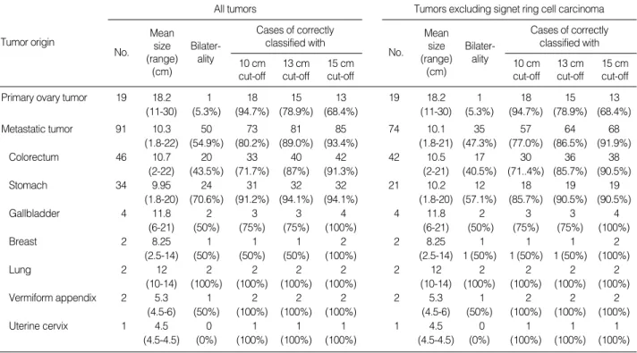
Metastatic mucinous adenocarcinoma cases included those from the colorectum in 46 cases, stomach in 34 cases, gallblad- der in 4 cases, breast in 2 cases, lung in 2 cases, vermiform appendix in 2 cases, and uterine cervix in 1 case. Among meta- static mucinous adenocarcinomas, 17 cases (18.7%) showed more than 50% of signet ring cell differentiation. These 17 cases of signet ring cell carcinoma (SRC) included 13 cases from the stomach and 4 cases from the colorectum.
Size and laterality
Primary mucinous adenocarcinomas were unilateral in 94.7% (18/19) of cases. Metastatic mucinous adenocarcino- mas were bilateral in 54.9% (50/91) of cases. Primary muci- nous adenocarcinomas had a mean size of 18.2 cm (range, 11- 30) and metastatic mucinous adenocarcinomas had a mean size of 10.3 cm (range, 1.8-22).
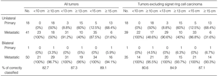
The distribution of primary and metastatic mucinous ade- nocarcinomas classified by tumor size (10 cm, 13 cm, and 15 cm of cut-off values) and laterality (unilateral and bilat- eral) is shown in Table 1. With the 10 cm size cut-off, the algorithm correctly classified primary vs. metastatic muci- nous adenocarcinoma in 82.7% of cases (91/110), including
All tumors
≥10 cm
<10 cm
No. <13 cm ≥13 cm <15 cm ≥15 cm
Tumors excluding signet ring cell carcinoma
≥10 cm
<10 cm
No. <13 cm ≥13 cm <15 cm ≥15 cm Unilateral
Primary 18 0 18 3 15 5 13 18 0 18 3 15 5 13
(0%) (50%) (8.8%) (60%) (12.5%) (68.4%) (0%) (50%) (8.8%) (60%) (12.5%) (68.4%)
Metastatic 41 23 18 31 10 35 6 39 22 17 29 10 33 6
(100%) (50%) (91.2%) (40%) (87.5%) (31.6%) (100%) (48.6%) (90.6%) (40%) (86.8%) (31.6%) Bilateral
Primary 1 0 1 0 1 0 1 1 0 1 0 1 0 1
(0%) (3.3%) (0%) (5%) (0%) (5.9%) (0%) (4.5%) (0%) (6.3%) (0%) (6.7%)
Metastatic 50 21 29 31 19 34 16 35 14 21 20 15 21 14
(100%) (96.7%) (100%) (95%) (100%) (94.1%) (100%) (95.5%) (100%) (93.7%) (100%) (93.3%)
% of correctly 82.7 87.3 89.1 80.6 84.9 87.1
classified
Table 1. Distribution of primary/metastatic mucinous adenocarcinomas based on size with 10 cm, 13 cm, and 15 cm cut off, respec- tively, and laterality
94.7% for primary and 80.2% for metastatic. With the 13 cm size cut-off, the algorithm correctly classified primary vs.
metastatic in 87.3% of cases (96/110), including 78.9% for primary and 89.0% for metastatic. With the 15 cm size cut- off, the algorithm correctly classified primary vs. metastatic in 89.1% of cases (98/110), including 68.4% for primary and 93.4% for metastatic (Tables 1, 2). Of 74 mucinous adenocar- cinomas with a size of <10 cm or with bilateral involvement, 73 cases (98.6%) were metastatic. Of 19 mucinous adenocar- cinomas with a size of ≥15 cm and unilateral involvement, 13 cases (68.4%) were primary. Of 17 mucinous adenocar- cinomas with a size between 10 cm and 15 cm and unilat- eral involvement, 5 cases (29.4%) were primary and 12 cases (70.6%) were metastatic (Table 3). We also evaluated these
algorithms with ovarian mucinous adenocarcinoma exclud- ing SRC, since SRC has characteristic histologic findings that can be easily diagnosed. With the 10 cm size cut-off, the algo- rithm correctly classified primary vs. metastatic mucinous adenocarcinoma in 80.6% of cases (75/93), including 94.7%
for primary and 77.0% for metastatic. With the 13 cm size cut-off, the algorithm correctly classified primary vs. metastat- ic in 84.9% of cases (79/93), including 78.9% for primary and 86.5% for metastatic. With the 15 cm size cut-off, the algorithm correctly classified primary vs. metastatic in 87.1%
of cases (81/93), including 68.4% for primary and 91.9% for metastatic (Tables 1, 2). Of 58 mucinous adenocarcinomas with a size of <10 cm or with bilateral involvement, 57 cases (98.3%) were metastatic. Of 19 mucinous adenocarcinomas
All tumors
No. size <10 cm size 10 to <15 cm size ≥15 cm
Tumors excluding signet ring cell carcinoma No. size <10 cm size 10 to <15 cm size ≥15 cm Unilateral
Primary 18 0 (0%) 5 (29.4%) 13 (68.4%) 18 0 (0%) 5 (31.3%) 13 (68.4%)
Metastatic 41 23 (100%) 12 (70.6%) 6 (31.6%) 39 22 (100%) 11 (68.7%) 6 (31.6%)
Total 59 23 (100%) 17 (100%) 19 (100%) 57 22 (100%) 16 (100%) 19 (100%)
Bilateral
Primary 1 0 (0%) 0 1 (5.9%) 1 0 (0%) 0 1 (6.7%)
Metastatic 50 21 (100%) 13 (100%) 16 (94.1%) 35 14 (100%) 7 (100%) 14 (93.3%)
Total 52 22 (100%) 13 (100%) 17 (100%) 37 15 (100%) 7 (100%) 15 (100%)
Table 3. Distribution of primary/metastatic mucinous adenocarcinomas classified by size <10 cm, 10 to <15 cm and ≥15 cm, and laterality
All tumors
No.
Tumor origin Mean
size (range)
(cm)
Bilater- ality
Cases of correctly classified with 10 cm
cut-off 13 cm cut-off
15 cm cut-off
Tumors excluding signet ring cell carcinoma
No.
Mean size (range)
(cm)
Bilater- ality
Cases of correctly classified with 10 cm
cut-off 13 cm cut-off
15 cm cut-off
Primary ovary tumor 19 18.2 1 18 15 13 19 18.2 1 18 15 13
(11-30) (5.3%) (94.7%) (78.9%) (68.4%) (11-30) (5.3%) (94.7%) (78.9%) (68.4%)
Metastatic tumor 91 10.3 50 73 81 85 74 10.1 35 57 64 68
(1.8-22) (54.9%) (80.2%) (89.0%) (93.4%) (1.8-21) (47.3%) (77.0%) (86.5%) (91.9%)
Colorectum 46 10.7 20 33 40 42 42 10.5 17 30 36 38
(2-22) (43.5%) (71.7%) (87%) (91.3%) (2-21) (40.5%) (71..4%) (85.7%) (90.5%)
Stomach 34 9.95 24 31 32 32 21 10.2 12 18 19 19
(1.8-20) (70.6%) (91.2%) (94.1%) (94.1%) (1.8-20) (57.1%) (85.7%) (90.5%) (90.5%)
Gallbladder 4 11.8 2 3 3 4 4 11.8 2 3 3 4
(6-21) (50%) (75%) (75%) (100%) (6-21) (50%) (75%) (75%) (100%)
Breast 2 8.25 1 1 1 2 2 8.25 1 1 1 2
(2.5-14) (50%) (50%) (50%) (100%) (2.5-14) 1 (50%) 1 (50%) 1 (50%) (100%)
Lung 2 12 2 2 2 2 2 12 2 2 2 2
(10-14) (100%) (100%) (100%) (100%) (10-14) (100%) (100%) (100%) (100%)
Vermiform appendix 2 5.3 1 2 2 2 2 5.3 1 2 2 2
(4.5-6) (50%) (100%) (100%) (100%) (4.5-6) (50%) (100%) (100%) (100%)
Uterine cervix 1 4.5 0 1 1 1 1 4.5 0 1 1 1
(4.5-4.5) (0%) (100%) (100%) (100%) (4.5-4.5) (0%) (100%) (100%) (100%) Table 2. Performance of the algorithm (size of 10 cm cut off and laterality) and modified algorithms (size of 13 cm and 15 cm cut off, respectively, and laterality)
with a size of ≥15 cm and unilateral involvement, 13 cases (68.4%) were primary. Of 16 mucinous adenocarcinomas with a size between 10 cm and 15 cm and unilateral involvement, 5 cases (31.3%) were primary and 11 cases (68.7%) were meta- static (Table 3).
Survival
With a median follow-up period of 11 months (range, 1- 122), 73 out of 110 (66.4%) patients died. Of 19 patients with primary mucinous adenocarcinoma, 17 patients were treated with surgery and adjuvant chemotherapy and two patients were treated with surgery only. The 5-yr overall sur- vival rates were 87% for primary mucinous adenocarcinoma and 6% for metastatic mucinous adenocarcinoma (Fig. 1).
DISCUSSION
Mucinous adenocarcinomas of the ovary are usually large, unilateral, smooth surfaced, multilocular or unilocular cystic masses containing watery or viscous mucoid materal. They are bilateral in 5% of cases (2, 11, 12). The proportion of pri- mary mucinous adenocarcinoma in primary ovarian epithe- lial tumors has been recently reported as 2.4-6.7% (4, 13), which is lower than the previously reported proportion of pri- mary mucinous adenocarcinoma (mean 12%, range 6-25%) (4). The low frequency of primary mucinous adenocarcinoma can be related to a wide variety in the appearance of metastatic mucinous adenocarcinomas of the ovaries. It has been recently recognized that many metastatic carcinomas have been mis- classified as primary. Some mucinous tumors that would have been diagnosed as mucinous adenocarcinomas are diagnosed as intraepithelial and microinvasive mucinous carcinomas and are categorized as mucinous borderline tumors. Pseu- domyxoma peritonei involving ovaries are known to be of
gastrointestinal origin (not ovarian) (4).
The prognosis and treatment modalities of primary muci- nous adenocarcinomas and metastatic mucinous adenocarci- nomas are quite different and an accurate diagnosis is essen- tial for an effective treatment. Because areas of malignancy may be limited, a generous sampling of all mucinous cystic tumors to include up to one histologic section per 1-2 cm of tumor diameter has been recommended to make a diagnosis (14). Sometimes, immunohistochemical staining for CK7, CK20, CDX2, and Dpc4 are helpful to differentiate metas- tatic from primary tumors (15-17). However, at the time of intraoperative consultation, it is not practical to examine mul- tiple sections or to perform immunohistochemical staining.
Therefore, a simple algorithm is useful that correctly classi- fies a high proportion of cases on the basis of easily assessible gross features (4).
A performance evaluation of the algorithm to distinguish primary from metastatic tumors using originally proposed criteria (bilateral tumors of any size, or unilateral tumor <10 cm=metastatic; unilateral tumor ≥10 cm=primary) demon- strated excellent diagnostic performance. Overall, 84-90%
of all tumors were correctly classified (4, 6, 13). Using the same criteria, we found that 91 out of 110 cases (82.7%) were correctly classified. In the subgroup of unilateral tumors ≥10 cm, the proportion of primary mucinous adenocarcinomas varied from 62% to 82% (4, 13). This discrepancy reflects differences in metastatic ovarian tumor sizes between the study populations. Delay prior to visiting hospital for detection of ovarian metastasis leads to larger sized tumors (13).
In our subgroup of unilateral tumors ≥10 cm, the propor- tion of primary mucinous adenocarcinomas was only 50%.
By adjusting the size criterion to 13 cm, and 15 cm, 87.3%
(96 out of 110 cases) and 89.1% (98 out of 110 cases) of cases, respectively, were correctly classified. In the unilateral tumor subgroup with cut-off values of ≥13 cm and ≥15 cm, the proportion of primary mucinous adenocarcinoma increased to 60% and 68.4%, respectively. In Korea, gastric cancer meta- stasis to the ovary is common. Many cases show characteris- tic signet ring cell features of which diagnosis is easily made.
Therefore we evaluated mucinous adenocarcinoma excluding SRC using the algorithms with cut-offs of 10 cm, 13 cm, and 15 cm. The results revealed similar trends with the results of total cases (Tables 1-3).
Using the algorithm with a size cut off of 15 cm, 89.1%
and 87.1% of total cases and SRC excluded cases were cor- rectly classified respectively. All unilateral tumors <10 cm were metastatic but unilateral tumor sizes of 10 cm to <15 cm were metastatic in 70.6% and 68.7% of total cases and SRC excluded cases, respectively. This result supports the need for the modification suggested by Khunamornpong et al. (13) (all bilateral tumors or unilateral tumors <10 cm are metastatic, unilateral tumors ≥15 cm are primary, and uni- lateral tumors with sizes between 10 cm and 15 cm are inde- terminate). Application of this modified algorithm resulted
Fig. 1. Overall survival curves of primary and metastatic mucinous adenocarcinomas.
Survival rate
1.0
0.8
0.6
0.4
0.2
0.0
0 20 40 60 80 100 120
Months
Primary tumor
P=0.000
Metastatic tumor
in correct classification of 86 out of 93 cases (92.5%) and 70 out of 77 cases (90.9%), respectively, and 17 cases (15.5%) and 16 cases (17.2%) remained undetermined. Misclassified cases included 1 case of bilateral primary mucinous adeno- carcinoma and 6 cases of ≥15 cm unilateral metastatic muci- nous adenocarcinomas (4 from the colorectum and 2 from the stomach). Among 6 misclassified and 12 indeterminat- ed metastatic tumors, primary malignancies were detected ahead of an ovarian mass in 5 cases and synchronously with an ovarian mass in 12 cases (Table 4). According to these data, a complete clinical history and a careful search for possible primary tumors in the operation field, especially in the gas- trointestinal tract, is important. When surgeons submit intra- operative frozen samples for examination, they should pro- vide pathologists a clinical history and intraoperative find- ings, such as the status of the opposite ovary.
In conclusion, an algorithm using size and laterality is a useful adjunct tool for differentiation of metastatic mucinous adenocarcinoma from primary mucinous adenocarcinoma of the ovary. However, clinicopathologic evaluation is impor- tant, especially when the tumor is unilateral with a size bet- ween 10 cm and 15 cm.
REFERENCES
1. Harrison ML, Jameson C, Gore ME. Mucinous ovarian cancer. Int J Gynecol Cancer 2008; 18: 209-14.
2. Lee KR, Young RH. The distinction between primary and metastat- ic mucinous carcinomas of the ovary: gross and histologic findings in 50 cases. Am J Surg Pathol 2003; 27: 281-92.
3. Riopel MA, Ronnett BM, Kurmann RJ. Evaluation of diagnostic criteria and behavior of ovarian intestinal-type mucinous tumors:
atypical proliferative (borderline) tumors and intraepithelial, microin- vasive, invasive, and metastatic carcinomas. Am J Surg Pathol 1999;
23: 617-35.
4. Seidman JD, Kurman RJ, Ronnett BM. Primary and metastatic muci- nous adenocarcinomas in the ovaries: incidence in routine practice with a new approach to improve intraoperative diagnosis. Am J Surg Pathol 2003; 27: 985-93.
5. Young RH, Scully RE. Differential diagnosis of ovarian tumors based primarily on their patterns and cell types. Semin Diagn Pathol 2001; 18: 161-235.
6. Yemelyanova AV, Vang R, Judson K, Wu LS, Ronnett BM. Distinc- tion of primary and metastatic mucinous tumors involving the ovary:
analysis of size and laterality data by primary site with reevaluation of an algorithm for tumor classification. Am J Surg Pathol 2008; 32:
128-38.
7. Baker P, Oliva E. A practical approach to intraoperative consultation in gynecological pathology. Int J Gynecol Pathol 2008; 27: 353-65.
8. Ministry of Health and Welfare. Annual report of the central cancer registry in Korea. 2002.
9. Koonings PP, Campbell K, Mishell DR Jr, Grimes DA. Relative fre- quency of primary ovarian neoplasm: a 10-year review. Obstet Gy- necol 1989; 74: 921-6.
10. Lee SJ, Bae JH, Lee AW, Tong SY, Park YG, Park JS. Clinical characteristics of metastatic tumors to the ovaries. J Korean Med Sci 2009; 24: 114-9.
11. Lee KR, Scully RE. Mucinous tumors of the ovary: a clinicopatho- logic study of 196 borderline tumors (of intestinal type) and carci- nomas, including an evaluation of 11 cases with ‘pseudomyxoma peritonei’. Am J Surg Pathol 2000; 24: 1447-64.
12. Guerrieri C, Ho_gberg T, Wingren S, Fristedt S, Simonsen E, Boeryd B. Mucinous borderline and malignant tumors of the ovary. A clini- copathologic and DNA ploidy study of 92 cases. Cancer 1994; 74:
2329-40.
13. Khunamornpong S, Suprasert P, Pojchamarnwiputh S, Na Chiang- mai W, Settakorn J, Siriaunkgul S. Primary and metastatic mucinous adenocarcinomas of the ovary: Evaluation of the diagnostic approach using tumor size and laterality. Gynecol Oncol 2006; 101: 152-7.
14. Lee KR, Tavassoli FA, Prat J, Dietel M, Gersell DJ, Karseladze AI, Hauptmann S, Rutgers J. Surface epithelial-stromal tumors. In: Tavas- soli FA, Devilee P, eds. Pathology & Genetics, Tumors of the breast and female genital organs. Lyon, France: IARC press 2003; 117-45.
15. Vang R, Gown AM, Barry TS, Wheelen DT, Yemelyanova A, Sei- dman JD, Ronnett BM. Cytokeratins 7 and 20 in primary and sec- ondary mucinous tumors of the ovary: analysis of coordinate immuno- histochemical expression profiles and staining distribution in 179 Age
(yr) of patients
Size (cm) Primary
Category site
Site diagnosed
ahead Laterality
Misclassified
Primary (n=1) 69 17 Bilateral
Metastatic Colorectum 49 15 Unilateral Primary focus (n=6) Stomach 43 15 Unilateral Primary focus Colorectum 48 17 Unilateral Primary focus Stomach 76 20 Unilateral Primary focus Colorectum 48 20 Unilateral Synchronous Colorectum 42 21 Unilateral Synchronous Indeterminated
Primary (n=5) 71 11 Unilateral
43 12 Unilateral 21 12 Unilateral 66 14 Unilateral 71 14 Unilateral
Metastatic Colorectum 65 10.3 Unilateral Primary focus (n=12) Colorectum 44 11 Unilateral Primary focus Colorectum 38 11 Unilateral Synchronous Colorectum 47 11.5 Unilateral Primary focus Colorectum 56 12 Unilateral Primary focus Colorectum 45 12 Unilateral Primary focus Colorectum 47 12 Unilateral Synchronous Colorectum 23 13 Unilateral Primary focus Colorectum 50 14 Unilateral Synchronous Stomach 52 11 Unilateral Primary focus Breast 41 14 Unilateral Primary focus Gallbladder 62 14 Unilateral Ovary Table 4. Cases of misclassified and indeterminate by modified algorithms with size <10 cm, 10-15 cm, ≥15 cm and laterality
cases. Am J Surg Pathol 2006; 30: 1130-9.
16. Ji H, Isacson C, Seidman JD, Ronnett BM. Cytokeratins 7 and 20, Dpc4 and MUC5AC in the distinction of metastatic mucinous carci- nomas in the ovary from primary ovarian mucinous tumors: Dpc4 assists in identifying metastatic pancreatic carcinomas. Int J Gynecol Pathol 2002; 21: 391-400.
17. Vang R, Gown AM, Wu LS, Barry TS, Sheeler DT, Yemelyanova A, Seidman JD, Ronnett BM. Immunohistochemical expression of CDX2 in primary ovarian mucinous tumors and metastatic mucinous carcinomas involving the ovary: comparison with CK20 and corre- lation with coordinate expression of CK7. Mod Pathol 2006; 19:
1421-8.