INTRODUCTION
Tissue ischemia was found to mobilize CD34+ cells from the bone marrow into the peripheral blood in an animal model (1), and circulating CD34+ counts significantly increased in patients with acute myocardial infarction (MI) (2). However, an optimum method for harvesting CD34+ cells in acute MI has not yet been determined.
After acute MI, cardiomyocytes are damaged and are grad- ually replaced by fibrosis and scar formation, which leads to the loss of regional contractile function (3). The source of cells used for transplantation into infarcted myocardium varies, e.g., cardiomyocytes (4), skeletal muscle cells (5), smooth muscle cells (6), and bone marrow mononuclear cells (7). A previous study reported that CD34+ cells were incorporated into the border of infarcted myocardium (1), which suggests that they may be used in cell transplantation for infarcted myocardium.
The purpose of this study was to propose the optimal me- thod of harvesting circulating CD34+ cells and to evaluate the potential roles of CD34+ cells in regeneration of infarct- ed myocardium.
MATERIALS AND METHODS Experimental Animals
All surgical procedures were performed under general anes- thesia with electrocardiographic monitoring. Anesthesia for ligating the left anterior descending coronary artery (LAD) was induced and maintained with isoflurane. The anesthetized rats were intubated, and were maintained on positive-pressure ventilation. Rat hearts were exposed through 2 cm left lateral thoracotomy. The rats for sacrifice were anesthetized with ke- tamine hydrochloride (25 mg/kg) and xylazine (3 mg/kg).
The muscle layer and skin incisions were closed with 5-0 Vicryl sutures. Penicillin G benzathine (150,000 U/mL) was given intramuscularly (0.25 mL per rat) once in every 3 days for 1 week postoperatively, and buprenorpine hydrochloride (0.02 mg/kg) was administered subcutaneously once in every 12 hr for the first 48 hr after the operation.
Experimental Donor Groups
In this study, donor rats were categorized into 5 groups: MI, sham, granulocyte-colony stimulating factor (GCSF), MI+
Yong-Hwan Kim
Department of Cardiothoracic Surgery, The Catholic University of Korea College of Medicine, Seoul, Korea
Address for correspondence Yong-Hwan Kim, M.D.
Department of Cardiothoracic Surgery, Uijongbu St.
Mary’s Hospital, The Catholic University of Korea College of Medicine, 65-1 Kumoh-dong, Uijongbu 480-130, Korea
Tel : +82.31-820-3586, Fax : +82.31-847-0301 E-mail : [email protected]
*This study was supported by grants from the Catholic University College of Medicine.
797
Intramyocardial Transplantation of Circulating CD34+ cells:
Source of Stem Cells for Myocardial Regeneration
This study was designed to investigate the increase in the number of circulating CD34+cells after acute myocardial infarction (MI) and the differentiation of these cells to cardiomyocytes after transplantation into infarcted myocardium. The study involved five donor groups: MI (n=27), sham (n=26), granulocyte-colony stimulating factor (GCSF) (n=26), MI+GCSF (n=25), and control (n=25). Acute MI was induced by ligating the left anterior descending coronary arteries (LAD) of donor rats, and LAD of recipient rats were ligated on the same day. Seven days after ligation, CD34+ cells in donor rats were counted and then were directly injected into the infarcted myocardium of recipient rats. Eight weeks after the transplantation, significant differ- ences (p<0.001) were observed in the CD34+cell counts among the 5 donor groups with the greatest increase in the MI+GCSF donor group. In rats receiving CD34+
cells, the size of the scar area smaller (p<0.001) and the thickness of the scar was greater (p=0.001) than in CD34- and saline-transplanted rats. The transplanted CD34+cells differentiated into cardiomyocytes in the scar. This study suggests that CD34+cells may be a potential source of stem cells and that they may be useful in strategies aimed at the regeneration of infarcted myocardium.
Key Words : Antigens, CD34; Cell Transplantation; Stem Cells; Myocardium; Myocardial Infarction;
Regeneration; Granulocyte Colony Stimulating Factor
Received : 26 May 2003 Accepted : 28 August 2003
GCSF, and control rats. Acute MI was induced by ligating the proximal LAD with 6-0 prolene sutures (MI group; n=27).
Seven days after ligation, the whole peripheral blood of each donor rat was obtained from the aorta through a mid-abdom- inal incision. Cells were suspended in phosphate-buffered saline (PBS) after red blood cell lysis and incubated for 15 min at 4℃with anti-CD34 antibody (Miltenyi Biotec, Auburn, CA, U.S.A.). After PBS wash, the cells were loaded onto a Midi-MACS column (Miltenyi) held within a magnetic field to retain CD34+ cells. Unbound cells were eluted with PBS and the retained CD34+ cells were eluted by washing with PBS after the column had been removed from the magnet.
After isolaton, CD34- cells were also collected for cell trans- plantation. CD34+ cells were counted by microscopic enu- meration with a cell-counting hematocytometer (Hausser scientific, Horsham, PA, U.S.A.). Investigators were unaware of which group the cells had been assigned. Rats that under- went the same procedure in the MI group, except for induc- tion of acute MI were assigned to the sham group (n=26). The rats in the GCSF group (n= 26) received GCSF subcutaneously at 40 g/kg/day for 4 days before cell counting. In the MI+
GCSF group (n=25), GCSF was administered as in the GCSF group after inducing MI. The LAD was not ligated, neither GCSF was administered in the control group (n=25).
Experimental Recipient Groups
The LAD of recipient rats was ligated on the same day as in the donor rats. Seven days after ligation, 3 different substances were transplanted into the acute infarcted myocardium of the recipients rats: CD34+ cells (n=20), CD34- cells (n=20), or saline (n=10). CD34+ cells and CD34- cells were obtained from the donor rats.
Cell Labeling and Cell Transplantation
To identify the transplanted cells, CD34+ cells and CD34- cells were labeled with PKH26 (Sigma-Aldrich, St. Louis, MO, U.S.A.) just before transplantation. CD34+ and CD34- cells were suspended in PBS (200 L, 2×10⁶cells) and were injected into several sites in the acute infarcted myocardium with a 28-gauge needle. Half of the suspension was delivered into the center of the infarct, and the other half was divided into 4 or 5 injections at the periphery of the infarct zone, to facilitate potential interaction between the transplanted cells and the normal myocardium. The transplanted zone was mac- roscopically identified by the pale color of the ischemic territory.
Measurement of Remodeling and Histologic Studies The recipient rats were killed 8 weeks after transplantation.
Their hearts were quickly excised, and the atria were removed.
The hearts were fixed with 10% phosphate-buffered formalin and then cut into 3 sections of 5 mm thickness. Each section
was then embedded in paraffin and sectioned to yield 10 m slices. The slices were then stained with hematoxylin and eosin.
For histological studies of each section, the thickened scar was measured at the center. The areas of normal tissue, scar tis- sue, and transplanted tissue in the left ventricular free wall (LVFW) were quantified by computed planimetry (Jandal Scientific Sigma-Scan, Corte Madera, CA, U.S.A.) (4, 8, 9).
The lengths of the LVFW and the scar tissue on both the endo- cardial and epicardial surface of each section were measured.
The surface areas of the epicardial and endocardial scar tissue and the LVFW were measured as the sum of the endocardial length and epicardial length multiplied by the thickness of section (5 mm). The percentage of surface area of scar tissue in the LVFW was calculated as follows:
(epicardial scar size+endocardial scar size)/(epicardial LVFW +endocardial LVFW)×100.
To calculate the percentage of the surface area in the scar tissue occupied by transplanted tissue, the transplanted tissue length in the scar tissue of each section multiplied by the thick- ness of the section (5 mm) was added and then divided by the total scar area multiplied by 100.
For immunohistochemical studies, tissue slices were serially rehydrated in 100%, 90%, 70%, and 50% ethanol after de- paraffinization with xylene. The samples were stained with antibodies against Troponin T-C (Santa Cruz Biotechnology, Santa Cruz, CA, U.S.A.) to identify transplanted cells that had differentiated into cardiomyocytes in the scar area (10).
Statistical Analysis
All data are presented as means±SD. SPSS for Windows (version 10.0) was used for all analyses. One way analysis of variance (ANOVA) followed by the Schffe’s test was used to compare the body weights and the CD34+ cell levels in the 5 donor groups. The comparisons between the recipient groups in terms of body weight, scar area, and scar thickness were conducted by ANOVA. A significance level of 0.05 was used for group comparison.
RESULTS Body Weight of Rats
The body weights of the 5 donor groups and 3 recipient groups are shown in Table 1. No significant differences were observed among the donor groups (p=0.995) or among the 3 recipient groups (p=0.399).
Number of Circulating CD34+ Cells
The median number of CD34+cells before MACS was 2.7
×108/mL of whole blood (range, 1.2-3.5) in MI, 2.3 (range,
0.9-3.4) in sham, 2.9 (1.2-3.5) in GCSF, 3.0 (1.4-3.8) in MI+
GCSF, and 2.2 (0.9-3.3) in control. The CD34+ cell count (106cells/mL of whole blood) demonstrated significant dif- ferences (p<0.001) among the 5 donor groups (Table 2): MI (mean±SD, 0.29±0.05, range, 0.20-0.40), sham (0.15± 0.04, 0.05-0.22), GCSF (0.50±0.06, 0.40-0.59), MI+GCSF (0.87±0.06, 0.74-0.98), and control (0.14±0.04, 0.06- 0.20). However, no significant differences were found between the control and sham groups in terms of cell count (p=0.98).
A greater increase was found in the number of CD34+ cells in the MI group than in the sham group or in the control group (p<0.001). In addition, circulating CD34+ cells increased more in the GCSF group than in the MI group (p< 0.001);
and the increase of the CD34+ cells was the greatest in the MI+GCSF group (MI+GCSF vs. GCSF, p<0.001), which was about 3 times higher than that in the MI group.
Morphometric studies
The comparisons of the scar size demonstrated significant
differences (p<0.001) among the 3 recipient group (Table 3).
The size of the scar was smaller in the CD34+ injection group than in the CD34- or saline injection group (CD34+ vs.
CD34-, p=0.002; CD34+ vs. saline, p=0.001). However, the size was not significantly different between the CD34- and saline injection groups (p=0.524).
The comparisons of the thickness of the scar also demonstrat- ed significant differences (p=0.001) among the 3 recipient groups (Table 4); the thickness of the scar was greater in the CD34+ injection group than in the CD34- or saline injection group (CD34+ vs. CD34-, p=0.003; CD34+ vs. saline, p=
0.011), though no significant difference was found between the CD34- and saline injection groups (p=0.976).
Histologic studies
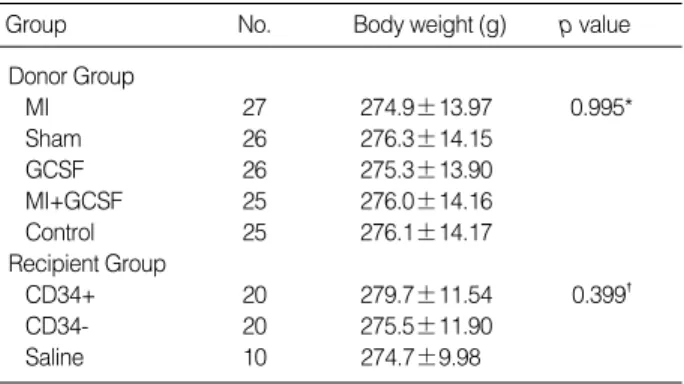
The scar in the control heart was larger and thinner than that in the heart after transplantation (Fig. 1A-D). Histologic evaluation of myocardial sections stained with hematoxylin and eosin demonstrated the transplanted CD34+ cells existed in islands in the myocardial scar tissue, suggesting that they might have differentiated into the cardiomyocytes (Fig. 1E).
Eight weeks after cell transplantation, the cells labeled with PKH were identified within the infarct zone of CD34+ cell transplanted hearts (Fig. 2A). In addition, a large portion of CD34+ cells labeled with PKH26 stained positively for Tro- ponin T-C (Fig. 2B). This finding indicates that the CD34+
cells may differentiate into cardiomyocytes in the scar of the recipient myocardium in vivo. However, no newly formed
MI, myocardial infarction; GCSF, granulocyte colony stimulating factor;
MI+GCSF, GCSF injection after induction of MI.
*,�Not significantly different among the 5 donor and 3 recipient groups.
Group No. Body weight (g) p value
Donor Group
MI 27 274.9±13.97 0.995*
Sham 26 276.3±14.15
GCSF 26 275.3±13.90
MI+GCSF 25 276.0±14.16
Control 25 276.1±14.17
Recipient Group
CD34+ 20 279.7±11.54 0.399�
CD34- 20 275.5±11.90
Saline 10 274.7±9.98
Table 1.Body weights in donor rats and recipient rats
MI, myocardial infarction; GCSF, granulocyte colony stimulating factor MI+GCSF, GCSF injection after inducing MI.
*Significantly different among the 5 donor groups. �Not significantly differ- ent between the sham and the control groups (p=0.98). �A greater in- crease was found in the MI group than in the sham group or in the con- trol group (p<0.001). �Significantly different between the MI and the GCSF groups (p<0.001). ‖Significantly different between the GCSF and the MI+GCSF groups (p<0.001).
Group No. Cell count (106cells/mL of
whole blood) p value*
MI 27 0.29±0.05�,� <0.001
Sham 26 0.15±0.04�,�
GCSF 26 0.50±0.06�,‖
MI+GCSF 25 0.87±0.06‖
Control 25 0.14±0.04�,�
Table 2.Comparisons of CD34+ cell counts among 5 donor groups
*Significantly different among the 3 recipient groups. �Significantly different between the CD34+ and the CD34- cell transplantation groups (p=0.002).
�Significantly different between the CD34+ and the saline transplantation groups (p=0.001). �Not significant between the CD34- and the saline transplantation groups (p=0.524).
Group No. Scar size (%) p value*
CD34+ 20 19.7±3.08�,� <0.001
CD34- 20 22.6±2.23�,�
Saline 10 23.7±1.25�,�
Table 3.Comparisons of the scar size in recipient rats after trans- plantation
*Significantly different among the 3 recipient groups. �Significantly differ- ent between the CD34+ and the CD34- transplantation groups (p=0.003).
�Significantly different between the CD34+ and the saline transplantation groups (p=0.011). �Not significantly different between the CD34- and the saline transplantation groups (p=0.976).
Group No. Scar thickness (mm) p value*
CD34+ 20 1.09±0.18�,� =0.001
CD34- 20 0.89±0.17�,�
Saline 10 0.87±0.19�,�
Table 4.The thickness comparisons of scar in transplanted re- cipient rats
Fig. 1.Photographs of rat hearts from the control (A) and transplan- tation (B) groups 8 weeks after the ligation of the left anterior des- cending artery. The transmural scar tissue in the control heart (C) is larger and thinner than that of the heart after transplantation (D).
Black arrow indicates the region of the microscopic examination.
The transplanted CD34+ cells might have differentiated into car- diomyocytes (T) in the scar tissue (S) (E) (E: H&E, ×100) (M=nor- mal myocardium).
A B
C
E
D
T
S
M
cardiac tissue was present in CD34- cell transplanted hearts.
DISCUSSION
A previous study reported that the number of circulating CD34+ cells was significantly increased in patients with acute MI, whereas it remained unchanged in control subjects who had no evidence of cardiac ischemia (2). Russel et al. (11) re- ported that the number of CD34+ cells was increased by 4- 5 times after mobilization with GCSF. However, the optimal method of harvesting CD34+ cells has not been determined.
To harvest CD34+ cells, this study involved 5 donor groups:
MI, sham, GCSF, MI+GCSF, and a controlas group. The num- ber of CD34+ cells was higher in the MI group than in the control or in the sham group (p<0.001). In addition, a greater number of the cells were present in the GCSF group than in the MI group (p<0.001), and in MI+GCSF group than in the GCSF group (p<0.001). The MI+GCSF cell count was about 3 times higher than the MI cell count.
Asahara and colleagues (12) reported that CD34+ cells, as putative endothelial precursor cells (EPCs), were isolated from human peripheral blood by magnetic bead selection by cell surface antigen expression and were differentiated into mature endothelial cells in vitro. In addition, Asahara et al. (1) demon- strated incorporation of EPCs into the foci of neovasculariza- tion at the border of the myocardial infarction. After tissue ischemia, CD34+ cells were mobilized from the bone marrow and were incorporated into neovascularization sites. These
findings are consistent with the fundamental notions in the postnatal vasculogenesis of CD34+ cells. In addition, the role of CD34+ cells in this pathophysiological response to tissue ischemia may provide strategies for the regeneration of infarct- ed myocardium.
Although the intravenous infusion of CD34+ cells may be systemically easier and simpler than direct myocardial injec- tion, a large number of CD34+ cells are required in the for- mer (13). Therefore, the present study was performed to deter- mine whether this shortcoming could be circumvented by delivering cells directly to the ischemic myocardium. In addi- tion, cryoinjury is often used experimentally to produce myo- cardial necrosis and subsequent scar formation because the transmural scar is more constant in size, and the myocardial dysfunction is less variable than that seen after coronary ligation (14, 15). However, myocardial injury resulting from cryoinjury has limited clinical relevance. Therefore, coronary ligation was selected in this study to induce myocardial ischemia.
Troponin T-C reacts with the cardiac muscle in mouse, rat, and human. However, no cross-reaction was observed in fast skeletal muscle or slow skeletal muscle (10). In the present study, a large portion of CD34+ cells labeled with PKH26 stained positively for Troponin T-C. This finding indicates that the CD34+ cells may differentiate into cardiomyocytes in the scar of recipient myocardium in vivo.
The size of scar was smaller in the CD34 injection group than in the CD34- or saline injection group (Table 3). The thickness of the scar was greater in the CD34+ injection group than in the CD34- or saline injection group (Table 4). The
Fig. 2.(A) Representative fluorescence microscopic findings of the recipient ischemic myocardium 8 weeks after CD34+ cell transplanta- tion. Red fluorescence indicates CD34+ cells labeled with PKH26 just before transplantation (×100). (B) Representative immunohistochem- ical findings for Troponin T-C in the recipient ischemic myocardium 8 weeks after CD34+ cell transplantation. Green fluorescence indicates Troponin T-C binding. A large portion of CD34+ cells labeled with PKH26 stained positively for Troponin T-C (×100).
A B
possible mechanism of the reducing the scar size and increas- ing the scar thickness in the CD34+ cell transplantation is that the CD34+ cells may differentiate into cardiomyocytes and these cardiomyocytes would prevent fibroblast stretching and ventricular enlargement.
According to manufacturer, the CD34+ cells in peripheral blood can be enriched to a purity of about 85-98%. The purity of the isolated hematopoietic progenitor cells can be confirmed by flow cytometry or fluorescence microscopy using an anti- body recognizing an epitope different from that recongnized by the CD34 monoclonal antibody.
AC133+ cells have been known as primitive cells like CD34+ cells. The CD34- cell fraction can contain primitive cells such as AC133+ cells. However, the CD34 antigen dis- appears as the differentiation process progresses (16, 17), CD34- cells rarely include the AC133+ cells. This might explain the observation of why no newly formed cardiac tis- sue in CD34- cell transplanted hearts in this study. However, incorporation of other immunocytochemical stains might show CD34- cells can differentiate into cardiomyocytes. And also some studies reported that CD34+ and AC133+ stem cells can efficiently be isolated to a high purity from GCSF- mobi- lized blood cells (18, 19), and whether CD34-/AC133+ cells can be mobilized using GCSF needs to be determined by fur- ther studies.
A previous study described that functional gap junctions exist between immature CD34+ cells and stromal cells of the microenvironment, and thus may provide an important regulatory pathway in hematopiesis. Further studies are needed to evaluate gap junctions between myocardial cells and trans- planted CD34+ cells.
This study suggests that CD34+ cells transplantation may be a feasible strategy in the management of MI. The number of circulating CD34+ cells may increase significantly, and much more CD34+ cells could be harvested for cell trans- plantation after GCSF administration in patients with MI.
The transplantation of CD34+ cells isolated from peripheral blood may successfully repopulate the ischemic myocardium.
Therefore, CD34+ cell transplantation at the time of coronary artery bypass grafting could reduce scar production, restore the ventricular function of the infarcted region, and ultimately reduce post-infarction mortality and morbidity.
REFERENCES
1. Asahara T, Masuda H, Takahashi T, Kalka C, Pastore C, Silver M, Kearne M, Magner M, Isner JM. Bone marrow origin of endothelial progenitor cells responsible for postnatal vasculogenesis in physiolog- ical and pathological neovascularization. Circ Res 1999; 85: 221-8 2. Shintani S, Murohara T, Ikeda H, Ueno T, Honma T, Katoh A, Sasaki
K, Shimada T, Oike Y, Imaizumi T. Mobilization of endothelial pro- genitor cells in patients with acute myocardial infarction. Circulation 2001; 103: 2776-9.
3. Chiu RCJ, Zibaitis A, Kao RL. Cellular cardiomyoplasty: myocardial regeneration with satellite cell implantation. Ann Thorac Surg 1995;
60: 12-8.
4. Li RK, Jia ZQ, Weisel RD, Mickle DAG, Zhang J, Mohabeer MK, Rao V, Ivanov J. Cardiomyocyte transplantation improves heart func- tion. Ann Thorac Surg 1996; 62: 654-61.
5. Van Meter CH Jr, Claycomb WC, Delcarpio JB, Smith DM, deGruiter H, Smart F, Ochsner JL. Myoblast transplantation in the porcine model: a potential technique for myocardial repair. J Thorac Car- diovasc Surg 1995; 110: 1442-8.
6. Li RK, Jia ZQ, Weisel RD, Merante F, Mickel DAG. Smooth muscle cell transplantation into myocardial scar tissue improves heart func- tion. J Mol Cell Cardiol 1999; 31: 513-22.
7. Kamihata H, Matsubara H, Nishiue T, Fujiyama S, Tsutsumi Y, Ozono R, Masaki H, Mori Y, Iba O, Tateishi E, Kosaki A, Shintani S, Muro- hara T, Imaizumi T, Iwasaka T. Implantation of bone marrow mononu- clear cells into ischemic myocardium enhances collateral perfusion and regional function via side supply of angioblasts, angiogenic lig- ands, and cytokines. Circulation 2001; 104: 1046-52.
8. Fletcher PJ, Pfeffer JM, Pfeffer MA, Braunwald E. Left ventricular diastolic pressure-volume relations in rats with healed myocardia infarction. Effects on systolic function. Circ Res 1981; 49: 618-26.
9. Jugdutt BI, Khan MI. Effect of prolonged nitrate therapy on left ven- tricular remodeling after canine acute myocardial infarction. Circu- lation 1994; 89: 2297-307.
10. Potter JD, Sheng Z, Pan BS, Zhao J. A direct regulatory role for tro- ponin T and a dual role for troponin C in the Ca2+regulation of mucle contraction. J Biol Chem 1995; 270: 2557-62.
11. Russell NH, Gratwohl A, Schmitz N. Developments in allogeneic pe- ripheral blood progenitor cell transplantation. Br J Haematol 1998;
103: 594-600.
12. Asahara T, Murohara T, Sullivan A, Silver M, van der Zee R, Li T, Witzenbichler B, Schatteman G, Isner JM. Isolation of putative pro- genitor endothelial cells for angiogenesis. Science 1997; 275: 964-7.
13. Kocher AA, Schuster MD, Szabolcs MJ, Takuma S. Burkhoff D, Wang J, Homma S, Edwards NM, Itescu S. Neovascularization of ischemic myocardium by human bone-marrow-derived angioblasts prevents cardiomyocyte apoptosis, reduces remodeling and improves cardiac function. Nat Med 2001; 7: 430-6.
14. Murry CE, Wiseman RW, Schwartz SM, Hauschka SD. Skeletal myoblast transplantation for repair of myocardial necrosis. J Clin Invest 1996; 98: 2512-23.
15. Taylor DA, Atkins BZ, Hungspreugs P, Jones TR, Reedy MC, Hutch- eson KA, Glower DD, Kraus WE. Regenerating functional myocardi- um: improved performance after skeletal myoblast transplantation.
Nat Med 1998; 4: 929-33.
16. D’Arena G, Savino L, Nunziata G, Cascavilla N, Matera R, Pistolese G, Carella AM. Immunophenotypic profile of AC133-positive cells in bone marrow, mobilized peripheral blood and umbilical cord blood. Leuk Lymphoma 2002; 43: 869-73.
17. Delfini C, Centis F, Falzetti F, Tabilio A. Expression of CD133 on a human cell line lacking CD34. Leukemia 2002; 16: 2174-5.
18. Gordon PR, Leimig T, Babarin-Dorner A, Houston J, Holladay M, Mueller I, Geiger T, Handgretinger R. Large-scale isolation of CD133+
progenitor cells from G-CSF mobilized peripheral blood stem cells.
Bone Marrow Transplant 2003; 31: 17-22.
19. Jung JT, Sung WJ, Chae YS, Seo KW, Kwon HE, Park SW, Lee NY, Kwak DS, Kim JG, Sohn SK, Suh JS, Lee KB. Peripheral blood stem
cell collection through large volume leukapheresis after mobilization with GM-CSF and/or G-CSF in normal healthy donors. Korean J Hematol 2001; 36: 154-61.