大韓獸醫學會誌 (2015) 第 55 卷 第 1 號 Korean J Vet Res(2015) 55(1) : 57~60 http://dx.doi.org/10.14405/kjvr.2015.55.1.57
57
<Short Communication>
Distribution and accumulation of
177Lu-labeled thermally cross-linked superparamagnetic iron oxide nanoparticles in the tissues of ICR mice
Jin Joo Hue
1,†, Hu-Jang Lee
2,†, Sang Yoon Nam
1, Jong-Soo Kim
1, Beom Jun Lee
1,*, Young Won Yun
1,*
1
College of Veterinary Medicine and Research Institute of Veterinary Medicine, Chungbuk National University, Cheongju 361-763, Korea
2
College of Veterinary Medicine and Institute of Life Science, Gyeongsang National University, Jinju 600-701, Korea (Received: November 10, 2014; Revised: December 11, 2014; Accepted: December 29, 2014)
Abstract : To investigate kinetics of free 177Lu and 177Lu-labeled thermally cross-linked superparamagnetic iron oxide nanoparticles (TCL-SPION), suspensions were intravenously injected into the tail vein of mice at a dose of 5µCi/
mouse or 15 mg/kg body weight, respectively. Free 177Lu radioactivity levels were highest in kidney followed by liver and lung 1 day post-injection. 177Lu-labeled TCL-SPION radioactivity in liver and spleen was significantly higher compared to that of other organs throughout the experimental period (p < 0.05). Radioactivity in blood, brain, and epididymis rapidly declined until 28 days. Based on these results, TCL-SPION could be a safe carrier of therapeutics.
Keywords : accumulation, biodistribution, 177Lu, nanotoxicity, TCL-SPION
Many previous studies were performed to investigate the potential of magnetic nanoparticles as the drug-delivery vehi- cle [1, 7]. This characteristic makes them attractive for many applications, ranging from contrast enhancing agents for MRI to drug-delivery systems [5].
Research on the superparamagnetic iron oxide nanoparti- cles (SPION) has already demonstrated that these nanoparti- cles have the potential becoming an important tool for enhancing magnetic resonance contrast [2, 8]. All of these biomedical applications require that the nanoparticles have high magnetization values, a size smaller than 100 nm, and a narrow particle size distribution [5]. The magnetic nanoparti- cles have many advantages including thermal therapy, guid- ing to the target site, and relatively non-toxic [15]. These applications also need peculiar surface coating of the mag- netic particles, which has to be nontoxic and biocompatible and must allow for a targetable delivery with particle local- ization in a specific area. Such magnetic nanoparticles can bind to drugs, proteins, enzymes, antibodies, or nucleotides and can be directed to an organ, tissue, or tumor using an external magnetic-field [3]. More importantly, iron oxide nanoparticles have a long blood retention time, biodegrad- ability and low toxicity [6, 7]. Recently, anti-biofouling poly- mer-coated TCL-SPION was reported as a novel diagnostic probe for in vivo cancer imaging [9, 10].
Lutetium 177 (
177Lu), a, γ and β emitter radionuclide, is
presently considered useful for a potential bone pain pallia- tion agent owing to its suitable nuclear decay characteristics [ T
1/2= 6.73 days, E
β(max)= 497 keV, E
γ= 113 keV (6.4%) and 208 keV (11%)] and the feasibility of large-scale production with adequate specific activity using moderate flux research reactors [3]. We investigated the distribution and accumula- tion of
177Lu-labeled TCL-SPION and the in vivo kinetics of
177
Lu-labeled TCL-SPION on tissues of mice.
177
Lu ( β = 495 keV, γ = 113 and 208 keV, t
1/2= 6.7 days) purchased from Nuclear Research and Consultancy Group (Netherland).
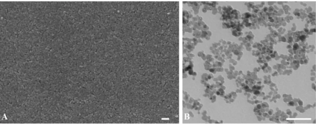
177Lu-labeled TCL-SPION (stability = 98% effi- ciency, period of efficiency = 21 days) was obtained from the Gwangju Institute of Science and Technology (Gwangju, Korea). SEM and TEM analysis of TCL-SPION were shown in Fig. 1. All other chemicals and reagents required for experiments were of analytical grade, and purchased from Sigma-Aldrich Chemical. (USA).
Sixty 5-week old male ICR mice were purchased from Koatec (Korea) at 21.3 ± 1.7 g in mass. Upon arrival, the mice were housed in a temperature- and humidity-controlled environment with a reversed 12/12 h light/dark cycle and free access to food and water. Animal experiments were per- formed in accordance with standard operation procedures of laboratory animals that were approved by the Institutional Animal Care and Use Committee of Chungbuk National Uni- versity (approval no. CBNUR-284-27).
*Corresponding authors
Tel: +82-43-261-2597, Fax: +82-43-267-2595
E-mails: [email protected] (Lee BJ), [email protected] (Yun YW)
†The first two authors equally contributed to this work.
58
Jin Joo Hue, Hu-Jang Lee, Sang Yoon Nam, Young Won Yun, Jong-Soo Kim, Beom Jun LeeAfter 1 week of acclimation, 50 μL of
177Lu-labeled TCL- SPION (15 mg/kg body weight, n = 55) and free
177Lu sus- pensions (5 μCi/mouse, n = 5) were intravenously injected in the tail vein of mice, respectively. At 1 day post-treatment, five mice injected with free
177Lu suspensions were sacri- ficed and collected the blood and various organs (liver, kid- ney, spleen, heart, lung, stomach, small intestine and large intestine). At 0.25, 0.5, 1, 2, 4 h, 1, 3, 7, 14, 21 and 28-day post-injection, five mice injected with
177Lu-labeled TCL- SPION were sacrificed by withdrawing blood from the heart under diethyl ether anesthesia. The various organs (liver, kid- ney, spleen, heart, small intestine, large intestine, lung, stom- ach, thymus, testis, epididymis, lymph node, brain) of each animal were also collected at 0.5, 2 h, 1, 7, 14, 21 and 28-day post-injection. The radioactivity in blood and organs was measured using a gamma counter (PerkinElmer, USA). Sta- tistical analyses were carried out using a one-way analysis of variance (ANOVA) and Student’s t-test. All data were expressed as the mean ± SD. p values < 0.05 were considered to be sta- tistically significant.
Results of the analytical free
177Lu radioactivity in blood and various organs of mice at one day-after treatment were shown in Fig. 1. In the average of free
177Lu radioactivity, the kidney (2.62 ± 1.14%) was first, followed by the liver (2.02
± 0.2%) and the lung (1.11 ± 0.56%), and the blood (0.19 ± 0.06%) was lowest. Due to partial reabsorption of radiola- belled peptides after glomerular filtration, the retention of free
177Lu radioactivity in the radiosensitive kidney is sub- stantial [11]. On the other hand, Persson et al. [12] reported that the mean of free
177Lu radioactivity in the blood was the highest, followed by the kidney at one day post-injection of
177
Lu-labeled pertuzumab into BALB/c mice with the tumor.
This result may be caused by the different of species and dosage of treatment. Fig. 2 shows the change of
177Lu-labeled TCL-SPION level in the blood of mice during 28-day post- injection. At 0.25 h,
177Lu-labeled TCL-SPION radioactivity in blood was 3.571 ± 0.679% injected dose (ID)/g and slowly declined in a time-dependent manner. Table 1 represents the biodistribution of
177Lu-labeled TCL-SPION in blood and
various organs of mice during 28-day post-injection.
177Lu- labeled TCL-SPION radioactivity of liver and spleen was significantly different compared to that of blood and other organs throughout the experimental period ( p < 0.05). The highest
177Lu-labeled TCL-SPION radioactivity in liver and spleen was 36.121 ± 8.239% ID/g at one day post-injection and 15.615 ± 2.225% ID/g at 0.5 h post-injection, respec- tively, and that in kidney, and small and large intestine was observed at 2 h. At 0.5 h post-injection, other organs showed the highest
177Lu-labeled TCL-SPION radioactivity.
In present study,
177Lu-labeled TCL-SPION was immedi- ately entered into the blood and rapidly eliminated during 0.25 h to one-day post-injection and then slowly eliminated thereafter (Fig. 3). This result suggests that the absorbed
177
Lu-labeled TCL-SPION was distributed from systemic cir- culation into the intracellular tissues. In the previous study, ferumoxides cleared very rapidly from plasma (elimination half-life of 6 min in rats) by uptaking of macrophages of the reticulo-endothelial system (mainly Kupffer cells in liver and spleen) [14]. Radioactivity of
77Lu-labeled TCL-SPION in liver and spleen was higher than that in other organs during
Fig. 1. (A) SEM analysis of thermally cross-linked superparamagnetic iron oxide nanoparticles (TCL-SPION). Nanoparticles had a smooth morphology. (B) TEM analysis of TCL-SPION. A: ×50,000, B: ×10,000. Scale bars = 100 nm (A), 50 nm (B).Fig. 2. Biodistribution of free 177Lu activation in blood and var- ious organs of mice at 1 day after treatment of free 177Lu sus- pensions at a dose of 5µCi/mouse. The bars were expressed as means± SD. SI: small intestine, LI: large intestine.
Distribution and accumulation of 177Lu-labeled TCL-SPION
59
experimental periods. Data were expressed as 15.1% ID/g in the liver and 9.1% ID/g in the spleen at 28 days. The iron contained in ferumoxtran-10 was incorporated into the body's iron store and was progressively found in hemoglobin [2]. A biodistribution study of MnMEIO-Herceptin conjugates labeled with radioactive
111In using γ-counter analysis showed that in addition to being distributed within the tumor (3.4% injected dose ID/g), nanoparticles were also found on the liver (12.8%
ID/g), spleen (8.7% ID/g) and muscle (1.0% ID/g) [8]. After intravenous injection, most SPIO nanoparticles were accu- mulated in the Kupffer cells in the liver and in the reticulo- endothelial system in the spleen [4]. Biodistribution of SPIO
(AMI-25) measured by
59Fe and relaxation time studies is consistent with a model of initial vascular distribution of the agent and specific uptake in reticulo-endothelial cells. These results from both techniques indicate similar initial distribu- tion of intravenous administered AMI-25, with 85-95% uptake in the reticulo-endothelial system [14]. In other studies, after SPIO injection approximately 1 day, increases in T
2and T
1values of the precontrast baseline represent the dissolution of the crystalline form necessary for super-paramagnetic behav- ior. Interestingly, spleen displays a second peak of radioactiv- ity after 60 days and clinical observation of reversal of liver and spleen signal intensity strengthened experimental evi- dence and suggested similar metabolism of iron oxide in humans [13]. These results were similar with the results from our study. In conclusion, the TCL-SPION was mainly accu- mulated in the liver and can be used for iron pool from the body iron stores.
Acknowledgments
This work was supported by the research grant of Chung- buk National University in 2012.
References
1. Alexiou C, Schmid RJ, Jurgons R, Kremer M, Wanner G, Bergemann C, Huenges E, Nawroth T, Arnold W, Parak FG. Targeting cancer cells: magnetic nanoparticles as drug carriers. Eur Biophys J 2006, 35, 446-450.
2. Bourrinet P, Bengele HH, Bonnemain B, Dencausse A, Idee JM, Jacobs PM, Lewis JM. Preclinical Safety and pharmacokinetic profile of ferumoxtran-10, an ultrasmall superparamagnetic iron oxide magnetic resonance contrast Table 1. Biodistribution of 177Lu-labeled TCL-SPION during 28 days in blood and various organs of mice intravenously injected with
a single dose of 15 mg/kg body weight
Tissue Time after treatment (% ID/g)
0.5 h 2 h 1 d 7 d 14 d 21 d 28 d
Blood
62.296
± 0.292e61.234
± 0.445de60.044
± 0.019b60.015
± 0.019ab60.010
± 0.028ab < 0.001 < 0.001 Brain60.037
± 0.030a60.018
± 0.005a60.009
± 0.005a60.004
± 0.002a60.002
± 0.001a60.002
± 0.001a < 0.001 Thymus60.564
± 0.167c60.404
± 0.141bc60.203
± 0.066d60.200
± 0.49ef60.053
± 0.029b60.216
± 0.070cd60.115
± 0.041bc Lymph node60.595
± 0.090c60.456
± 0.112bc60.155
± 0.045cd60.142
± 0.040de60.160
± 0.056de60.176
± 0.067c60.181
± 0.052c Heart60.607
± 0.143c60.491
± 0.106bc60.177
± 0.040d60.129
± 0.030de60.108
± 0.030cd60.149
± 0.057c60.087
± 0.035ab Lung61.043
± 0.186d60.893
± 0.109d60.473
± 0.180e60.237
± 0.006f60.206
± 0.055e60.173
± 0.048c60.461
± 0.178d Liver 31.283± 1.087g 35.549± 2.093g 36.121± 8.240h 33.540± 2.740i 24.521± 3.867h 21.507± 1.648f 15.147± 3.518f Spleen 15.615± 2.225f 13.033± 1.280f 14.622± 1.363g 11.446± 1.320h69.269
± 1.320g69.237
± 1.350e69.094
± 0.739e Kidney61.189
± 0.310d61.702
± 0.334e61.066
± 0.291f60.531
± 0.076g60.525
± 0.133f60.335
± 0.063d60.365
± 0.108d Stomach61.286
± 0.690c60.914
± 0.452cde60.187
± 0.057d60.160
± 0.010e60.158
± 0.010e60.132
± 0.068c60.044
± 0.010a Small intestine60.347
± 0.184bc60.487
± 0.130b60.175
± 0.068cd60.080
± 0.028cd60.054
± 0.033bc60.020
± 0.023b60.073
± 0.035ab Large intestine60.327
± 0.078b61.055
± 0.295de60.142
± 0.015d60.047
± 0.022bc60.042
± 0.027b60.027
± 0.013b60.079
± 0.010b Testis60.311
± 0.092b60.291
± 0.089b60.065
± 0.023bc60.053
± 0.007c60.047
± 0.030b60.049
± 0.014b60.051
± 0.016a Epididymis60.661
± 0.130c60.511
± 0.167bc60.101
± 0.021c60.081
± 0.023cd60.003
± 0.001a < 0.001 < 0.001 Means with the different letter in the same column are significantly different (p < 0.05).Fig. 3. Biodistribution of 177Lu-labeled TCL-SPION during 28 days in the blood of ICR mice intravenously injected with sin- gle dose of TCL-SPION 15 mg/kg TCL. The bars were expressed as means± SD.
60
Jin Joo Hue, Hu-Jang Lee, Sang Yoon Nam, Young Won Yun, Jong-Soo Kim, Beom Jun Leeagent. Invest Radiol 2006, 41, 313-324.
3. Chastellain M, Petri A, Gupta A, Rao KV, Hofmann H.
Super paramagnetic silica-iron oxide nanocomposites for application in hyperthermia. Adv Eng Mater 2004, 6, 235- 241.
4. Ferrucci JT, Stark DD. Iron oxide-enhanced MR imaging of the liver and spleen: review of the first 5 years. AJR Am J Roentgenol 1990, 155, 943-950.
5. Gupta AK, Gupta M. Synthesis and surface engineering of iron oxide nanoparticles for biomedical applications.
Biomaterials 2005, 26, 3995-4021.
6. Harisinghani MG, Barentsz J, Hahn PF, Deserno WM, Tabatabaei S, van de Kaa CH, de la Rosette J, Weissleder R. Noninvasive detection of clinically occult lymph-node metastases in prostate cancer. N Engl J Med 2003, 348, 2491-2499.
7. Jain TK, Morales MA, Sahoo SK, Leslie-Pelecky DL, Labhasetwar V. Iron oxide nanoparticles for sustained delivery of anticancer agents. Mol Pharm 2005, 2, 194-205.
8. Lee JH, Huh YM, Jun YW, Seo JW, Jang JT, Song HT, Kim S, Cho EJ, Yoon HG, Suh JS, Cheon J. Artificially engineered magnetic nanoparticles for ultra-sensitive molecular imaging. Nat Med 2007, 13, 95-99.
9. Lee H, Lee E, Kim DK, Jang NK, Jeong YY, Jon S.
Antibiofouling polymer-coated superparamagnetic iron oxide nanoparticles as potential magnetic resonance contrast agents
for in vivo cancer imaging. J Am Chem Soc 2006, 128, 7383-7389.
10. Lee H, Yu MK, Park S, Moon S, Min JJ, Jeong YY, Kang HW, Jon S. Thermally cross-linked superparamagnetic iron oxide nanoparticles: synthesis and application as a dual imaging probe for cancer in vivo. J Am Chem Soc 2007, 129, 12739-12745.
11. Melis M, Krenning EP, Bernard BF, Barone R, Visser TJ, de Jong M. Localisation and mechanism of renal retention of radiolabelled somatostatin analogues. Eur J Nucl Med Mol Imaging 2005, 32, 1136-1143.
12. Persson M, Gedda L, Lundqvist H, Tolmachev V, Nordgren H, Malmström PU, Carlsson J. [177Lu]Pertuzumab:
experimental therapy of HER-2-expressing xenografts. Cancer Res 2007, 67, 326-331.
13. Weissleder R, Bogdanov A, Neuwelt EA, Papisov M.
Long-circulating iron oxides for MR imaging. Adv Drug Deliv Rev 1995, 16, 321-334.
14. Weissleder R, Stark DD, Engelstad BL, Bacon BR, Compton CC, White DL, Jacobs P, Lewis J. Superpara- magnatic iron oxide: pharmacokinetics and toxicity. AJR Am J Roentgenol 1989, 152, 167-173.
15. Yu MK, Jeong YY, Park J, Park S, Kim JW, Min JJ, Kim K, Jon S. Drug-loaded superparamagnetic iron oxide nanoparticles for combined cancer imaging and therapy in vivo. Angew Chem Int Ed Engl 2008, 47, 5362-5365.