Copyright © 2014 The Korean Society of Plastic and Reconstructive Surgeons
This is an Open Access article distributed under the terms of the Creative Commons Attribution Non-Commercial License (http://creativecommons.org/
licenses/by-nc/3.0/) which permits unrestricted non-commercial use, distribution, and reproduction in any medium, provided the original work is properly cited. www.e-aps.org
Original Article
INTRODUCTION
The primary goals in the treatment of zygomatic fractures in- clude the restoration of the projection and the height of the zy- goma by accurate reduction and the restoration of the aesthetic
appearance. Adequate exposure and reduction by multiple inci- sions and strong fixation by plates are believed to be essential to achieving satisfactory results. However, these conventional ap- proaches require a long operation time and may lead to unnec- essary scarring. To overcome these limitations, several new at-
An Anthropometric and Three-Dimensional
Computed Tomographic Evaluation of Two-Point Fixation of Zygomatic Complex Fractures
Taehee Jo, Junhyung Kim
Department of Plastic and Reconstructive Surgery, Keimyung University Dongsan Medical Center, Daegu, Korea
Background Maintaining stability and restoring the aesthetic appearance are the funda
mental goals when managing zygomatic fractures. We aimed to evaluate the stability and anthropometric outcomes of zygomatic fracture patients who underwent twopoint fixation involving the infraorbital rim and zygomaticomaxillary buttress via the transconjunctival and gingivobuccal approaches without any skin incisions.
Methods We examined 15 zygomatic fracture patients who underwent twopoint fixation during a 3year period. Stability was evaluated using threedimensional facial bone computed tomography. Superoinferior and anteroposterior displacement of the zygoma was quantified.
The aesthetic appearance of the periorbital region was evaluated using indirect anthropometry with standardized clinical photographs. The ratios between the eye fissure height and width, and lower iris coverage ratio were used to evaluate aesthetical changes. The bony displacement and aesthetic ratios were analyzed using Wilcoxon or Friedman tests. The correlation between the preoperative zygoma position and anthropometric values was analyzed.
Results The positions of the zygoma were similar to those on the contralateral side at the longterm followup. The preoperative anthropometric measurements on the fractured side differed from those on the contralateral side, although these values were close to the normal values at the longterm followup. Furthermore, we noted that the anteroposterior displace
ment strongly positively correlated with the lower iris coverage rate (Spearman’s coeffici
ent= 0.678, P= 0.005).
Conclusions Twopoint fixation of zygomatic fractures achieved stable outcomes on long
term followup, and also appeared to be reliable in restoring the aesthetic appearance of the periorbital region.
Keywords Zygomatic fractures / Fracture fixation / Anthropometry
Correspondence: Junhyung Kim Department of Plastic and Reconstructive Surgery, Keimyung University School of Medicine, 56 Dalseong-ro, Jung-gu, Daegu 700-712, Korea Tel: +82-53-250-7635 Fax: +82-53-255-0632 E-mail: [email protected]
This article was presented at the 71st congress of Korean society of plastic and reconstructive surgeons, Nov 1–3, 2013, in Seoul, Korea.
No potential conflict of interest relevant to this article was reported.
Received: 28 Mar 2014 • Revised: 24 Jun 2014 • Accepted: 8 Jul 2014
pISSN: 22346163 • eISSN: 22346171 • http://dx.doi.org/10.5999/aps.2014.41.5.493 • Arch Plast Surg 2014;41:493499
tempts have been made in the management of zygomatic frac- tures [1,2].
Previous studies on the appearance of zygomatic fractures were limited to reporting the rates of gross complications such as lower lid retractions, ectropions, and entropions. Smaller changes to the periorbital structures were neglected. Moreover, previous studies on the stability of zygomatic fractures primarily involved experiments on cadaveric heads [3,4], and did not cor- respond with the actual clinical situations [5,6]. The recent widespread use of three-dimensional computed tomography (3D CT) made it easier to verify the position and displacement of the zygomatic body in vivo. Consequently, less aggressive methods to obtain stability were developed [7,8].
Recently, the main concern of patients who experience zygo- matic fractures is whether they can recover an aesthetically pleasing appearance. To minimize scar formation, our institu- tion has been performing two-point fixations involving the in- fraorbital rim and the zygomaticomaxillary buttress for zygo- matic fractures via the transconjunctival and gingivobuccal inci- sions. In the present study, we aimed to evaluate the stability of the zygoma with 3D CT and to evaluate the periorbital anthro- pometric changes using standardized photographs in patients who underwent zygomatic fracture surgeries with the two-point fixation technique.
METHODS
After institutional review board approval, our study included patients who underwent surgical repair of zygomatic fractures at our institution between January 2009 and December 2012.
Two-point fixation of the fractures was accomplished via trans- conjunctival and gingivobuccal incisions. Preoperative and 6-month postoperative 3D CT images were obtained in all pa- tients. Under general anesthesia, the infraorbital rim was ex- posed via a preseptal approach after a transconjunctival incision.
The fracture was fixed using 0.4-mm-thick plates (micro plate, Matrix midface plate, Synthes CMF, West Chester, PA, USA).
In patients with associated orbital floor fractures, a porous poly- ethylene implant (SynPOR, Synthes CMF) was placed after re- duction [9]. The zygomaticomaxillary buttress was exposed by gingivobuccal incision, and fixed using a 0.5-mm-thick plate (mini plate, Matrix midface plate, Synthes CMF) after adequate reduction. Preoperative 3D CT and standardized photographs were obtained for every patient. At the 6-month follow-up, 3D CT was performed and standardized photographs were retaken after consent was obtained from the patients.
After performing maxillofacial tomography scans, the 3D CT images were reconstructed by using three-dimensional recon-
struction software (Definition Flash, Siemens Healthcare, Forchheim, Germany). The craniofacial structures in the three- dimensional images were reoriented according to the reference planes—the Frankfurt horizontal plane and the infraorbital rims.
Frontal view photographs were obtained with a DSLR camera (D300, Nikon, Tokyo, Japan). Patients were consistently posi- tioned according to the Frankfurt line and the intercanthal axis using the cephalostat [10]. Photographic analysis was performed using the Adobe Photoshop CS2 (Adobe Inc., San Jose, CA, USA) measurement software tool by a single plastic surgeon.
The height and the projection of the zygoma complex were evaluated on preoperative and postoperative 3D CT images.
The height was assessed using the frontal view images and was quantified as the vertical difference (VD) ratio of the vertical distance from the supraorbital rim to the infraorbital rim on the fracture side compared to that on the contralateral side (Fig. 1).
When the fractured zygoma is inferiorly displaced, the VD ratio would be greater than 1. The projection of the zygoma was also evaluated in the axial view images and was quantified as the hor- izontal difference (HD) ratio of the distance between the ante- rior margin and the posterior margin of the fossa temporalis on the fractured side compared to that on the contralateral side (Fig. 2). If the fractured zygoma was posteriorly displaced, the HD value would be less than 1.
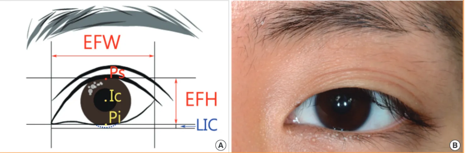
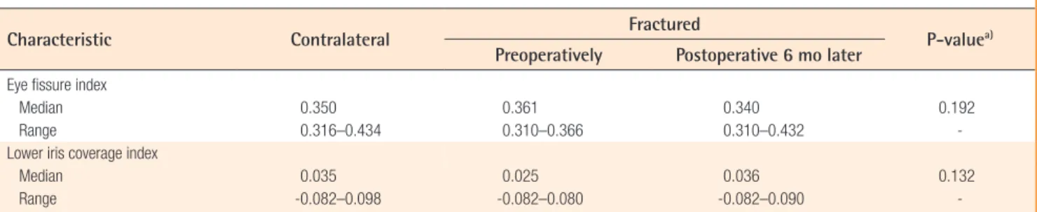
The eye fissure index (EFI) and lower iris coverage index (LIC) were estimated for both the fractured and the contralat- eral sides using the frontal view of standardized photographs.
EFI was defined as the ratio of eye fissure width (EFW) to eye fissure height (EFH). LIC was defined as the percentage of the
The line connecting the supraorbital rim margins on the fractured and on the contralateral side was defined as the horizontal base line. The lowermost point of the infraorbital rim margin was set as point A on the contralateral side, and the distance from the base line was defined as distance a. On the fractured side, these compo- nents were defined as point B and distance b, respectively. The ver- tical difference was defined as the ratio of b to a (b/a).
Fig. 1. Vertical difference on frontal view
iris that was covered by the lower eyelid (Fig. 3).
To evaluate the inter-rater reliability of indirect anthropometry by photogrammetric analysis, we assessed 5 additional patients with zygomatic fractures, before they underwent surgery. Two plastic surgery residents individually photographed the patients, and calculated the EFI and LIC of the fractured and contralater- al sides. The measurements were analyzed by intra-class correla- tion coefficient for interobserver reliability.
To analyze the changes in VD and HD, nonparametric Wil-
coxon test was conducted using the PASW (ver. 18.0, SPSS Inc., Chicago, IL, USA) for Windows. For EFI and LIC, Friedman test was conducted. Moreover, to determine the correlation be- tween the preoperative values of VD and HD and the preopera- tive values of EFI and LIC, correlation analysis was performed using the Spearman’s coefficient value as the correlation coeffi- cient. A P-value of < 0.05 was considered to be statistically sig- nificant. Data are reported as medians and ranges.
RESULTS
Subjects
We included 15 patients (male, 9; female, 6) in the present study. The median age of subjects was 33.5 years (range, 19–64 years).
Measurement reliability
In total, 10 eyes in 5 subjects were analyzed to evaluate the mea- surement reliability of the photogrammetry analysis. The intra- class correlation coefficient was 0.969 for EFI and 0.981 for LIC (P< 0.05). Good reliability was observed for both these mea- surements.
Outcomes
Both the height and the projection on the fractured side were restored to a point close to that on the contralateral side. The VD value was 1.019 (1.000–1.104) preoperatively and was re- stored to 1.003 (0.987–1.010) at the 6-month follow-up exami- nation (P= 0.002). The HD value was 0.912 (0.841–0.973) The line connecting the posterior margins of the fossa temporalis
on the fractured and the contralateral sides was defined as the horizontal base line. The most distant point on the anterior margin was considered as point A on the contralateral side, and the dis- tance from the base line was defined as distance a. On the frac- tured side, these components were defined as point B and distance b, respectively. The horizontal difference was defined as the ratio of b to a (b/a).
Fig. 2. Horizontal difference on axial view
(A) Eye fissure index (EFI) was defined as the ratio of the eye fissure width (EFW) to the eye fissure height (EFH) (EFI = EFW/EFH). EFH was defined as the distance from the margin of the superior palpebra to the margin of the inferior palpebral (Ps-Pi), whereas EFW was defined as the distance from the medial and lateral margin. Moreover, lower iris coverage index (LIC) was defined as the percentage of the iris that was covered by the lower eyelid. The distance from the center point of the iris to the lower eyelid margin (Ic-Pi) is distracted from the radius to calculate the length covered by the eyelid. This value is then divided by the total iris diameter (ID) to yield the LIC value (LIC= [ID/2-(Ic-Pi)]/ID). (B) Partial close-up im- age of the periorbital region on a standardized photograph.
Fig. 3. Diagram indicating the EFI and LIC
A B
preoperatively, and was restored to 0.989 (0.950–1.002) at the 6-month follow-up examination (P= 0.001).
Although not statistically significant, the EFI on the fractured side did not greatly differ from that on the contralateral side be- fore the surgery but the difference between the EFI on the frac- tured side and that on the contralateral side was slightly lower at the final follow-up examination (P= 0.192). The LIC on the fractured side preoperatively was somewhat different to that on the contralateral side. However, at the 6-month follow-up exam- ination, the LIC was restored to a value close to the initial value on the contralateral side (P= 0.132) (Table 1).
The VD preoperatively was not significantly correlated with the EFI or LIC. Moreover, the HD did not significantly corre- late with the EFI; however, the HD indicted a strongly signifi- cant positive correlation with the LIC (Table 2, Fig. 4).
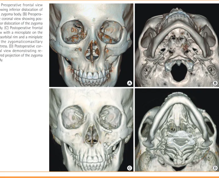
Case
A 32-year-old woman who experienced a zygomatic fracture on the left side underwent two-point fixation. The preoperative CT image indicated inferior and posterior dislocation of the zygoma body along with small zygomaticofrontal suture diastasis. At 6 months postoperatively, CT images indicated that the zygoma was restored to a point adjacent to the normal position (Fig. 5).
Although periorbital swelling was observed preoperatively, the patient indicated that the aesthetic appearance of the periorbital
region was satisfactory at the long-term follow-up (Fig. 6).
DISCUSSION
Zygomatic fractures have traditionally been treated using 3-point fixations involving the zygomaticomaxillary buttress, in- fraorbital rim, and zygomaticofrontal suture [3]. Rohner et al.
Characteristic Contralateral Fractured
P-value
a)Preoperatively Postoperative 6 mo later
Eye fissure index
Median 0.350 0.361 0.340 0.192
Range 0.316–0.434 0.310–0.366 0.310–0.432 -
Lower iris coverage index
Median 0.035 0.025 0.036 0.132
Range -0.082–0.098 -0.082–0.080 -0.082–0.090 -
a)