• 검색 결과가 없습니다.

Increased SOX2 expression in three-dimensional sphere culture of dental pulp stem cells

N/A
N/A
Protected

Academic year: 2021

Share "Increased SOX2 expression in three-dimensional sphere culture of dental pulp stem cells"

Copied!
7
0
0

로드 중.... (전체 텍스트 보기)

전체 글

(1)

Introduction Introduction

Mesenchymal stem cells (MSCs) are adult stem cells residing in the organ-specific supporting tissue and regarded to con- tribute to the organ repair from the injury [1,2]. MSCs display the differentiation potential toward several lineages including adipocytes, osteoblasts, chondrocytes, and organ-specific adult cell types [3]. Dental pulp stem cells (DPSCs) are MSCs residing in dental pulp and can be differentiated to odontoblast and restore the damaged dentin [4,5]. Though the recent lin- eage tracing experiments and the single cell analysis revealed the developmental origin and the constituting cell types of dental pulp [6-8], the exact nature of DPSCs still remains elu-

sive.

Organoid culture has been replacing the sphere culture, both of which have provided the platform for the long-term cul- ture of adult stem cells [9]. In the sphere culture, serum-free minimal condition drives the formation of three-dimensional (3D) cell aggregates in the suspension resulting in the gradual increment of stem cell population during serial passaging whereas the heterogeneity and cellular arrangement of in vivo origin are well-preserved in the organoid culture [10]. Organoid is advantageous in studying the morphogenesis and the loca- tion of adult stem cells, but sphere culture provides the edge in enriching the stem cell pool for the further analysis [11]. As the major types of organoids have been derived from epithelial Int J Oral Biol 45:197-203, 2020

pISSN: 1226-7155 • eISSN: 2287-6618 https://doi.org/10.11620/IJOB.2020.45.4.197

Increased SOX2 expression in three-dimensional sphere culture of dental pulp stem cells

Eun Jin Seo

1

and Il Ho Jang

1,2

*

1

Dental and Life Science Institute, Pusan National University School of Dentistry, Yangsan 50612, Republic of Korea

2

Department of Oral Biochemistry, Pusan National University School of Dentistry, Yangsan 50612, Republic of Korea

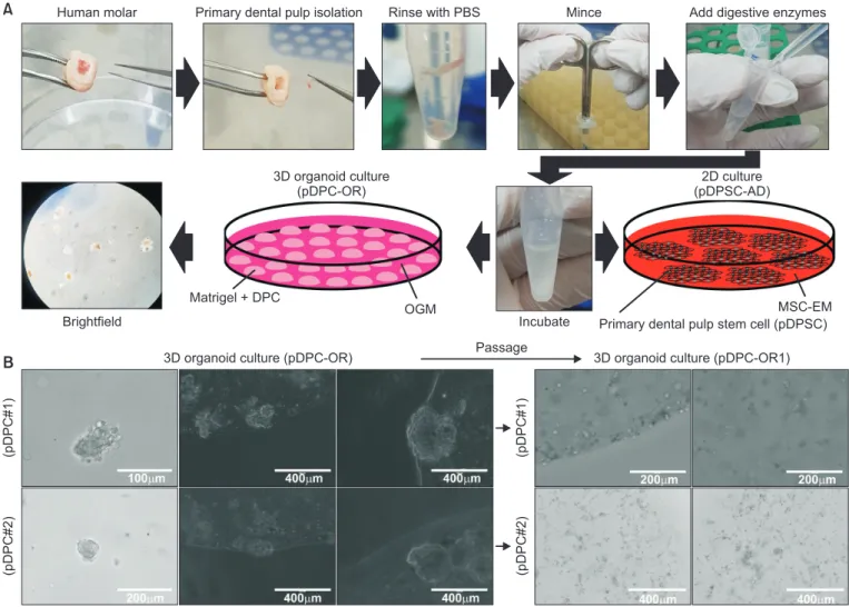
Mesenchymal stem cells in the dental pulp exhibit a tendency for differentiation into various dental lineages and hold great potential as a major conduit for regenerative treatment in dentistry. Although they can be readily isolated from teeth, the exact characteristics of these stem cells have not been fully understood so far. When compared to two- dimensional (2D) cultures, three-dimensional (3D) cultures have the advantage of enriching the stem cell population.

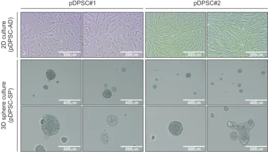
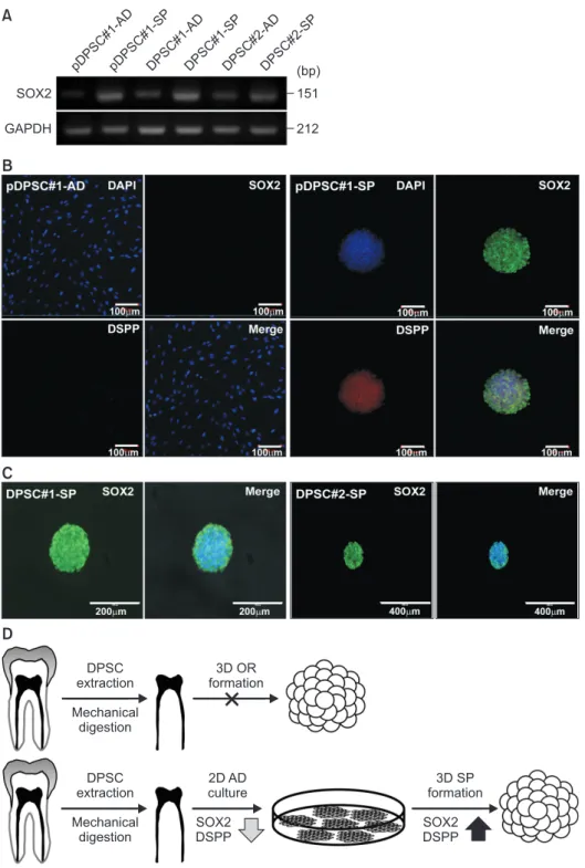
Hence, 3D-organoid culture and 3D-sphere culture were applied to dental pulp cells in the current study. Although the establishment of the organoid culture proved unsuccessful, the 3D-sphere culture readily initiated the stable generation of cell aggregates, which continued to grow and could be passaged to the second round. Interestingly, a significant increase in SOX2 expression was detected in the 3D-spheroid culture compared to the 2D culture. These results indicate the enrichment of the stemness-high population in the 3D-sphere culture. Thus, 3D-sphere culture may act as a link between the conventional and 3D-organoid cultures and aid in understanding the characteristics of dental pulp stem cells.

Keywords: Dental pulp stem cell, SOX2, Three-dimensional culture, Sphere, Organoids

Received November 30, 2020; Revised December 15, 2020; Accepted December 15, 2020

*Correspondence to: Il Ho Jang, E-mail: ilho.jang@pusan.ac.kr https://orcid.org/0000-0002-0820-3035 Copyright © The Korean Academy of Oral Biology

CC

This is an open-access article distributed under the terms of the Creative Commons Attribution Non-Commercial License (http://creativecommons.org/licenses/by- nc/4.0/), which permits unrestricted non-commercial use, distribution, and reproduction in any medium, provided the original work is properly cited.

Original Article IJOB

(2)

tissue, sphere culture may bridge the gap between the con- ventional two-dimensional (2D) culture and in vivo structure- recapitulating organoid culture [12].

Sox2 is a critical transcription factor in pluripotent stem cells and adult stem cells regulating self-renewal and differentiation [13,14]. Sox2 directs the differentiation and the maintenance of neural stem/progenitor cells [15,16]. Sox2 expression is well-documented in the epithelium of cervical loop in mouse incisor where dynamic stem cell activities are detected, but the expression in the dental pulp has not been reported [17]. In MSCs derived from human umbilical cord blood or bone mar- row, SOX2 was shown to regulate stemness and differentiation especially at a low density [18,19]. In DPSCs, overexpression of SOX2 augmented the cellular proliferation, migration and adhesion, which were abolished by siRNA-mediated SOX2 knockdown [20].

In the present study, 3D culture, including organoid and sphere, of DPSCs were tested with an attempt to enrich and characterize the core-stemness population from the conven- tional culture. As a result, the sphere culture of DPSCs was established and exhibited the higher expression of SOX2.

Materials and Methods Materials and Methods

1. Cell culture

Human DPSCs isolated from the third molar of an anony- mous adult male donor and cryopreserved at a primary pas- sage were purchased (PT-5025; Lonza, Basel, Switzerland).

DPSCs were maintained and expanded in MSC expansion media (MSC-EM, Miltenyi Stem MACS MSC Expansion Media Kit XF; Miltenyi Biotec GmbH, Bergisch Gladbach, Germany) with 100 U/mL penicillin-G, 100 µg/mL streptomycin in 15 cm cell culture plates. At passage completion, cells were detached from culture plates using TrypLE

TM

Express (Life Technologies, Carlsbad, CA, USA) for 3 minutes and replaced at a density of 5,000–6,000 cells/cm

2

.

2. Isolation and culture of primary dental pulp stem cells

Molar teeth were obtained from a male (age 17 years) and a female (age 24 years) donors under IRB protocol (PNUDH-2020-003). The current study was performed in ac- cordance with the gender equality guideline of International Journal of Oral Biology. Immediately after extraction, the teeth

were placed in basic media (Dulbecco’s modified Eagle’s me- dia, Gibco, Invitrogen, Carlsbad, CA, USA), transported to the laboratory, and washed with phosphate-buffered saline (PBS, Invitrogen). The tooth surfaces were cleaned, and the pulp chamber was revealed by cutting around the cementoenamel junction with sterilized dental fissure burs. The pulp tissue was gently separated from the teeth and divided into frag- ments approximately 1 mm × 1 mm × 2 mm in size. Primary DPSCs (pDPSCs) were then isolated and cultured by MSC-EM after digestion of fragmented dental pulp tissue in a solution of 3 mg/mL collagenase type I and 4 mg/mL dispase (Sigma, St. Louis, MO, USA) for 30–60 minutes at 37℃ and passing through a 70 µm cell strainer (Falcon; Becton, Dickinson and Company, Franklin Lakes, NJ, USA). Single cell suspensions (1 × 10

5

cells/flask) were seeded in MSC-EM supplemented with 100 U/mL penicillin-G, 100 µg/mL streptomycin, and 0.25 µg/mL Amphotericin B (Fungizone; GIBCO, Grand Island, NY, USA). Cells were maintained at 37℃ in a 5% CO

2

atmosphere.

3. Organoid culture of primary dental pulp cells

Prior to establishing pDPSC culture, dental pulp cells (DPCs) freshly isolated from dental pulp were subjected to organoid culture by resuspending in Growth factor reduced Matrigel (GFR Matrigel, BD Biosciences, Bedford, MA, USA; Catalog No. 354230) and human organoid growth media (OGM, Intes- tiCult

TM

; STEMCELL Technologies, Vancouver, BC, Canada) fol- lowed by drop-plating onto 60 mm Nunclon

TM

Sphera

TM

Dishes (ThermoFisher Scientific, Waltham, MA, USA). OGM was re- placed every 3–4 days.

4. Sphere culture of DPSCs and pDPSCs

Single DPSCs or pDPSCs were resuspended in sphere cul- ture media which consisted of the following: Neurobasal media (Life Technologies) supplemented with 20 ng/mL bFGF, 10 ng/mL EGF, 2.5 µg/mL amphotericin, 100 IU/mL penicil- lin, 100 µg/mL streptomycin, and B-27 Supplement (50×) (Life Technologies, without serum) in Ultra-Low Attachment six- well plates. Fresh media was added every two or three days.

Spheres were transferred to the next generation by dissocia-

tion into single cells with Accutase followed by filtering through

a 40- µm cell strainer and plating at 10

4

cells/mL.

(3)

5. Immunofluorescence staining

For immunofluorescence staining, cells or spheres were fixed in 4% paraformaldehyde in PBS for 10 minutes, washed twice with PBS, and blocked with 1% fetal bovine serum in PBS for 30 minutes. All procedures were performed at 4℃

or room temperature. The fixed specimens were incubated with anti-SOX2 antibody (rabbit polyclonal antibody, Cat No.

ab79351) or anti-Dentin sialophosphoprotein (DSPP) (mouse monoclonal, Cat No. sc-73632) at 4℃ overnight, followed by incubation with secondary antibodies at room temperature for 1 hour. Primary antibodies (1:100) were detected by Alexa Fluor 568-labeled goat anti-rabbit (1:1,000, Invitrogen, Cat No. A11011) or Alexa Fluor 488-labeled donkey anti-mouse

secondary antibody (1:1,000, Invitrogen, Cat No. A32766). The specimens were finally washed and mounted in Vectashield medium (Vector Laboratories, Burlingame, CA, USA) with 4’,6-diamidino-2-phenylindole for visualization of nuclei. The stained sections were visualized using Invitrogen EVOS

TM

FL Auto 2 Imaging System.

6. RNA isolation and quantitative reverse transcription polymerase chain reaction (RT-PCR)

Total RNA was extracted using TRIzol reagent (Sigma-Aldrich, St. Louis, MO, USA) and reverse transcribed into cDNA us- ing the Reverse Transcription cDNA Kit (#RT50KN; NanoHelix, Daejeon, Korea). cDNA in 1 µL of the reaction mixture was am-

A

(pDPC#2) (pDPC#1)

B

MSC-EM Primary dental pulp stem cell (pDPSC) Incubate

3D organoid culture (pDPC-OR)

2D culture (pDPSC-AD)

Matrigel + DPC

OGM Brightfield

Human molar Primary dental pulp isolation Rinse with PBS Mince Add digestive enzymes

3D organoid culture (pDPC-OR) Passage 3D organoid culture (pDPC-OR1)

(pDPC#2) (pDPC#1)

Fig. 1. Isolation of dental pulp cells (DPCs) for establishing two-dimensional (2D) and three-dimensional (3D) culture. (A) Human dental pulp was isolated from tooth, followed by mechanical and enzymatic digestion to single cells. Primary DPCs (pDPCs) were subjected either to 3D organoid culture in organoid growth media (OGM) (pDPC-OR) or to 2D culture (pDPSC-AD) in mesenchymal stem cell expansion media (MSC-EM) for the establishment of dental pulp stem cells. (B) Bright field images of 3D organoid culture from pDPCs are shown (left panels, representative of n = 9 cultures). Bright field images of the sec- ond round of 3D organoid culture after passaging (pDPC-OR1) are shown in the right panels.

PBS, phosphate-buffered saline.

(4)

plified using the Ready-2×-Go pre-mix PCR kit (#PMD008L;

NanoHelix) and 10 pmol each of sense and antisense primers.

The thermal cycle profile was as follows: denaturation at 95℃

for 30 seconds, annealing at 54℃ for 30 seconds depending on the primers used, and extension at 72℃ for 30 seconds.

Each PCR reaction was carried out for 25–30 cycles and PCR products were analyzed by 1% agarose gel electrophoresis.

The following primer pairs were used: SOX2: 5′-CAACAT- GATGGAGACGGAGC-3′, 5′-GTG CATCTTGGGGTTCTCCT-3′;

GAPDH: 5′-TCACCATCTTCCAGGAGCG-3′, 5′-CTGCTTCAC- CACCTTCTTGA-3′.

Results Results

1. Organoid culture of human DPCs

DPCs were isolated from human molars by mechanical dis- section and enzyme digestion as single cells. Cell pellets were suspended and subjected either to 2D culture for the establishment of pDPSCs or to 3D organoid culture (Fig. 1A).

In organoid culture (pDPC-OR), morphologically-distinct cell aggregates were identified within 3 days after initial plating.

However, the formation of cell aggregates eventually disap- peared in the following passaging, yielding non-proliferating

3D sphere culture (pDPSC-SP) 2D culture (pDPSC-AD)

pDPSC#1 pDPSC#2

Fig. 2. Sphere generation from primary dental pulp stem cells in adherent culture (pDPSC- AD). pDPSCs were established from two- dimensional (2D) culture of primary dental pulp cells in mesenchymal stem cell expan- sion media after 4 days (upper panels). pDP- SCs were switch to sphere culture media and subsequent sphere formation was observed (lower panels).

3D organoid culture (DPSC-OR)

2D culture (DPSC-AD)

3D sphere culture (DPSC-SP)

3D sphere culture (DPSC-SP1) Passage

A

B Fig. 3. Sphere generation from conventional

dental pulp stem cells (DPSCs). (A) Con-

ventional DPSCs were subjected to three-

dimensional (3D) organoid culture. Bright field

images are shown. (B) Conventional DPSCs

were maintained in two-dimensional (2D) cul-

ture with mesenchymal stem cell expansion

media (left panel) and switched to 3D sphere

culture (DPSC-SP) (middle panel). Sphere

generated from DPSCs were passaged to the

second round of 3D sphere culture (DPSC-

SP1) (right panel). Bright filed images are

shown.

(5)

individual cells (pDPC-OR1) (Fig. 1B). These results show the unsuccessful maintenance of DPCs in the organoid culture.

2. Sphere formation of pDPSCs in serum-free suspension culture

When DPCs from Fig. 1A were subjected to the conventional 2D-culture, adherent cells proliferated and pDPSC culture was

established (pDPSC-AD). In the attempt to enrich the stem cell population in pDPSC-AD, cells were switched to serum- free minimal media. Spontaneous formation of compact mul- ticellular sphere was observed on day 1 and spheres reached the maximum size on day 7 in the suspension culture (pDPSC- SP) (Fig. 2). These results suggest that sphere-forming cells arise spontaneously from a minor population of pDPSCs and continue to grow as a sphere.

B

D

DPSC extraction

3D OR formation

Mechanical digestion

DPSC extraction

2D AD culture

Mechanical digestion

SOX2 DSPP

3D SP formation

SOX2 DSPP

A

SOX2 GAPDH

151 212 pDPSC#1-AD pDPSC#1-SP DPSC#1-AD DPSC#1-SP DPSC#2-AD DPSC#2-SP (bp)

C

Fig. 4. Increased SOX2 expression in spheres generated from primary dental pulp stem cells (pDPSCs) and DPSCs. (A) Re- verse transcription polymerase chain reaction results of adherent cells (AD) and spheres (SP) with indicated probes are shown. (B) Immunocytochemistry images of primary dental pulp stem cells in adherent culture (pDPSC-AD) and sphere culture (pDPSC-SP) are shown. Samples were probed with anti- SOX2 antibody (upper panels) or anti-dentin sialophosphoprotein (DSPP) antibody (lower panels). Nuclei were stained with 4′,6-diamid- ino-2-phenylindole (DAPI). (C) Immunocyto- chemistry images of spheres generated from conventional DPSCs are shown. Sphere were probed with anti-SOX2 antibody and nuclei were stained with DAPI. (D) Graphical sum- mary of experimental results is shown. Or- ganoid culture (OR) was not established from primary dental pulp cells or DPSCs. Spheres were spontaneously generated from pDPSCs and conventional DPSCs in sphere culture media.

2D, two-dimensional; 3D, three-dimensional.

(6)

3. Sphere formation of conventional DPSCs

To address whether the difficulty in establishing organoid culture and the spontaneous formation of spheres from DPSCs are broadly applicable, conventional DPSCs purchased from Lonza were subjected to the organoid culture and to the sphere culture. As shown in Fig. 3A, DPSCs did not proliferate or gen- erate a distinct 3D structure in the organoid culture (DPSC- OR). DPSCs properly proliferated in the conventional 2D culture and started to form spheres as switched to serum-free culture condition. Small-size spheres appeared in the suspension on day 2–3 and continued to grow to become larger spheres. The average diameter of DPSC-SP was 130 µm. When spheres were passaged to the second round of sphere culture (DPSC- SP1), numerous small-size spheres were generated (Fig. 3B).

These results suggest that self-renewing spheres spontane- ously arise from conventional DPSCs.

4. pDPSC Spheres and DPSC spheres express SOX2

When spheres generated from pDPSCs and conventional DPSCs were subjected to RT-PCR to examine the expres- sion of stemness-related markers, SOX2 expression was significantly upregulated in spheres compared with adherent cells (Fig. 4A). Immunostaining of adherent cells and spheres confirmed the little expression of SOX2 in the adherent cell and the significant increase of SOX2 in the sphere of pDPSCs.

DSPP expression also increased in the pDPSC-sphere com- pared with adherent cells (Fig. 4B). Spheres generated from conventional DPSCs showed the high expression of SOX2 (Fig. 4C). These results suggest that, though organoid culture was unsuccessful, stemness-high population can be enriched through the sphere culture of DPSCs (Fig. 4D).

Discussion Discussion

Sox2 expression has been well-documented in the oral epi- thelium of developing tooth and the dental epithelium at the cervical loop of mouse incisor [21]. Dental mesenchyme re- ceives a stimulatory signal from the epithelium and develops to dental pulp wherein DPSCs reside [22]. Identification of SOX2

expression in spheres generated from pDPSCs and DPSCs was unexpected. Co-expression of DSPP and SOX2 may indi- cate the enrichment of DPSCs during the sphere culture. In the previous report, addition of Desert Hedgehog during the organ culture of mouse incisor pushed the expression of Sox2 from the outer enamel epithelium to the inner enamel epithelium and the transit amplifying cell zone in the pulp mesenchyme [23]. Another possibility includes the mesenchymal to epithe- lial transition of DPSCs toward epithelial phenotype. Examina- tion of the differentiation potential of DPSC spheres toward odontogenic lineages or the increase of Sox2 expression in dental pulp during dentin-pulp damage repair may provide the further clue in the understanding of SOX2 function in DPSCs.

The 3D organoid culture was attempted with primary DPCs or conventional DPSCs, both of which resulted the unsuccess- ful establishment. Though cells were residing inside the ex- tracellular matrix, the surrounding media was not optimized for DPSC culture. Replacing the current organoid media with MSC expansion media or starting with the intact dental pulp instead of single cells may lead to novel observations aiding the suc- cessful establishment of organoid culture with DPSCs. How- ever, as organoid recapitulates in vivo 3D-cellular organization, the arrangement of niche and stem cells should be clarified by imaging in advance.

In conclusion, spheres were generated from adherent culture of DPSCs in the minimal media with increased expression of SOX2. Our study suggests that stemness-high population in DPSCs can be enriched by spheroid culture, which may bridge the gap between 2D culture and organoid culture in the pursuit for achieving regenerative treatment in dentistry.

Acknowledgements Acknowledgements

This study was financially supported by the 2019 Post-Doc.

Development Program of Pusan National University.

Conflicts of Interest Conflicts of Interest

No potential conflict of interest relevant to this article was reported.

References References

1. Nombela-Arrieta C, Ritz J, Silberstein LE. The elusive nature and function of mesenchymal stem cells. Nat Rev Mol Cell

Biol 2011;12:126-31. doi: 10.1038/nrm3049.

2. Pittenger MF, Mackay AM, Beck SC, Jaiswal RK, Douglas R,

(7)

Mosca JD, Moorman MA, Simonetti DW, Craig S, Marshak DR. Multilineage potential of adult human mesenchymal stem cells. Science 1999;284:143-7. doi: 10.1126/science.284.

5411.143.

3. Uccelli A, Moretta L, Pistoia V. Mesenchymal stem cells in health and disease. Nat Rev Immunol 2008;8:726-36. doi: 10.

1038/nri2395.

4. Sharpe PT. Dental mesenchymal stem cells. Development 2016;143:2273-80. doi: 10.1242/dev.134189.

5. Liu J, Yu F, Sun Y, Jiang B, Zhang W, Yang J, Xu GT, Liang A, Liu S. Concise reviews: characteristics and potential applica- tions of human dental tissue-derived mesenchymal stem cells. Stem Cells 2015;33:627-38. doi: 10.1002/stem.1909.

6. Zhao H, Feng J, Seidel K, Shi S, Klein O, Sharpe P, Chai Y.

Secretion of shh by a neurovascular bundle niche supports mesenchymal stem cell homeostasis in the adult mouse in- cisor. Cell Stem Cell 2014;14:160-73. doi: 10.1016/j.stem.

2013.12.013.

7. Kaukua N, Shahidi MK, Konstantinidou C, Dyachuk V, Kaucka M, Furlan A, An Z, Wang L, Hultman I, Ahrlund-Richter L, Blom H, Brismar H, Lopes NA, Pachnis V, Suter U, Clevers H, Thesleff I, Sharpe P, Ernfors P, Fried K, Adameyko I. Glial origin of mesenchymal stem cells in a tooth model system.

Nature 2014;513:551-4. doi: 10.1038/nature13536.

8. Krivanek J, Soldatov RA, Kastriti ME, Chontorotzea T, Herdina AN, Petersen J, Szarowska B, Landova M, Matejova VK, Holla LI, Kuchler U, Zdrilic IV, Vijaykumar A, Balic A, Marangoni P, Klein OD, Neves VCM, Yianni V, Sharpe PT, Harkany T, Metscher BD, Baj énoff M, Mina M, Fried K, Kharchenko PV, Adameyko I. Dental cell type atlas reveals stem and differ- entiated cell types in mouse and human teeth. Nat Commun 2020;11:4816. doi: 10.1038/s41467-020-18512-7.

9. Simian M, Bissell MJ. Organoids: a historical perspective of thinking in three dimensions. J Cell Biol 2017;216:31-40. doi:

10.1083/jcb.201610056.

10. Zhao H, Yan C, Hu Y, Mu L, Huang K, Li Q, Li X, Tao D, Qin J. Sphere-forming assay vs. organoid culture: determining long-term stemness and the chemoresistant capacity of pri- mary colorectal cancer cells. Int J Oncol 2019;54:893-904.

doi: 10.3892/ijo.2019.4683.

11. Lancaster MA, Knoblich JA. Organogenesis in a dish: model- ing development and disease using organoid technologies.

Science 2014;345:1247125. doi: 10.1126/science.1247125.

12. Huch M, Koo BK. Modeling mouse and human development using organoid cultures. Development 2015;142:3113-25.

doi: 10.1242/dev.118570.

13. Schaefer T, Lengerke C. SOX2 protein biochemistry in stem- ness, reprogramming, and cancer: the PI3K/AKT/SOX2 axis and beyond. Oncogene 2020;39:278-92. doi: 10.1038/

s41388-019-0997-x.

14. Zhang S, Cui W. Sox2, a key factor in the regulation of pluri- potency and neural differentiation. World J Stem Cells 2014;6:

305-11. doi: 10.4252/wjsc.v6.i3.305.

15. Bylund M, Andersson E, Novitch BG, Muhr J. Vertebrate neu- rogenesis is counteracted by Sox1-3 activity. Nat Neurosci 2003;6:1162-8. doi: 10.1038/nn1131.

16. Graham V, Khudyakov J, Ellis P, Pevny L. SOX2 functions to maintain neural progenitor identity. Neuron 2003;39:749-65.

doi: 10.1016/s0896-6273(03)00497-5.

17. Sanz-Navarro M, Seidel K, Sun Z, Bertonnier-Brouty L, Amendt BA, Klein OD, Michon F. Plasticity within the niche ensures the maintenance of a Sox2

+

stem cell population in the mouse incisor. Development 2018;145:dev155929. doi:

10.1242/dev.155929.

18. Park SB, Seo KW, So AY, Seo MS, Yu KR, Kang SK, Kang KS.

SOX2 has a crucial role in the lineage determination and pro- liferation of mesenchymal stem cells through Dickkopf-1 and c-MYC. Cell Death Differ 2012;19:534-45. doi: 10.1038/cdd.

2011.137.

19. Yoon DS, Kim YH, Jung HS, Paik S, Lee JW. Importance of Sox2 in maintenance of cell proliferation and multipotency of mesenchymal stem cells in low-density culture. Cell Prolif 2011;44:428-40. doi: 10.1111/j.1365-2184.2011.00770.x.

20. Liu P, Cai J, Dong D, Chen Y, Liu X, Wang Y, Zhou Y. Effects of SOX2 on proliferation, migration and adhesion of human dental pulp stem cells. PLoS One 2015;10:e0141346. doi: 10.

1371/journal.pone.0141346.

21. Sun Z, Yu W, Sanz Navarro M, Sweat M, Eliason S, Sharp T, Liu H, Seidel K, Zhang L, Moreno M, Lynch T, Holton NE, Rogers L, Neff T, Goodheart MJ, Michon F, Klein OD, Chai Y, Dupuy A, Engelhardt JF, Chen Z, Amendt BA. Sox2 and Lef-1 interact with Pitx2 to regulate incisor development and stem cell renewal. Development 2016;143:4115-26. doi: 10.1242/

dev.138883.

22. Li J, Parada C, Chai Y. Cellular and molecular mechanisms of tooth root development. Development 2017;144:374-84. doi:

10.1242/dev.137216.

23. Binder M, Chmielarz P, Mckinnon PJ, Biggs LC, Thesleff I,

Balic A. Functionally distinctive Ptch receptors establish mul-

timodal Hedgehog signaling in the tooth epithelial stem cell

niche. Stem Cells 2019;37:1238-48. doi: 10.1002/stem.3042.

수치

Fig. 1.  Isolation of dental pulp cells (DPCs) for establishing two-dimensional (2D) and three-dimensional (3D) culture
Fig. 2.  Sphere generation from primary dental  pulp stem cells in adherent culture  (pDPSC-AD)
Fig. 4.  Increased  SOX2  expression  in  spheres generated from primary dental pulp  stem cells (pDPSCs) and DPSCs

참조

관련 문서

Effect of amino acid addition in culture media on the mycelial growth and g-aminobutyric acid (GABA) production of Sparassis latifolia..

Meterials and Methods: Our study involved three stem cell sources-subacromial bursal tissue, bone marrow, and umbilical cord-derived stem cells. Under consent we

Recent stem cell studies have reported that cultured hematopoietic stem cells (HSCs) are reactivated through fetal hemoglobin expression by treatment with

In Chapter 3, we looked at teaching and teaching models using video media, which can be the basis of the YouTube culture and learning model, and

During culture, culture aliquots were withdrawn to monitor bacterial growth and to meas- ure β -galactosidase activity, and culture supernatants and

Sometimes called a direct neural interface or a brain-machine interface, is a direct communication pathway between a brain (or brain cell culture) and an external device..

Approved clinical use of bone marrow stem cells for myocardial infarction treatment... Cardiac

In the tissue culture using leaf tissue of Orostachys japonicus , callus were induced most efficiently in 5-week-after culture in the MS medium supplemented with 10 μ BAP and 4