NMDA (n-methyl-d-aspartate) Change Expression Level of Transcription Factors (Egr-1, c-jun, Junb, Fosb) mRNA in the Cerebellum Tissue of Balb/c Mouse
Jong-Su Ha
1,2Jae-Wha Kim
3and Jae-Chan Song
1*
1College of Veterinary Medicine, Kyungpook National University, Daegu 41566, Korea
2Present address; SAMHWA GPS BREEDING AGRI., INC. Chungcheongnam-do 33407, Korea
3Medical Genomic Reach Center KRIBB, Daejeon 34141, Korea
Received August 18, 2015 /Revised September 10, 2015 /Accepted September 10, 2015
Glutamate is one of the principle transmitters in the CNS. Ionotropic receptors of glutamate, se- lectively activated by N-methyl-D-aspartate (NMDA), play an important role in the processes of cell development, learning, memory, and etc. On the other hand, many studies discovered that over-acti- vation of glutamate receptors leads to neurodegeneration and are known to be implicated in major areas of brain pathology. Any sustained effect of a transient NMDA receptor activation is likely to involve signaling to the nucleus and to trigger coordinated changes in gene expression. Classically, a set of immediate-early genes are induced first; some of genes are by themselves transcription factors that control expression of other target genes. This study provides understanding of changes of in- ducible transcription factors mRNA levels with RT-PCR by inducing over-activation of NMDA re- ceptor with intraperitoneal NMDA injection. The experimental conditions were varied by 1, 5, 25, and 125 g/ of body weight NMDA and measured transcription factors mRNA levels are Egr-1, c-Jun, JunB, and FosB. Based on result obtained, inducible transcription factors mRNA in NMDA injection to mice with 5 g/body weight showed the greatest change. And ITF mRNA showed greatest change 24 hr after injection. The expression level of JunB mRNA was markedly changed. Up to the present days, no study clearly understood how ITF mRNA affected the apoptosis of purkinje cells in the cerebellum.
The current study improves the understanding of the mechanism of apoptosis of purkinje cells in the cerebellum.
Key words :
Inducible transcription factors, mRNA level, neurodegeneration, NMDA, over-activation
*Corresponding author
*Tel : +82-53-950-5958, Fax : +82-53-950-5955
*E-mail : [email protected]
This is an Open-Access article distributed under the terms of the Creative Commons Attribution Non-Commercial License (http://creativecommons.org/licenses/by-nc/3.0) which permits unrestricted non-commercial use, distribution, and reproduction in any medium, provided the original work is properly cited.
Journal of Life Science 2015 Vol. 25. No. 9. 1043~1050 DOI : http://dx.doi.org/10.5352/JLS.2015.25.9.1043
서 론
음성전하를 갖는 아미노산인 glutamate와 Asparate는 소뇌 의 오름신경로(ascending tract)에서 작용하는 흥분성 신경전 달물질로서[23] 중추신경계에서 다른 종류의 아미노산보다 높 은 농도를 유지하면서 세포의 사멸을 조절하거나 신경전달에 있어서 중요한 역할을 하고 있으며, 정상 이상의 농도에서는 신경과흥분을 유발한다[11, 29]. 신경과흥분은 신경세포의 수 지돌기의 말단부에 있는 NMDA, Quis, KA로 구성되는 흥분 성 수용체[32]에 대한 과도한 자극으로 인하여 신경세포가 손 상을 받는 것을 지칭하는 것으로[25, 26], 그 중에서 NMDA 수용체는 신경 전달을 담당하는 수용체로서 glutamate 수용체 의 ionotropic glutamate 수용체 중 하나이며[20], 리간드, 전압
그리고 Ca
2+의 투과성에 의존하여 수용체의 채널이 열리는 특 성을 가지고 있다[21]. 이 수용체는 GluR1 (NR1 in rat)와 GluR (NR2)의 두 개의 subunit family로 구성되어있으며[35], 뇌에서 분포는 neocortex에는 주로 GluR1과 GluR2 subunit가 분포되어 있다. prefrontal cortex에는 모든 layer에서 GluR1이 가장 많이 분포하고 있으며[8], 소뇌에서는 과립세포층에 많이 분포하고 있다[32]. 이들 수용체의 발현시기는 성장과정의 특 정시기에 정해져 있지 않고 다양한 시기에 발현한다. 특히 소 뇌의 Purkinje cell에서 GluR1 subunit mRNA는 출생 후부터 1년 동안 모든 Purkinje cell clusters에서 발현되지만 GluR subunit의 발현시기는 출생 후에 처음 나타나 1년까지 발현이 지속되고, GluR2, 4 subunit는 출생 시에 일시적으로 발현되었 다가 사라지며, GluR3 subunit의 경우에는 발현이 전혀 관찰 되지 않는다[24]. NMDA 수용체가 과흥분 되면 뇌에서 저산 소증에 의한 국소빈혈, 발작성 경련, 괴사 등을 유발하여 신경 변성을 일으키고[2, 28], NMDA 수용체의 과흥분에 의한 일시 적인 활성은 NMDA 수용체와 L-voltage-sensitive calcium channel의 활성화에 의해 유도되는 transcription factor에 의 해서 신호전달과 유전자 발현의 변화를 초래하는 것으로 보고 되어 있다[32].
Transcription factor 중의 하나인 inducible transcription
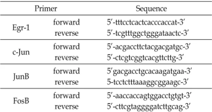
Table 1. PCR primer sequences of inducible transcription factors (Egr-1, C-Jun, JunB, FosB)
Primer Sequence
Egr-1 forward
reverse
5’-tttcctcactcacccaccat-3’
5’-tcgtttggctgggataactc-3’
c-Jun forward
reverse
5’-acgaccttctacgacgatgc-3’
5’-ctcgtcggtcacgttcttg-3’
JunB forward
reverse
5’gacgacctgcacaagatgaa-3’
5-tcctctttaaaggcggaagc-3’
FosB forward
reverse
5’-aaccaccagtggacctgtgt-3’
5’-cttcgtaggggatcttgcag-3’
유도되는 것이 확인되었으며, c-fos의 발현은 NMDA 주입 후 일시적으로 나타났지만 Kianate를 주입한 경우에는 c-fos의 발현이 NMDA를 주입한 경우보다 더 오래 지속되는 것으로 나타났다[36].
Inducible transcription factors 발현은 glutamate receptors 의 활성에 의해서 유도되지만 아직까지 NMDA receptor와 연관관계는 아직 밝혀지지 않았다. MK-801, CPP, APH 등의 NMDA receptor antagonist는 전체적인 단백질 합성에는 영 향을 미치지 않지만 위에서 언급한 다양한 자극 후에 Fos-like 면역활성이 나타나지 않고, c-fos의 경우에는 glutamate의 자 극에 의해서 mRNA의 합성이 억제되지만, 다른 자극 후에는 합성의 억제가 부분적이거나 전혀 억제가 되지 않는다. 이러 한 결과는 NMDA receptor에 의한 inducible transcription factors의 발현이 유도 될 수 있다는 현상을 제시할 뿐만 아니 라 신경전달과 inducible transcription factors가 유도되는 연 관관계가 gene transcription 수준에 한정되지 않음을 의미하 고 있다[31].
본 연구에서는 EAA 수용체의 하나인 NMDA 수용체의 과 흥분에 의해서 뇌조직이 손상을 받는 원인이 inducible tran- scription factor의 발현변화에 의한 뇌세포 내의 유전자의 발 현이 변화의 주요 요인일 것으로 판단하여, NMDA를 mouse 의 복강 내로 투여하여 NMDA 수용체의 과흥분을 유도한 후, 뇌 조직에서 mRNA의 발현에 영향을 주는 transcription fac- tor (Egr-1, c-jun, JunB, FosB)의 mRNAs 발현을 RT-PCR 방법 으로 비교, 분석하였다.
재료 및 방법
공시동물
공시동물은 한국생명공학연구원에서 분양 받은 52마리의 생후 56일령의 Balb/C mouse를 실험에 사용하였으며, 실험 기간 동안 무균실에서 고형사료와 수돗물을 자유롭게 공급하 였다(KRIBB-AEC-13056).
RT-PCR은 RT-PCR Kit (Stratagene. U.S.A)에 제시된 방법 에 따라 수행하였으며, PCR반응에 사용한 primers (β-actin 및 Inducible transcription factors)는 Primer-3 program을 이 용하여 제작한 primer (Table 1)를 Genotech (Korea.
Daegeon)에 의뢰하여 제작하였다. PCR 반응은 GeneAmp PCR system 2700 (Perkin-Elmer/Centus, Norwalk, CT)을 사 용 하였으며 50 μl의 반응액 (Templete DNA 1 μl, 10 pmole forward primer 1 μl, 10pmole reverse primer 1 μl, Taq poly- merase(5U/μl), 0.3 μl (Superbio. Korea), 10×buffer Ⅰ 2.5 μl (Superbio. Korea), 10×buffer Ⅱ, 2.5 μl (Superbio. Korea), 10mM dNTP 1.5 μl, DW 40.2 μl)으로 95℃, 5분에서 1 cycle, 95℃, 1분, 54℃, 1분, 72℃, 1분에서 각 15, 20, 25, 30 cycles, 72℃에서 10분 1 cycle로 수행하였다.
NMDA 처리 후의 mRNA의 발현 양상에 대한 비교는 PCR 반응산물을 전기영동 후, 실험군의 mRNA로부터 증폭된 DNA의 강도를 β-actin mRNA로부터 증폭된 DNA의 강도로 나눈 값을 1로 정하고, 각 농도 및 시간 별로 NMDA를 투여한 군의 mRNA로부터 증폭된 DNA의 강도를 β-actin mRNA로 부터 증폭된 DNA의 강도로 나눈 값과 비교하였다.
결 과
NMDA 투여군과 정상 대조군의 임상증상
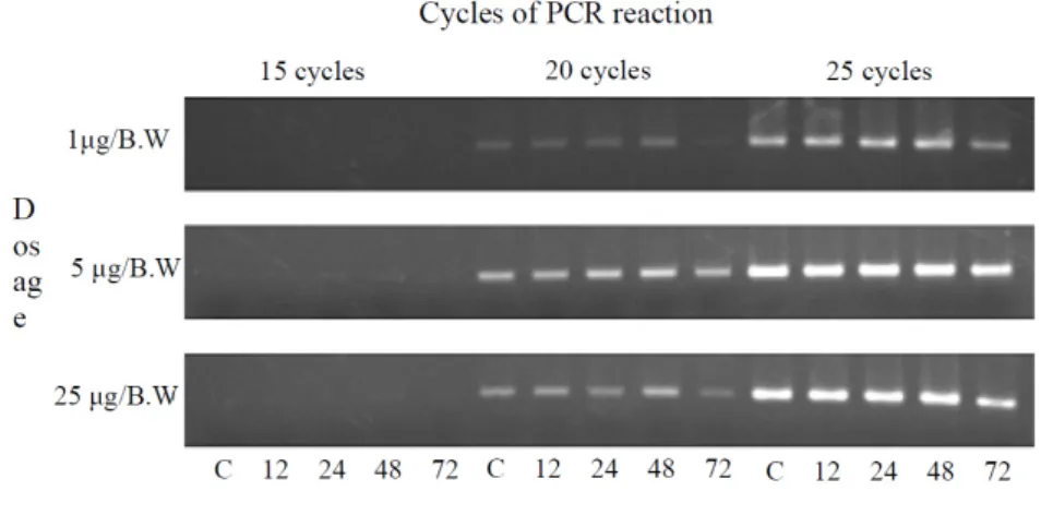
Fig. 1. β-actin mRNA sensitivity in each mRNA sample from RT-PCR reaction cycle. PBS(C) or NDNA was injected into mice abdomen and mRNA samples from cerebrum were extracted after each 12, 24, 48, 72 hr after injection. A; agarose electro- phoresis pattern of RT-PCR reaction. B; ratio changes of mRNA expression based on β-actin expression, C; total mRNA that extracted from cerebrum injected 300 μl PBS into abdomen, 1 μg/B.W: total mRNA that extracted from cerebrum injected 1 μg/B.W NMDA into abdomen, 5 μg/B.W: total mRNA that extracted from cerebrum injected 5 μg/B.W NMDA into abdo- men, 25 μg/B.W: total mRNA that extracted from cerebrum injected 25 μg/B.W NMDA into abdomen
NMDA를 투여한 후 12, 24, 48, 72시간 경과 후 NMDA 투여군에서 나타난 임상증상은 체중 g 당 125 μg의 NMDA를 투여한 군에 대한 3회의 실험에서 총 12마리 중 9마리가 10분 이내에 발작성 경련을 일으키고 즉사하였으며, 그 외의 군에 서는 NMDA 투여 후 뇌를 적출하기 전까지 이상이 있는 임상 증상을 나타내지 않았다.
Transcription factor의 상대적 발현량의 비교
실험대상이 되는 mRNA의 상대적 발현량을 보정하기 위해 서 먼저 정상 대조군과 NMDA 투여군의 시료에서 추출된 to- tal mRNA를 주형으로 하여 β-actin 유전자에 대한 RT-PCR의 감도를 측정하였다(Fig. 1). RT-PCR에 의한 감도를 비교한 결 과, 15 cycles의 PCR 반응에서는 모든 시료에서 RT-PCR 반응 결과물을 확인 할 수 없었으며, 25 cycles의 RT-PCR 반응에서 는 반응산물의 포화에 의해 각 시료간에 증폭된 DNA의 양의 차이를 판별할 수 없었다. 그러나 20 cycles의 PCR 반응에서는 각 시료간의 증폭된 DNA의 양의 차이를 유의성 있게 측정할 수 있어, transcription factor mRNA의 발현 량을 비교하기 위해서 20 cycles의 RT-PCR 반응을 사용하여 β-actin mRNA 의 발현량을 기준으로 각 transcription factor의 mRNA 발현 에 대한 양적인 차이를 보정하였다.
Egr-1 mRNA의 발현 양상
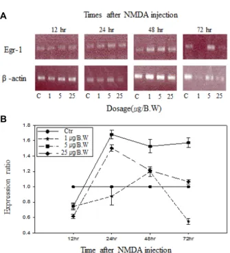
(Fig. 2A)는 NMDA 투여량과 투여 후 경과시간에 따른 Egr-1 mRNA의 RT-PCR 반응을 agarose 전기영동으로 확인 한 결과이며, (Fig. 2B)는 각 시료에 따른 mRNA의 발현량의 상대적인 비율의 변화를 나타낸 그래프로 나타내었다. 본 논 문에서 조사된 inducible transcription factor에 대해서 동일한
방법으로 결과를 나타내었다.
Erg-1 mRNA의 발현량은 NMDA 투여량이 일정할 때 투여 경과시간에 따라서, 또한 투여 후 일정한 시간에서 투여 량에 따라 발현량의 차이를 나타내었다. NMDA 투여 12시간 후에 는 모든 NMDA 투여량에서 감소를 나타내었으며 이후 NMDA의 투여량에 따라 변화를 보였다. 체중 g 당 1 μg의 NMDA를 투여하였을 때, 투여 후 24시간까지는 정상 대조군 에 비하여 mRNA의 발현이 감소하였으나, 48 시간 경과 후에 는 mRNA의 발현이 다른 투여군과 비교하여 소폭 증가한 다 음 72시간까지 급격한 감소를 나타내었다. 체중 g 당 5 μg의 NMDA를 투여했을 경우에는 24시간 경과 후에 가장 많은 증 가율을 보였으며, 72시간까지 높은 증가율이 지속되었다. 체 중 g 당 25 μg의 NMDA를 투여했을 경우에는, 투여 후 12시간 경과 이후 급격히 증가하여 24시간 후에 가장 높은 증가율을 나타낸 후에 감소하여 72시간 후에는 대조군과 거의 같은 정 도로 감소하였다. 반면, NMDA 투여 후 일정한 시간에서 NMDA 투여량에 따른 변화는 체중 g 당 5 μg의 NMDA를 투여하였을 경우에 가장 많은 변화를 나타내었으며, NMDA 의 투여량에 비례하여 mRNA의 발현량이 증가하는 양상은 나타내지 않았다.
c-Jun mRNA의 발현 양상
NMDA 투여 12시간 경과 후 모든 투여군에서 c-Jun mRNA
의 발현량의 증가가 나타났으며, 체중 g 당 1 μg의 NMDA를
투여했을 경우에 다른 투여군 중에서 가장 높은 증가율을 보
였다. 투여 후 48시간까지는 증가율을 감소하였으나 정상 대
조군에 비하여 mRNA의 발현이 증가한 상태가 유지되었으며,
이후 72시간 경과 후까지 급속히 감소하여 정상 대조군보다
Fig. 2. Expression pattern of Erg-1 mRNA in total mRNA sam- ples by RT-PCR reactions. mRNA sample were extracted after 12 hr, 24 hr, 48 hr, 72 hr from cerebrum of mouse that injected PBS(C) or NMDA into abdomen. A; agarose electrophoresis pattern of RT-PCR reaction, B; ratio changes of mRNA expression based on β-actin ex- pression, C; total mRNA that extracted from cerebrum injected 300 μl PBS into abdomen, 1 μg/B.W: total mRNA that extracted from cerebrum injected 1 μg/B.W NMDA into abdomen, 5 μg/B.W: total mRNA that ex- tracted from cerebrum injected 5 μg/B.W NMDA into abdomen, 25 μg/B.W: total mRNA that extracted from cerebrum injected 25 μg/B.W NMDA into abdomen.
Fig. 3. Expression pattern of c-Jun mRNA in total mRNA sam- ples by RT-PCR reactions. mRNA sample were extracted after 12 hr, 24 hr, 48 hr, 72 hr from cerebrum of mouse that injected PBS(C) or NMDA into abdomen. A; agarose electrophoresis pattern of RT-PCR reaction, B; ratio changes of mRNA expression based on β-actin ex- pression, C; total mRNA that extracted from cerebrum injected 300 μl PBS into abdomen, 1 μg/B.W: total mRNA that extracted from cerebrum injected 1 μg/B.W NMDA into abdomen, 5 μg/B.W: total mRNA that extracted from cerebrum injected 5 μg/B.W NMDA into abdomen, 25 μg/B.W: total mRNA that extracted from cerebrum injected 25 μg/B.W NMDA into abdomen
발현이 감소하였다. 체중 g 당 5 μg의 NMDA를 투여했을 경우 에는 투여 후 72시간까지 지속적으로 증가하는 경향을 나타내 었다. 체중 g 당 25 μg의 NMDA를 투여 했을 경우에는 24시간 경과 후까지 정상 대조군에 비하여 mRNA의 발현량에 거의 변화를 나타내지 않았으나 이후 mRNA의 발현이 증가하여 72시간 경과 후에는 투여군 중에서 가장 높은 증가율을 나타 내었다. 반면, NMDA 투여 후 일정한 시간에서 투여량에 따른 변화는 48시간 후까지 모든 투여군에서 증가하였으며, 이후에 는 투여 량에 따라 감소 혹은 증가하는 경향을 나타내었다.
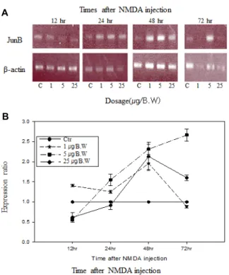
JunB의 mRNA의 발현 양상
체중 g 당 1 μg의 NMDA를 투여 했을 경우에는 투여 후 12시간부터 24시간까지 증가한 상태를 유지하다가 48시간에 서 정상 대조군에 비하여 mRNA의 발현이 가장 많이 증가하 였고 이후 감소하여 72시간 경과 후에는 정상 대조군에 비하 여 mRNA의 발현이 소폭 감소하였다. 체중 g 당 5 μg의 NMDA를 투여 했을 경우 투여 후 12시간 경과 후에는 정상
대조군에 비하여 mRNA의 발현이 감소하였으나 이후 증가하 여 48시간까지 급격한 증가율을 나타내고, 72시간까지 증가 폭은 감소하나 지속적으로 발현율이 증가하는 것으로 나타났 다. 체중 g 당 25 μg의 NMDA를 투여했을 경우에는 투여 후 24시간까지 정상 대조군보다 발현이 감소되어 나타났지만, 그 이후 지속적으로 증가하여 48시간에서 최고의 증가율을 나타 내었고, 이후 72시간까지 증가율은 감소하나 정상 대조군보다 발현이 증가하는 것으로 나타났다. JunB의 경우 NMDA 투여 후 일정한 시간에서 투여량에 따른 변화는 체중 g 당 5 μg의 NMDA를 투여하였을 경우에 본 실험에서 조사 대상으로 한 inducible transcription factors (Egr-1, c-Jun, JunB, FosB) 중에 서 mRNA의 발현이 가장 급격하고 가장 높은 증가율을 보였 다.
FosB의 mRNA의 발현 양상
FosB의 mRNA를 대상으로 한 RT-PCR 반응에서는 비특이
A
B
Fig. 4. Expression pattern of JunB mRNA in total mRNA sam- ples by RT-PCR reactions. mRNA sample were extracted after 12 hr, 24 hr, 48 hr, 72 hr from cerebrum of mouse that injected PBS(C) or NMDA into abdomen. A; agarose electrophoresis pattern of RT-PCR reaction, B; ratio changes of mRNA expression based on β-actin ex- pression, C; total mRNA that extracted from cerebrum injected 300 μg PBS into abdomen, 1 μg/B.W: total mRNA that extracted from cerebrum injected 1 μg/B.W NMDA into abdomen, 5 μg/B.W: total mRNA that ex- tracted from cerebrum injected 5 μg/B.W NMDA into abdomen, 25 μg/B.W: total mRNA that extracted from cerebrum injected 25 μg/B.W NMDA into abdomen.
A
B
Fig. 5. Expression pattern of FosB mRNA in total mRNA sam- ples by RT-PCR reactions. mRNA sample were extracted after 12 hr, 24 hr, 48 hr, 72 hr from cerebrum of mouse that injected PBS(C) or NMDA into abdomen. A; agarose electrophoresis pattern of RT-PCR reaction, ; RT-PCR product of FosB, B; ratio changes of mRNA expression based on β-actin expression, C; total mRNA that ex- tracted from cerebrum injected 300 μl PBS into abdomen, 1 μg/B.W: total mRNA that extracted from cerebrum in- jected 1 μg/B.W NMDA into abdomen, 5 μg/B.W: total mRNA that extracted from cerebrum injected 5 μg/B.W NMDA into abdomen, 25 μg/B.W: total mRNA that ex- tracted from cerebrum injected 25 μg/B.W NMDA into abdomen.
적 반응에 의한 산물이 나타났으나, 증폭된 DNA 단편 중에서 FosB의 mRNA에서 유래하는 DNA산물을 명확히 구별할 수 있고, 투여한 NMDA의 용량에 따른 변화를 반영하고 있어 FosB로부터 증폭된 DNA를 대상으로 β-actin의 DNA와 비교 하여 FosB mRNA의 변화를 비교하였다. g 당 1 μg의 NMDA 를 투여 했을 경우에는 발현이 48시간까지 정상 대조군보다 증가하였으나, 이후 감소하여 72시간에는 정상 대조군보다 감 소하였다. 체중 g 당 5 μg의 NMDA를 투여했을 경우에는 투여 후 12시간이 경과하였을 때는 정상 대조군에 비하여 mRNA의 발현이 감소하나 이후 증가하여 48시간 경과 후에 mRNA의 발현이 최고치에 이른 후, 72시간까지 증가율이 감소하면서 정상 대조군보다 발현이 다소 증가하였다. 체중 g 당 25 μg의 NMDA를 투여했을 경우에는 투여 후 12시간 경과 후에는 정 상 대조군에 비하여 mRNA의 발현의 변화가 감소하였지만 이후 48시간 경과 후까지 지속적으로 증가하여 48시간 경과 후에 증가율이 최고치에 이른 후 72시간 경과 후에는 정상
대조군에 비하여 감소하였다. 반면, NMDA투여 후 일정한 시 간에서 투여량에 따른 변화는 체중 g 당 5 μg의 NMDA를 투 여하였을 경우에 가장 급격한 변화를 나타내었다.
고 찰
NMDA (N-methyl-D-asparrate)는 신경세포에 있는 수용체
로 도파민 D1 수용체와 직접적인 상호작용을 하면서 상호작
용의 결과에 따라 세포의 사멸을 조절하거나, 정상적인 세포
사이의 통신을 유도하는 기능을 한다. 포유동물의 뇌에서
NMDA 수용체는 성장하는 동안에 신경접합부의 형성 또는
long term potentiation (LTP), 기억, 학습 과정에서 중요한
역할을 하지만 NMDA receptor가 과흥분 되면 저산소증에
의한 국소빈혈, 발작성 경련, 괴사 등을 유발하여 신경변성을
일으킨다[10, 22]. 본 실험에서는 NMDA 수용체가 과흥분 될
경우에 나타나는 증상들에 관련된 기전을 해석하기 위해서
을 확인하였고, 이러한 결과는 동일한 양의 total RNA에 포함 된 각 transcription factor mRNA의 양이 다르다는 것을 의미 하고 있기 때문에 이러한 결과를 근거로 각 실험대상이 되는 NMDA를 투여한 후의 transcription factor mRNA에 대해서 양적인 변화를 비교할 수 있었다. NMDA투여군에서 in- ducible transcription factors (Egr-1, c-Jun, JunB, FosB) mRNA의 발현의 변화는 NMDA 투여량이 일정할 때 투여 후 경과 시간에 따른 변화는 NMDA투여 후 24시간에서 가장 현저하게 나타났으며, NMDA의 투여량에 따른 변화는 체중 g 당 5 μg의 NMDA를 투여하였을 때 inducible transcription factors의 발현의 변화가 현저하게 나타났으며, JunB mRNA 의 발현이 현저히 증가하였다. 본 실험의 결과는 NMDA의 투여에 의해 NMDA 수용체가 활성화되어 본 실험에서 조사 된 inducible transcription factors (Egr-1, c-Jun, JunB, FosB) mRNA의 발현의 변화가 일어나는 것을 나타낸다. 이러한 실 험결과는 FosB mRNA의 발현의 변화가 배양된 cerebellar granule neurons에서 glutamate 또는 NMDA에 의해서 c-fos mRNA와 Fos protein의 발현을 유도한다는 실험결과[30, 33]
와 일치하고 있으며, Egr-1 mRNA의 발현의 변화는 gluta- mate에 의한 NMDA 수용체의 활성에 의해 소뇌에서 Egr-1 mRNA의 발현이 유도된다는 보고와[5, 30], NMDA의 복강 내 투여에 의해 소뇌, 대뇌피질, 해마에서 Egr-1 mRNA의 발 현이 증가한다는[36] 보고와 일치하고 있다. 또한 JunB mRNA 의 발현의 변화는 소뇌의 granule cell에 다량의 NMDA를 투 여하였을 때 JunB의 발현에 영향을 준다는 보고와도[9, 15]
일치하고 있다. 현재까지 밝혀진 바에 의하면 NMDA re- ceptor와 L-voltage-sensitive calcium channel이 활성상태가 되면 Ca
2+과 calmodulin이 결합하여 핵 내로 이동하여 핵 내의 CaM kinase (Ca
2+/calmodulin-dependentkinases) Ⅳ의 활성 화와 CREB(cAMP-response element-binding protein)의 Ser133을 인산화시키며, 또한 Ca
2+은 Ras/ERK/RSK pathway 를 활성화시켜, 활성화된 ERK kinase와 RSK kinase는 protein kinase A의 활성에 의하여 핵 내로 이동한다. 핵 내에서, ERK 는 Elk-1을 인산화시키는 반면에 RSK kinase는 constitutive
scription factors로부터 발현되는 protein은 AP1 (activator
protein 1)으로 알려져 있는 actual transcription factor를 형성
하기 위하여 leucine zipper motifs를 통하여 heterodimer를
형성하며 TRE (TPA response element)로 불려지는
TGACTCA의 DNA 염기서열을 인식하고 결합한다[31]. 즉
Fos/c-Jun, FosB/c-Jun, Fos/JunD 등의 AP1는 활성적이지만
c-Jun/JunB, JunB/JunB, FosB/JunB은 비활성적이다. 이들
AP1이 TRE와 결합하면 transcription의 repressors로 작용하
며[12], 또한 Fos protein과 Jun protein은 인산화에 의하여 복
합적인 조절을 받는다. 따라서 세포 내에서 c-Jun이 존재하고
있지만 c-Jun의 TRE에 결합은 c-Jun의 C-terminal region의
인산화에 의해서 억제된다. 그러나 protein kinase C는 c-Jun
의 인산화된 C-terminal region를 신속하게 탈인산화시켜
c-Jun을 활성화시킨다[13, 31]. Inducible transcription factors
는 통증을 유발하는 자극, 다양한 신경전달물질, 호르몬, neu-
rotrophins, long-term potentiation, 학습, 발작, 흥분, 신경변
성, 저산소성 국소빈혈, 뇌신경손상, 신경절제, 약제 내성 등
다양한 요인에 에 의해서 유도되는데[37, 21], 이러한 현상에
관해서는 Egr-1 protein 이 유도되면 c-Jun을 활성화시켜서 신
경의 apoptosis를 일으키는 것으로 알려져 있다[16]. 특히, 소
뇌에서 Egr-1 protein는 granule cell에서 발현이 증가하며[4],
c-Jun이 활성화되면 potassium deprivation에 의해서 granule
cell의 사멸을 일으킨다. c-Jun의 발현은 beta-amyloid-medi-
ated apoptosis를 유도하고 Alzheimer's disease (AD)에서 증
가되는 것으로 보고되었다[1, 19]. 그리고 Purkinje cell의 재생
및 변성은 c-Jun protein은 단독으로는 발생할 수 없고 세포
내 신호전달의 상호작용에 의한 것으로 추측된다[3]. Fos와
Jun transcription factor protein은 Purkinje cell의 사멸에 관련
된 신호전달과 cell death repressor gene인 bcl-2 발현의 조절
에 관여하는 것으로 추측되며[6], Fos protein은 homodimer를
형성할 수 없기 때문에 FosB/JunD heterodimer가 신경과흥
분에 의한 신경세포의 사멸에 특히 중요한 원인으로 추측된다
[18]. 본 연구의 대상이 된 Inducible transcription factors
(Egr-1, C-Jun, JunB, FosB)가 어떤 기전으로 소뇌의 purkinje
cell의 사멸과정에 관여하는지는 아직 알려져 있지 않다. 본 연구의 결과는 NMDA 수용체의 과흥분에 의해서 나타나는 Egr-1, C-Jun, JunB, FosB transcription factors에 의한 소뇌에 서의 유전자 발현의 변화를 2D-gel 또는 microarray와 같은 방법을 이용하여 세포 내의 전체 단백질 혹은 유전자의 변화 [17]를 관찰함으로써 NMDA 수용체의 과흥분에 의한 뇌세포 의 사멸에 관련된 기전을 밝힐 수 있는 좋은 자료가 될 수 있을 것으로 기대된다.
감사의 글
본 연구는 2012년 경북대학교 학술진흥기금에 의한 연구임.
References
1. An derson, A. J., Su, J. H. and Cotman, C. W. 1996. DNA damage and apoptosis in Alzheimer's disease:colocalization with c-Jun immunoreactivity, relationship to brain area, and effect of postmortem. J. Neurosci. 16, 1710-1719.
2. Bazhenov, A. V. and Kleshchevnikov, A. M. 1999. Reciprocal inhibition of the AMPA and NMDA components of ex- citatory postsynaptic potentials in field CA1 of the rat hip- pocampus in vitro. Neurosci. Behav. Physiol. 29, 719-725.
3. Carulli, D., Buffo, A., Botta, C., Altruda, F. and Strata, P.
2002. Regenerative and survival capabilities of Purkinje cells overexpressing c-Jun. Eur. J. Neurosci. 16, 105-118.
4. Catania, M. V., Copani, A., Calogero, A., Ragonese, G. I., Condorelli, D. and Nicoletti, F. 1999. An enhanced ex- pression of the immediate early gene, Egr-1, is associated with neuronal apoptosis in culture. Neuroscience 91, 1529-1538.
5. Cheyou, E. R., Youreva, V. and Srivastava, A. K. 2014.
Involvement of the early growth response protein 1 in vas- cular pathophysiology: an overview. Indian J. Biochem.
Biophys. 51, 457-66
6. Gillardon, F., Baurle, J., Wickert, H., Grusser-Cornehls, U.
and Zimmermann, M. 1995. Differential regulation of bcl-2, bax, c-fos, junB, and krox-24 expression in the cerebellum of Purkinje cell degeneration mutant mice. J. Neurosci. Res.
41, 708-715.
7. Hashimoto, M. and Hibi, M. 2012. Development and evolu- tion of cerebellar neural circuits. Dev. Growth Differ. 54, 373-389.
8. Hirai, H., Kirsch, J., Laube, B., Betz, H. and Kuhse, J. 1996.
The glycine binding site of the N-methyl-D-aspartate re- ceptor subunit NR1: identfication of novel determinants of co-agonist potentiation in the extracellular M3-M4 loop region. Proc. Natl. Acad. Sci. USA 93, 6031-6036.
9. Hou, Y. N., Cebers, G., Terenius, L. and Liljequist, S. 1997.
Characterization of NMDA- and AMPA-induced enhance- ment of AP-1 DNA binding activity in rat cerebellar granule cells. Brain Res. 754, 79-87.
10. Hume, R. I., Digledine, R. and Heinemann, S. F. 1991.
Identification of a site in glutamate receptor subunits that controls calcium permeability. Sciences 253, 1028-1031.
11. Ishizaki, Y. 2006. Control of proliferation and differentiation of neural precursor cells: focusing on the developing cerebellum. J. Pharmacol. Sci. 101, 183-188.
12. Järlestedt, K., Rousset, C. I., Ståhlberg, A., Sourkova, H., Atkins, A. L., Thornton, C., Barnum, S. R., Wetsel, R. A., Dragunow, M., Pekny, M., Mallard, C., Hagberg, H. and Pekna, M. 2013. Receptor for complement peptide C3a: a therapeutic target for neonatal hypoxic-ischemic brain injury. FASEB J. 27, 3797-3804.
13. Karin, M. 1995. The regulation of AP-1 activity by mi- togen-activated protein kinases. J. Biol. Chem. 270, 16483- 16486.
14. Kleven, G. A., Booth, H. M., Voogd, M. and Ronca, A. E.
2014. L-dopa reverses behavioral deficits in the Pitx3 mouse fetus. Behav. Neurosci. 128, 749-59
15. Lerea, L. S., Butler L. S. and Mcnamara, J. O. 1992. NMDA and non-NMDA receptor-mediated increase of c-fos mRNA in dentate gyrus neurons involves calcium influx via differ- ent routes. J. Neurosci. 12, 2973-2981.
16. Levkovitz, Y. and Baraban, J. M. 2001. A dominant negative inhibitor of the Egr family of transcription regulatory factors suppresses cerebellar granule cell apoptosis by blocking c-Jun activation. J. Neurosci. 21, 5893-5901.
17. Li, H., Wang, Z. X. and Wu J. W. 2014. Purification, charac- terization and docking studies of the HIN domain of human myeloid nuclear differentiation antigen (MNDA). Biotechnol.
Lett. 36, 899-905.
18. Lidwell, K. and Griffiths, R. 2002. Possible role for the FosB/
JunD AP-1 transcription factor complex in glutamate-medi- ated excitotoxicity in cultured cerebellar granule cells. J.
Neurosci. Res. 62, 427-439.
19. Marcus, D. L., Strafaci, J. A, Miller, D. C., Masia, S., Thomas, C. G., Rosman, J., Hussain, S. and Freedman, M. L. 1998.
Quantitative neuronal c-fos and c-jun expression in Alzheimer's disease. Neurobiol. Aging 19,393-400.
20. Mayer, M. L. and Westbrook, G. L. 1987. The physiology of excitatory amino acids in the vertebrate central nervous system. Prog. Neurobiol. 28, 197-206.
21. Mayer, M. L., Westbrook, G. L. and Guthrie, P. B. 1984.
Voltage-dependent block by Mg2+ of NMDA Responses in spinal cord neurons. Nature 309, 261-263.
22. Mcdonald, J. W. and Johnston, M. V. 1990. Physiological and pathophysiological roles of excitatory amino acids dur- ing central nervous system development. Brain Res. Rev. 15, 41-70
23. Meldrum, B. S. 2000. Glutamate as a neurotransmitter in the brain: review of physiology and pathology. J. Nutr. 130, 1007-1015.
24. Michisuke, Y., Douglas, F., Lynne, M. V., Shaiu, C. S., Curran, T. and Connor, J. A. 1996. Functional NMDA Receptors are transiently active and support the survival of purkinje cells in culture. J. Neurosci. 16, 4651-4661.
25. Olney, J. W. 1974. Toxic effects of glutamate and relate ami- no acids on the developing central nerve system, pp.
초록:NMDA투여에 의한 transcription factor (Egr-1, C-Jun, JunB, FosB)의 발현 변화 양상 하종수
1,2․김재화
3․송재찬
1*
(
1경북대학교 수의과대학,
2삼화종계,
3의생명중계연구센터 한국생명공학연구원)
신경과흥분은 신경세포의 수지돌기 말단부에 있는 흥분성 수용체에 대한 과도한 자극에 의해서 신경세포가 손 상을 받는 현상으로 transcription factor의 발현을 유도하여 통증을 유발하는 자극, 학습, 발작, 흥분, 신경변성, 저산소성 국소빈혈, 뇌신경손상, 신경절제, 약제내성 등의 원인이 된다. 신경과흥분은 정상농도 이상의 NMDA에 의해서도 유발되는데 본 논문에서는 mouse의 복강으로 과량의 NMDA를 투여하여 소뇌에서 RT-PCR 방법으로 Inducible transcription factors (Egr-1, c-jun, JunB, FosB) mRNAs의 상대적 발현량을 비교하였다. NMDA를 투여 한 군에서 inducible transcription factors (Egr-1, C-Jun, JunB, FosB)가 투여량과 시간의 경과에 따라 다양한 발현 의 변화를 보였으며, NMDA투여 후 일정한 시간에서 투여한 양에 대한 변화는 체중 g 당 5 μg의 NMDA투여한 경우에 현저한 변화가 나타났다. 조사한 transcription factor 중에서 JunB의 발현 변화가 다른 transcription factor 보다 두드러지게 나타났다. NMDA 투여량이 일정할 때 투여 후 경과 시간에 따른 발현양상은 투여 후 24시간이 경과한 후에 발현의 변화가 두드러지게 증가하는 경향을 나타내었고 대부분 이 48시간 경과 후 발현이 최고치에 도달하였다. 이러한 결과는 과흥분이 유도된 소뇌에서의 유전자 발현의 변화를 2D-gel 또는 microarray와 같은 방법을 이용하여 세포 내의 전체 단백질 혹은 유전자의 변화를 관찰함으로써 NMDA 수용체의 과흥분에 의한 뇌세포의 사멸에 관련된 기전을 밝힐 수 있는 좋은 자료가 될 수 있을 것으로 기대된다.
Transcriptional program coordination by N-methyl-D-as- partate-sensitive glutamate receptor stimulation in primary cultures of cerebellar neurons. Mol. Pharmacol. 38, 624-633.
31. Tönges, L., Planchamp, V., Koch, J. C., Herdegen, T., Bähr, M. and Lingor, P. 2011. JNK isoforms differentially regulate neurite growth and regeneration in dopaminergic neurons in vitro. J. Mol. Neurosci. 45, 284-293.
32. Watanabe, M., Moise, I. M. and Inoue, Y. 1996. Modified
tor protein-1 complex with DNA binding activity following systemic administration of N-methyl-D-aspartate in dentate granule cells of murine hippocampus. Neuroscience 93, 19-31.
37. Zheng, S., Yang, H., Chen, Z., Zheng, C., Lei, C. and Lei, B. 2015. Activation of liver X receptor protects inner retinal damage induced by N-methyl-D-aspartate. Invest. Ophthal- mol. Vis. Sci. 22, 1168-1180.