Anti-oxidant and Anti-inflammatory Potentials of Sasa quelpaertensis Leaf Residue Extracts
Ju-Yeop Lee
1, Hana Song
1, Hee-Chul Ko
1, Mi-Gyeong Jang
2and Se-Jae Kim
1,2*
1Jeju Sasa Industry Development Agency, Jeju National University, 102 Jejudaehakno, Jeju-si, Jeju Special Self-Governing Province 690-756, Korea
2Department of Biology, Jeju National University, 102 Jejudaehakno, Jeju-si, Jeju Special Self-Governing Province 690-756, Korea
Received January 31, 2018 /Revised March 19, 2018 /Accepted March 26, 2018
Sasa quelpaertensis Nakai is a native Korean plant that grows only on Mt. Halla of Jeju Island. Its leaf
is used for a popular bamboo tea due to its various health-promoting properties, and it has been in- creasingly used as food and cosmetic ingredients. To utilize the S. quelpaertensis leaf efficiently, the preparation method for phytochemical-rich extract (PRE) using the leaf’s residue was previously re- ported, which was produced after hot water extraction. This study was undertaken to evaluate the anti-oxidant and anti-inflammatory potential of PRE and its solvent fractions. The ethyl acetate frac- tion of PRE (EPRE) showed higher DPPH, ABTS, and superoxide radical scavenging activities, and it effectively inhibited intracellular reactive oxygen species (ROS) and nitric oxide (NO) production in lipopolysaccharides (LPS)-stimulated RAW 264.7 cells. EPRE also induced the expression of heme oxy- genase-1 (HO-1) by increasing the level of nuclear factor E2-related factor 2 (Nrf2) in a nuclear fraction. The inhibiting effect of EPRE on LPS-induced NO production was partially reversed by the HO-1 inhibitor (zinc protoporphyrin, ZPP), suggesting that HO-1 is involved in suppressing NO production. Taken together, the results suggest that EPRE has potential as a promising anti- oxidant and anti-inflammatory agent.
Key words : Anti-inflammatory, anti-oxidant, heme oxygenase-1, Sasa quelpaertensis
*Corresponding author
*Tel : +82-64-754-3529, Fax : +82-64-751-4406
*E-mail : [email protected]
This is an Open-Access article distributed under the terms of the Creative Commons Attribution Non-Commercial License (http://creativecommons.org/licenses/by-nc/3.0) which permits unrestricted non-commercial use, distribution, and reproduction in any medium, provided the original work is properly cited.
Journal of Life Science 2018 Vol. 28. No. 6. 738~744 DOI : https://doi.org/10.5352/JLS.2018.28.6.738
서 론
염증(inflammation)은 세포상해, 자극제 혹은 병원균에 대 응하는 숙주의 방어기작으로 염증반응에는 대식세포와 T 세 포들과 같은 면역세포와 전염증 사이토카인, prostaglandin (PG), nitric oxide (NO) 등을 포함하는 염증매개인자들이 관 여한다[13]. 대부분의 경우 염증은 조직에 침투한 대식세포에 서 생성되는 활성산소종(reactive oxygen species, ROS)이 핵 전사인자(nuclear transcription factor kB, NFkB)의 경로를 통 해 전염증 사이토카인을 암호화하는 유전자들의 발현을 조절 함으로써 염증반응을 매개한다고 알려져 있다[7]. 호기성 생물 체에서 산소 대사산물로 지속적으로 생성되는 ROS는 다양한 세포과정에 신호전달 물질로서 작용한다. 그러나 산화제 혹은 활성화된 염증세포들에 의해 과도하게 생성되는 ROS는 생체 내 산화환원 불균형을 유발하여 다양한 질환을 일으킨다고 알려져 있다[22].
생물체는 산화제와 항산화제의 균형을 유지하기 위해 효소 적 및 비효소적 항산화 방어기전을 가지고 있다. 항산화 효소 중에서 heme oxygenase-1 (HO-1)은 산화적 스트레스 상태에 서 세포를 보호해주는 항산화 효소로 heme을 biliverdin, car- bon monoxide (CO)와 iron으로 전환한다[19]. 스트레스나 자 극에 의해 유도되는 HO-1 단백질은 심혈관 질환이나 염증 반 응과 같은 다양한 질병 상태에서 건강유지에 도움이 된다고 보고되었으며[1, 17], 활성화된 RAW 264.7 세포에서 HO-1의 발현은 NO의 생성과 inducible nitric oxide synthase (iNOS) 유전자 발현을 저해함으로써 염증반응을 억제한다고 알려져 있다[3,16]. 그리고 CO와 biliverdin은 cyclooxygenase 2 (COX-2)의 발현과 NO의 생성을 통제함으로써 염증반응을 예 방한다고 알려져 있다[21]. 따라서 ROS 소거능과 더불어 HO-1의 발현을 적절한 수준으로 유도할 수 있는 천연물 소재 에 대한 관심이 증가하고 있다.
조릿대 속(Sasa sp.) 식물은 작은 대나무(dwarf bamboo)라 고 불리는 다년생 식물로 한국, 중국, 일본, 러시아 등 아시아 국가에 여러 종들이 분포하고 있다[18]. 이 식물의 잎은 예로부 터 항염, 해열, 이뇨 작용이 있어 민간약재로 이용되어 왔으며 [2], in-vitro 및 in-vivo 에서 항산화, 항염 및 항암 활성 등 다양 한 건강증진 효과들이 보고 된 바 있다[5, 10, 20]. 이 중에 제주 조릿대(Sasa quelpaertensis Nakai)는 제주도 한라산에 자생하 는 특산식물로 그 잎은 차로 음용되어 왔다. 최근 들어서는 제주조릿대 잎 추출물은 항염, 항균, 항바이러스, 항암, 항산
- Note -
화, 항당뇨, 항비만 등의 활성[8, 9, 11, 12, 26]을 가지고 있다고 알려지고 있어, 건강증진 식품 및 화장품 소재로서 활용이 증 대되고 있다. 우리 연구팀은 제주조릿대 자원의 효율적인 활 용방안을 마련하기 위하여 열수 추출물 잔사로부터 식물화합 물을 다량으로 함유하는 추출물(phytochemical-rich extract, PRE)의 제조방법을 보고한 바 있다[14]. 본 연구에서는 항산화 혹은 항염 소재로서의 PRE 활용가능성을 평가하기 위하여 PRE 에틸아세테이트 분획물(EPRE)의 항산화 및 항염 활성을 조사하였다.
재료 및 방법
제주조릿대 추출물 제조
제주조릿대는 2015년 9월 제주 한라산에서 채집하여 세척 하고 60℃에서 24시간 건조한 후 분쇄하여 추출 시료로 사용 하였다. 식물화합물을 다량으로 함유한 제주조릿대 잎 잔사 추출물(PRE)은 Lee 등[14]의 방법에 따라 준비하였다. 그리고 PRE 및 에탄올 추출물은 용매분획을 통해 n-hexane, chloro- form (CHCl
3), ethyl acetate (EtOAc), n-butanol (BuOH) 층으 로 분리한 후 동결 건조하여 실험에 사용하였다.
HPLC 분석
PRE와 에탄올 분획물의 식물화합물의 함량을 비교하기 p- coumaric acid와 tricin을 지표성분으로 선정하여 HPLC (Waters Corp., Milford, MA) 방법으로 분석하였다[14]. 지표 성분의 분리는 Sunfire
TMC
18column (4.6×250 mm I.D., 5μm particle size)을 사용하였고 이동상은 Acetonitrile (A)와 1%
acetic acid (B)을 사용하여 0.8 ml/min 유속 조건으로 정량분 석을 실행하였다. 이동상 조건은 기울기용리법을 사용하여 초 기조건으로 이동상 A를 15%로 시작하여 40.0분 동안 42.5%로 변화시키고 40.1분부터 45분 동안 100%로 용리한 후 초기조건 으로 약 10분간 유지하였다. 시료는 50 mg/ml의 농도로 DMSO/MeOH (v/v, 1/1)로 용해하여 0.45 μm syringe filter 로 여과한 후 자동시료주입기를 이용하여 10 μl씩 주입하였다.
지표물질의 표준용액은 0.05, 0.1, 0.5 mg/ml의 농도로 제조하 여 시료와 동일한 조건 하에 머무름 시간과 흡광도를 비교하 여 분석하였다.
항산화 활성
DPPH (1,1-diphenyl-2-picrylhydrazy) 소거활성은 시료를 96 well plate에 100 μl씩 분주하고 0.4 mM DPPH 용액을 동량 첨가 하여 실온에서 10분간 방치한 후 517 nm에서 흡광도를 측정하였다. ABTS (2,2'-azino-bis-3-ethylbenzothiazoline-6- sulphonic acid) 소거활성은 7 mM ABTS와 2.45 mM ammo- nium persulphate를 동량 혼합하여 100 ul씩 시료에 첨가한 후 실온에서 10분간 방치한 후 735 nm에서 흡광도를 측정하여
산출하였다. Superoxide 소거활성은 phenazin methosulfate (PMS), nicotinamide adenine dinucleotide (NADH), nitro- blue tetrazolium (NBT)이 함유한 Tris-HCl buffer와 시료를 반응시킨 후 560 nm에서 흡광도를 측정하여 산출하였다.
Xanthine oxidase 억제 활성은 시료와 0.5 mM xanthine와 1 mM EDTA를 함유한 200 mM sodium phosphate buffer (pH7.5) 100 μl에 30 mU/ml xanthine oxidase를 첨가하여 ur- ic acid 생성을 유도하였고290 nm에서 흡광도를 측정하여 산 출하였다.
ROS, NO 생성 억제 활성 및 세포독성
RAW264.7 세포를 96 well plate에 최종농도가 3×10
5cells/
ml 가 되도록 분주한 뒤 LPS (200 ng/ml)와 시료를 첨가하여 24시간 배양하였다. 세포 내에서 생성된 ROS는 세포를 2’, 7’-dichlorofluorescin diacetate (DCF-DA)로 30분간 염색한 후 공초점 현미경으로 관찰하였다. NO 생성은 세포배양액 100 μl와 Griess reagent (1% sulfanilamide and 0.1% naphthyle- thylenediamine dihydrochloride in 5% phosphoric acid) 100 μl를 혼합하여 실온에서 15분 동안 반응시킨 후 microplate reader (Epoch, BioTek, USA)를 이용하여 540 nm에서 흡광도 를 측정하였다. NO 생성량은 sodium nitrate로 표준곡선을 작성하여 산출하였다. 세포독성은 MTT (5 mg/ml) 시약 10 μl를 넣고 2시간 동안 배양한 후 DMSO에 용해하여 micro- plate reader (Epoch, BioTek, USA)를 이용하여 595 nm에서 흡광도를 측정하여 산출하였다.
Nuclear protein extraction
RAW264.7 세포를 100 mm dish에 최종농도가 2×10
5cells/
ml가 되도록 분주한 뒤 37℃, 5% CO
2incubator에서 24시간 배양한 후 시료를 처리하였다. 시료처리 후 1시간 후 LPS (200 ng/ml)를 처리하여 30분 동안 배양한 후, PBS로 세척하고 세 포를 원심분리하여 pellet을 얻었다. 핵 및 세포질 단백질은 nuclear extract kit (Active motif, USA)를 사용하여 분리하였 다.
Western blot analysis
단백질 20-30 μg을 sample buffer와 섞어 95℃에서 5분간
끓인 후, 10% SDS-PAGE로 분리한 다음, PVDF membrane으
로 transfer 한 후 5% skim milk에서 1시간 동안 blocking하였
다. 1차 항체(iNOS, COX-2, HO-1, Nrf2, β-actin, Lamin B1)는
4℃에서 overnight 반응시킨 후 TBS-T (Tris-buffered saline
and tween-20) buffer로 10분간 3회 세척하였고, 2차 항체는
1:10,000으로 희석하여 1시간 동안 상온에서 반응시켰다. 단백
질은 TBS-T로 10분간 3회 세척한 후 membrane에 ECL (WEST-
ZOL Plus, iNtRON, Korea) 시약을 처리하여 검출하였다.
Fig. 1. HPLC chromatogram of PRE fractions (upper) and comparison of two phytochemical contents in PRE and ethanol fractions (low). EtOH extract, 70% ethanol extract; RT, retention time; %RSD, relative standard deviation.
통계분석
모든 실험결과는 평균 ± 표준편차로 나타내었으며, 통계적 유의성에 대한 검증은 Microsoft excel program의 Student's t-test를 사용하여 실시하였으며, 조사 항목들 간의 유의성 검 정은 p<0.05 수준에서 실시하였다.
결과 및 고찰
EPRE의 항산화 및 항염 활성
조릿대 식물은 다양한 종류의 당류 화합물들이 함유하고 있다고 알려져 있다. Hayashi 등[6]은 당류 화합물을 다량으로 추출하기 위해서 알코올을 처리하여 비극성의 특성을 갖는 화합물들을 제거한 후 열수 추출하여 획득하였다고 보고한 바 있다. 이와는 대조적으로 우리 연구팀은 제주조릿대 자원 을 효율적으로 활용할 수 있도록 제주조릿대 잎을 열수 추출 하고 남은 잔사에 알코올 가하여 식물화합물이 풍부한 추출물 (PRE)을 제조하는 방법을 보고한 바 있다[14]. 본 연구에서는 항산화 혹은 항염 소재로서의 PRE의 활용가능성을 탐색하였
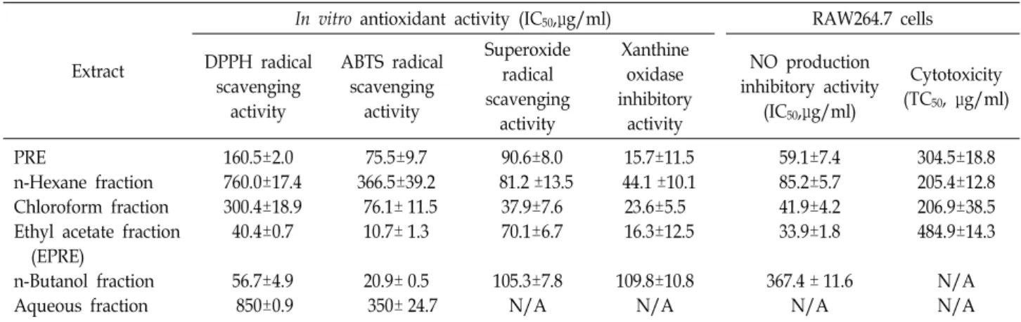
다. 우선 제주조릿대 잔사 추출물(PRE)과 기존 에탄올 추출물 의 용매 분획물을 대상으로 지표성분인 p-coumaric acid와 tri- cin의 함량을 비교하였다(Fig. 1). PRE의 EtOAc 분획물(EPRE) 이 두 지표성분을 기준으로 볼 때 식물화학물 함량이 가장 높을 것이라 판단되었다.
항산화제는 수소 혹은 전자를 줄 수 있는 능력, 비공유 전자
를 안정화시키거나 탈취하는 능력, 혹은 금속원소를 제거할
수 있는 능력을 가진 물질을 일컫는다. PRE 및 그 분획물의
DPPH radical 소거활성, ABTS radical 소거활성, superoxide
radical 소거활성, 그리고 xanthine oxidase 억제 활성을 IC
50값
으로 비교하였다(Table 1). 분석한 PRE 분획물 중에서 에틸아
세테이트 분획물(EPRE)이 DPPH 소거활성(IC
50= 40.4±0.7 μ
g/ml), ABTS 소거활성(IC
50= 10.7±1.3 μg/ml)이 가장 우수하
였다. PRE 클로로포름 분획물은 세포독성을 나타냈지만(data
not shown), EPRE는 세포독성을 나타내지 않으면서 강력한
superoxide 소거활성(IC
50= 70.1±6.7 μg/ml), xanthine oxi-
dase 억제 활성(IC
50= 16.3±12.5 μg/ml)을 나타내었다. EPRE
가 LPS 유도된 ROS 생성에 어떠한 영향을 미치는지 분석한
Table 1. Anti-oxidant activity, NO production inhibitory activity, and cytotoxicity of PRE and its fractions
Extract
In vitro antioxidant activity (IC50,μg/ml) RAW264.7 cells DPPH radical
scavenging activity
ABTS radical scavenging
activity
Superoxide radical scavenging
activity
Xanthine oxidase inhibitory
activity
NO production inhibitory activity
(IC50,μg/ml)
Cytotoxicity (TC50, μg/ml)
PRE
n-Hexane fraction Chloroform fraction Ethyl acetate fraction (EPRE)
n-Butanol fraction Aqueous fraction
160.5±2.0 760.0±17.4 300.4±18.9 40.4±0.7 56.7±4.9 850±0.9
75.5±9.7 366.5±39.2 76.1± 11.5 10.7± 1.3 20.9± 0.5 350± 24.7
90.6±8.0 81.2 ±13.5
37.9±7.6 70.1±6.7 105.3±7.8 N/A
15.7±11.5 44.1 ±10.1 23.6±5.5 16.3±12.5 109.8±10.8
N/A
59.1±7.4 85.2±5.7 41.9±4.2 33.9±1.8 367.4 ± 11.6
N/A
304.5±18.8 205.4±12.8 206.9±38.5 484.9±14.3
N/A N/A Cell were treated with LPS (200 ng/ml) in the presence of various concentration of sample for 24 hr. The amount of nitrite in the culture medium was determined with Griess reagent. The IC50 values were calculated from regression lines using five different concentrations in triplicate experiments. Each value is the average ± S.D. of triplicate determinations.IC50 is the concentration produc- ing 50% inhibition of NO production in RAW 264.7 cells. TC50 is the concentration producing 50% toxicity in RAW 264.7 cells.
* : Can’t calculate the value of IC50.
Fig. 2. Effect of EPRE on intracellular ROS production in LPS- stimulated RAW 264.7 cells. Cell were treated with LPS (200 ng/ml) alone or LPS plus the indicated concen- trations of sample for 24 hr. The cells were stained with oxidation sensitive dye, 2',7'-dichlorofluorescin diacetate (DCF-DA) for 30 mins in the dark. The cells were exam- ined by confocal microscopy.
결과, 예상되는 바와 같이 세포내 ROS 생성이 현저히 감소되 는 것을 확인할 수 있었다(Fig. 2).
대식세포는 외부에서 침입한 세균들의 세포구성 성분이나 면역세포에서 분비된 cytokines에 의해 활성화되어 TNF-α, NO, PGE
2등과 같은 물질의 분비를 조절한다. NO는 활성 산소종의 하나로, 정상적으로는 세포 내에서 세포신호 전달을 통한 항상성 기능 조절에 관여한다. 그러나 염증과정에서는 iNOS에 의해 과량의 NO가 생성되어 주변조직의 손상을 유도
A
B
C
Fig. 3. Effect of EtOAc fraction (EPRE) on the expression of iNOS and COX-2 in LPS-stimulated RAW 264.7 cells. (A) Cell were treated with LPS (200 ng/ml) alone or LPS plus the indicated concentrations of sample for 24 hr. iNOS, COX-2 protein levels were determined by western blotting.
Quantification of iNOS (B), COX-2 (C) protein expression was performed by densitometric analysis. The relative level was calculated as the ratio of iNOS, COX-2 protein expression to β-actin protein expression. The lower panel shows the quantified results (mean ± SEM, n=3, *p< 0.05,
**p<0.01, ***p<0.001, comparison is between sample value and previous LPS alone-treated cells).
A B
Fig. 4. Effect of EPRE on HO-1 mRNA (A) and protein expression (B) in LPS-stimulated RAW 264.7 cells. (A) Real-time PCR.
(B) Western blot analysis. Quantification of HO-1 protein expression was performed by densitometric analysis. The relative level was calculated as the ratio of HO-1 protein expression to β-actin protein expression. The lower panel shows the quantified results (mean SEM, n=3, *p<0.05, **p<0.01, ***p<0.001, comparison is between sample value and previous LPS alone-treated cells).
A B
Fig. 5. Effect of EPRE on nuclear translocation of Nrf2 (A) and effects of ZPP on NO production (B) in LPS-stimulated RAW 264.7 cells. (A) Cell were treated with LPS (200 ng/ml) alone or LPS plus the indicated concentrations of sample for 6 hr. Nrf2 levels were determined using western blotting method. (B) HO-1 mediates the suppressive effect of EPRE on LPS-stimulated NO production. Macrophages were pre-treated for 1 hr with EPRE (100 μg/ml) in the presence or absence of ZPP (10 μM), and stimulated for 24 hr with LPS (200 ng/ml). Data represent mean values of 3 experiments S.D. **p<0.01.
하고 만성염증을 유발한다고 알려져 있다[4]. EPRE는 LPS로 자극된 RAW264.7 세포에서 NO 생성을 효과적으로 억제 (IC
50= 33.9 μg/ml) 함과 동시에 세포독성이 가장 낮았다. 또 한, EPRE는 iNOS와 COX-2 단백질 발현을 효과적으로 억제하 였다(Fig. 3).
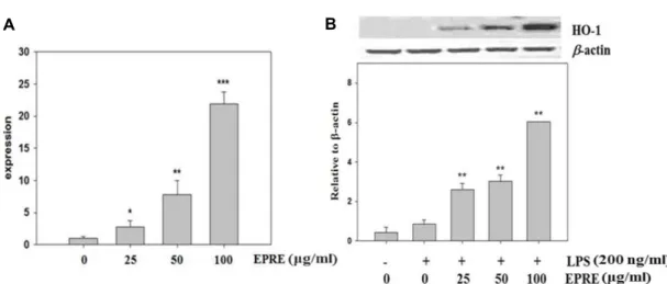
EPRE의 heme oxygenase-1 (HO-1) 발현 촉진 효과 Heme oxygeanse-1 (HO-1)은 heme을 분해하여 biliverdin, free iron, CO 등을 생성하는데 관여하는 효소로서 산화적 스 트레스에 대한 보호 작용을 통해 염증 반응을 약화시킨다[15].
HO-1에 의해 생성되는 CO는 혈관을 확장시켜 장기 손상을 억제하며 항염증 및 항세포사멸 기능이 있으며, bilirubin은 출혈 쇼크 시 정맥 내 백혈구 이동을 억제시켜 염증을 감소시 킨다[24]. LPS로 처리된 대식세포에 EPRE를 처리하여 HO-1
발현에 미치는 영향을 분석한 결과, EPRE은 농도 의존적으로 HO-1의 발현량을 유의적으로 증가시킴을 확인할 수 있었다 (Fig. 4).
항산화 효소의 발현을 조절하는 Nrf2는 Keap1에 결합한 비활성화 상태로 세포질에 존재하지만, 산화적 손상을 받으면 Keap1로부터 분리되어 핵내로 이동하여 HO-1 유전자의 an- ti-oxidant response element (ARE)에 결합함으로써 HO-1 유 전자 발현을 조절한다고 알려져 있다[23]. EPRE가 Nrf2를 활 성화에 미치는 영향을 조사한 결과, EPRE은 농도 의존적으로 Nrf2 단백질이 세포질에서부터 핵으로 이동을 증가시키는 것 을 확인 할 수 있었다(Fig. 5A). 이 결과는 EPRE가 Nrf2 경로를 통해 HO-1의 발현을 유도한다는 것을 암시해 준다.
EPRE에 의한 HO-1 발현이 NO 생성에 미치는 영향을 확인
하기 위하여 HO-1 inhibitor인 ZPP를 처리하여 HO-1 발현을
억제시켰다. HO-1 발현을 억제시켰을 때 EPRE에 의해 감소되 었던 NO 생성량이 다시 증가됨을 확인할 수 있었다(Fig. 5B).
따라서 EPRE가 HO-1 발현을 유도하여 NO 생성을 억제함으 로서 항염증 효과를 발휘하고 있다고 사료된다. 스트레스나 자극에 의해 유도되는 HO-1 단백질은 심혈관 질환이나 염증 반응과 같은 다양한 질병 상태에서 건강유지에 도움을 주며 [1, 17], 또한 활성화된 대식세포에서 HO-1의 발현은 NO의 생성과 iNOS 유전자 발현을 저해함으로써 염증반응을 차단한 다고 알려져 있다[3, 16]. 본 연구는 HO-1 발현을 유도하는 작용을 가진 EPRE가 항산화 및 항염 소재로서의 개발 가능성 을 제시해준다.
감사의 글
본 연구는 2017년도 제주대학교 학술진흥연구비의 지원에 의해 이루어진 것입니다.
References
1. Abraham, N. G. and Kappas, A. 2008. Pharmacological and clinical aspects of heme oxygenase. Pharmacol Rev. 60, 79- 127.
2. Bae, K. 2000. The Medicinal plants of Korea, pp.565-567, Kyo-Hak Publishing Company, Seoul, Republic of Korea.
3. Bornman, L., Baladi, S., Richard, M. J., Tyrrell, R. M. and Polla, B. S. 1999. Differential regulation and expression of stress proteins and ferritin in human monocytes. J. Cell Physiol. 178, 1-8.
4. Coleman, J. W. 2001. Nitric oxide in immunity and in- flammation. Int. Immnopharmacol. 1, 1397-1406.
5. Hasegawa, T., Tanaka, A., Hosoda, A., Takano, F. and Ohta, T. 2008. Antioxidant C-glycosylflavones from the leaves of Sasa kurilensis var. gigantea. Phytochemistry 69, 1419-1424.
6. Hayashi, K., Lee, J. B., Kurosaki, Y., Nozawa, M., Asai, S., Takeshita, K. and Hayashi, T. 2014. Evaluation of fractions and isolated polysaccharides from Sasa veitchii for their pre- ventive effects on influenza A virus infection. J. Funct. Foods 10, 25-34.
7. Hensley, K., Robinson, K. A., Gabitta, S. P., Salsman, S. and Floyd, R. A. 2000. Reactive oxygen species, cell signaling, and cell injury. Free Rad. Biol. Med. 28, 1456-1462.
8. Hwang, J. H., Choi, S. Y., Ko, H. C., Jang, M. G., Jin, Y.
J., Kang, S. I., Park, J. G., Chung, W. S. and Kim, S. J. 2007.
Anti-inflammatory effect of the hot water extract from Sasa quelpaertensis leaves. Food Sci. Biotechnol. 16, 728-733.
9. Jang, M. G., Park, S. Y., Lee, S. R., Choi, S. Y., Hwang, J.
H., Ko, H. C., Park, J. G., Chung, W. S. and Kim, S. J. 2008.
Sasa quelpaertensis leaf extracts induce apoptosis in human leukemia HL-60 cells. Food Sci. Biotechnol. 17, 188-190.
10. Jeong, Y. H., Chung, S. Y., Han, A. R., Sung, M. K., Jang, D. S., Lee, J., Kwon, Y., Lee, H. J. and Seo, E. K. 2007. P-gly- coprotein inhibitory activity of two phenolic compounds,
(-)-syringaresinol and tricin from Sasa borealis. Chem. Biodiv- ers. 4, 12-16.
11. Kang, H. and Lee, C. 2015. Sasa quelpaertensis Nakai extract suppresses porcine reproductive and respiratory syndrome virus replication and modulates virus-induced cytokine production. Arch. Virol. 160, 1977-1988.
12. Kang, S. I., Shin, H. S., Kim, H. M., Hong, Y. S., Yoon, S.
A., Kang, S. W., Kim, J. H., Ko, H. C. and Kim, S. J. 2012.
Anti-obesity properties of a Sasa quelpaertensis extract in high-fat diet-induced obese mice. Biosci. Biotechnol. Biochem.
76, 755-761.
13. Laskin, D. L. and Pendino, K. J. 1995. Macrophages and in- flammatory mediators in tissue injury. Annu. Rev. Pharmacol.
Toxicol. 35, 655-677.
14. Lee, J. Y., Ko, H. C., Jang, M. K. and Kim, S. J. 2016.
Preparation and characterization of phytochemical-rich ex- tract from Sasa quelpaertensis leaf. J. Life Sci. 26, 1330- 1335.
15. Li, L., Grenard, P., Nhieu, J. T., Julien, B., Mallat, A., Habib, A. and Lotersztajn, S. 2003. Heme oxygenase-1 is an anti- fibrogenic protein in human hepatic myofibroblasts. Gas- troenterology 125, 460-469.
16. Lin, H. Y., Juan, S. H., Shen, S. C., Hsu, F. L. and Chen, Y.C. 2003. Inhibition of lipopolysaccharide-induced nitric oxide production by flavonoids in RAW264.7 macrophages involves heme oxygenase-1. Biochem. Pharmacol. 66, 1821- 1832.
17. Loboda, A. Jazwa, A., Grochot-Przeczek, A., Rutkowski, A.
J., Cisowski, J., Agarwal, A., Jozkowicz, A. and Dulak, J.
2008. Heme oxygenase-1 and the vascular bed: from molec- ular mechanisms to therapeutic opportunities. Antioxid.
Redox Signal. 10, 1767-1812.
18. Okabe, S., Takeuchi, K., Takagi, K. and Shibata, M. 1975.
Stimulatory effect of the water extract of bamboo grass (Folin solution) on gastric acid secretion in pylorus-ligated rats. Jpn. J. Pharmacol. 25, 608-609
19. Otterbein, L. E. and Choi, A. M. 2000. Heme oxygenase: col- ors of defense against cellular stress. Am. J. Physiol. Lung Cell. Mol. Physiol. 279, 1029-1037.
20. Ren, M., Reilly, R. T. and Sacchi, N. 2004. Sasa health exerts a protective effect on Her2/NeuN mammary tumorigenesis.
Anticancer Res. 24, 2879-2884
21. Ryter, S. W., Otterbein, L. E., Morse, D. and Choi, A. M. 2002.
Heme oxygenase/carbon monoxide signaling pathways:
regulation and functional significance. Mol. Cell Biochem.
235, 249-263.
22. Sies, H. 2015. Oxidative stress: a concept in redox biology and medicine. Redox. Biol. 4, 180-183.
23. Srisook, K., Kim, C. and Cha, Y. N. 2005. Molecular mecha- nisms involved in enhancing HO-1 expression: de-repre- ssion by heme and activation by Nrf2, the "one-two" punch.
Antioxid. Redox Signal. 7, 1674-1687.
24. Tamion, F., Richard, V., Bonmarchand, G., Leroy, J., Lebre- ton, J. P. and Thuillez, C. 2001. Induction of heme-oxygen- ase-1 prevents the systemic responses to hemorrhagic shock.
Am. J. Respir. Crit. Care Med. 164, 1933-1938.
25. Wojcik, M., Burzynska-Pedziwiatr, I. and Wozniak, L. A.
초록:제주조릿대 잎 잔사 추출물의 항산화 및 항염 활성
이주엽
1․송하나
1․고희철
1․장미경
2․김세재
1,2*
(1제주대학교 제주조릿대 RIS사업단, 2제주대학교 생물학과)
제주조릿대는 한라산 일대에서 자생하는 제주특산식물이다. 예로부터 조릿대 잎은 다양한 약리효과를 가지고 있어 전통의약에서 사용되어 왔을 뿐만 아니라 최근에는 식품 및 화장품 소재로서 활용되고 있다. 우리 연구팀은 제주조릿대 잎의 효율적인 산업적 활용을 촉진하기 위하여 열수 추출한 후 남는 잔사로부터 식물화합물을 다량으 로 함유한 잔사 추출물(PRE)을 제조하는 방법을 보고한 바 있다. 본 연구는 PRE 및 분획물이 항산화 혹은 항염 소재로서 활용 가능성을 평가하기 위하여 수행하였다. PRE 에틸아세테이트 분획물(EPRE)은 DPPH, ABTS, su- peroxide 자유기 소거활성이 우수하였고, LPS로 자극한 RAW 264.7 세포에서 활성 산소종(ROS)과 nitric oxide (NO) 생성을 효과적으로 억제하였다. EPRE는 nuclear factor E2-related factor 2 (Nrf 2)의 핵내 이동을 증가시킴 으로써 heme oxygenase-1 (HO-1)의 발현을 유도하였다. HO-1 저해제인 zinc protoporphyrin (ZPP)은 EPRE에 의한 NO 생성 저해 능을 감소시키기 때문에 HO-1이 NO 생성 억제에 관여함을 알 수 있었다. 본 연구결과는 PRE와 EPRE가 항산화 및 항염 소재로서의 활용 가능성을 제시해 준다.
2010. A review of natural and synthetic antioxidants im- portant for health and longevity. Curr. Med. Chem. 17, 3262- 3688.
26. Yoon, S. A., Kang, S. I., Shin, H. S., Kang, S. W., Kim, J.
H., Ko, H. C. and Kim, S. J., 2013. p-Coumaric acid modu- lates glucose and lipid metabolism via AMP-activated pro- tein kinase in L6 skeletal muscle cells. Biochem. Biophys. Res.
Commun. 432, 553-557.