홍삼의 분획에 따른 면역활성 비교
현선희1․김은선1․이상민1․경종수1․이상명1․이종원1․김미리2․홍진태3․김영숙1
1한국인삼연구원
2충남대학교 생활과학대학
3충북대학교 약학대학
Comparative Study on Immuno-Enhancing Effects of Red Ginseng Fractions
Sun Hee Hyun
1, Eun Sun Kim
1, Sang Min Lee
1, Jong Soo Kyung
1, Sang Myung Lee
1, Jong Won Lee
1, Mee Ree Kim
2, Jin Tae Hong
3, and Young Sook Kim
11
The Korea Ginseng Research Institute, Korea Ginseng Corporation
2
Department of Food & Nutrition, Chungnam National University
3
College of Pharmacy and Medical Research Center, Chungbuk National University
ABSTRACT The objective of this study was to compare the compositions and immuno-enhancing effects of 6-year-old red ginseng powder (RGP) with those of its fractions. RGP was subjected to extraction with 100% ethanol to obtain an ethanol fraction (E) and residue 1 (R1). Then, R1 was subjected to extraction with distilled water to obtain water fraction (W) and residue 2 (R2). Chemical compositions were as follows: 4.94% acidic polysaccharides and 1.56%
ginsenosides (amounts of Rg1, Re, Rf, Rg2, Rb1, Rc, Rd, and Rg3) in RGP, 0.11% acidic polysaccharides and 6.99%
ginsenosides in E, 4.93% acidic polysaccharides and 0.40% ginsenosides in R1, 0.50% acidic polysaccharides and 0.30% ginsenosides in R2, and 7.46% acidic polysaccharides and 0.61% ginsenosides in W. Immuno-enhancing effects of fractions from RGP were examined based on suppression of immune responses by cyclophosphamide. In the first fraction test, the antibody response to SRBCs increased significantly in the R1-treated group, but not the E-treated group. In the second fraction test, W showed higher immuno-enhancing effect than R1 and R2. W, which contained the highest amount of acidic polysaccharides, restored numbers of T and B cells, macrophages, as well as CD4
+and CD8
+T cells in the spleen suppressed by cyclophosphamide. These results suggest that acidic polysaccharides from red ginseng may be more effective than saponin in enhancing immune functions and reducing immunotoxicity of cyclophosphamide.
Key words: Korea red ginseng, acidic polysaccharides, antibody forming cells, T cells, B cells
Received 25 September 2014; Accepted 6 November 2014 Corresponding author: Young Sook Kim, The Korea Ginseng Research Institute, Korea Ginseng Corporation, Daejeon 305-805, Korea
E-mail: [email protected], Phone: +82-42-870-3030
서 론
면역반응은 외부 감염물질의 침입으로 유도된 질병환경 을 제거하고 수복하는 중요한 생체적 방어 작용의 하나이다.
외부로부터 인체에 들어온 이물질에 대한 방어기전은 선천 성 면역(innate immunity)반응과 후천성 면역(adaptive immunity)반응으로 구분한다. 후천성 면역반응은 획득성 면역이라고도 불리고, 특이성, 다양성, 기억, 자기/비자기의 인식이라는 네 가지 특징을 가지고 있으며, 외부 유입물질을 제거하는 반응에 따라 체액성 면역반응(humoral immune response)과 세포성 면역반응(cell-mediated immune re-
sponse)으로 구분된다. 체액성 면역은 침입한 항원의 구조 특이적으로 생성된 B cell 유래 항체와의 반응과 간이나 대 식세포 등에서 합성되어 분리된 혈청 내 보체 등에 의해 매 개되는 반응으로 구성되어 있다. 세포성 면역반응은 helper T cells(CD4+), cytotoxic T cells(CD8+), B cells 및 anti- gen presenting cells의 중개를 통한 세포 간 상호 작용에 의해 발생되는 면역반응이다(1,2). 면역조절 물질은 비특이 적으로 면역세포들을 자극하여 생체의 면역기능을 증진시 킴으로써 질병요인으로부터 생체의 방어력을 증강시키는 것이다. 따라서 면역조절 물질로 화학 합성물질, 미생물 조 성물, 생물제제 등이 연구되고 있지만 상기의 면역조절 물질 의 대부분은 부작용 또는 독성으로 인하여 실제 생체에 적용 하기에는 한계가 있다. 이러한 문제점을 해결하기 위하여 식품소재 또는 천연물로부터 추출한 유효성분이나 기존의 한방생약재의 효능검정을 통해 면역조절물질 개발의 연구 가 수행되고 있다.
Powdered 6-year old red ginseng root (RGP) Extraction with 100% EtOH
Ethanol fraction (E) Residue 1 (R1)
Extraction with distilled water
Water fraction (W) Residue 2 (R2)
Fig. 1. Ginseng sample preparation. First fractions: ethanol frac-
tion (E) and residue 1 (R1), Second fractions: water fraction (W) and residue 2 (R2).고려인삼(
Panax ginseng
C. A. Meyer) 성분은 전분 등의 다당체가 60∼70% 차지하고 있으며, 이외에도 방향족 화합 물, polyacetylene, 산성펩타이드(acidic peptide) 등의 성 분과 인삼의 주요 약리 성분인 인삼사포닌(ginsenosides) 이 함유되어 있다(3-5). 홍삼에는 백삼에 없는 새로운 사포 닌을 함유하고 있으며 폴리아세틸렌, 페놀성 성분 등의 비극 성 성분의 함량이 백삼에 비해 높게 존재한다는 것이 보고되 었다(6-10). 홍삼 사포닌은 암 예방 작용(11), 혈압 강하 작용(12,13), 뇌신경세포 보호 및 학습능력 개선작용(14), 항혈전 작용(15,16), 항산화 작용(17,18) 등이 보고되고 있 다. 인삼의 비사포닌계 성분, 특히 가장 많은 함량을 차지하 는 다당체는 구조적 복잡성으로 인해 사포닌에 비해 성분과 효능연구가 매우 늦게 추진되었다. Tomoda 등(19)과 Konno 등(20)이 고려인삼으로부터 다당체 성분인 Panaxans A-U 21종을 분리하였고 이들 다당체의 혈당 저하 작용을 보고하 였다(20-24). 홍삼으로부터 분리된 산성 다당체 성분은 macrophage부터 iNOS 발현 증가와 NO 생성에 의한 면역 조절작용이 보고되었다(25). 홍삼 산성 다당체는 B16 mel- anoma와 sarcoma 180을 이식한 마우스에서 항암작용을 나타내었으며, 산성 다당체의 항암작용은 macrophage로부 터 NO와 TNF-α의 분비 및 자연살해세포의 활성 촉진에 기인됨을 나타내었다(26). 최근 홍삼 산성 다당체는 NF-κB 와 AP-1과 같은 전사인자와 ERK와 JNK와 같은 상위 신호 전달을 활성화하여 macrophage를 활성화시킴이 보고되었 다(27). 그러나 현재까지 인삼 또는 홍삼 추출물, 사포닌, 다당류 등이 면역활성을 증진시킨다는 결과가 보고되었으 나 이들 중 어느 성분이 면역 증진에 효과적인가는 확실하게 구명되지 않았다. 그러므로 본 연구에서는 홍삼의 면역증진 작용에 보다 더 효과적인 성분을 구명하고자 홍삼을 에탄올 또는 물로 추출하여 분획을 제조하였고 각 분획의 사포닌과 다당 성분을 분석하였다. 이들 분획의 면역 증강 효과를 비 교하기 위하여 면역억제제인 cyclophosphamide(CY)로 면 역을 저하시킨 동물에서 항체 생성능과 비장 세포 아형을 분석하였다.재료 및 방법
시약 및 기기
면양혈액은 South Pacific Sera Ltd.(Timaru, New Zealand)에서 구입하였다. Earle's Balanced Salt Solution (EBSS), DEAE-dextran, agar, 2-mercaptoethanol, cy- clophosphamide 시약은 Sigma-Aldrich Co.(St. Louis, MO, USA)에서 구입하였으며, RPMI 1640, fetal bovine serum(FBS), penicillin-streptomycin은 Gibco Co.
(Rockville, MD, USA), guinea pig complement는 Invi- trogen Co.(Carlsbad, CA, USA)에서 구입하여 사용하였 다. 비장 세포 아형분석은 Flow cytometry(FC500-2 laser 5 color, Beckman Coulter, Fullerton, CA, USA) 장비를
이용하여 측정하였다. Purified anti-mouse CD16/CD32 Fc receptor, peridinin chlorophyll-a protein(PerCP)- conjugated anti-mouse CD3e(clone: 145-2C11), R- phycoerythrin(R-PE)-conjugated anti-mouse CD45R/
B220(clone: RA3-6B2), FITC-conjugated anti-mouse CD4(clone: GK1.5) 및 R-PE-conjugated anti-mouse CD8a(clone: 53-6.7), FITC-conjugated anti-mouse CD11b(clone, M1/70)는 BD Pharmingen Co.(San Diego, CA, USA)에서 구입하여 사용하였다. 그 외 일반시약들은 최상급을 사용하였다.
분획 제조
본 실험에 사용한 홍삼은 2007년 경기도 이천에서 수확 한 6년근 수삼으로 (주)KGC 인삼공사 부여제조창에서 제조 된 양삼급 홍삼을 제공받아 사용하였다. 균질화된 6년근 홍 삼분말(RGP)을 100% 에탄올로 추출하여 에탄올 분획(E) 및 1차 잔사(R1)를 분리하여 1차 분획이라 하였고, 1차 잔사 로부터 물로 추출하여 물분획(W)과 2차 잔사(R2)를 분리하 여 2차 분획이라 하였으며 분획 제조 방법을 Fig. 1에 나타 내었다. 1차 분획은 홍삼근 분말 1 kg을 스테인리스 추출용 기에 넣고 100% 에탄올 3 L를 가하여 40°C에서 2시간 초음 파 추출하였다. 추출 후 12시간 정치하여 잔사를 침전시킨 후 상층액을 수집하였다. 에탄올 추출 후 생성된 잔사에 에 탄올 2 L를 가하고 초음파로 2시간 추출한 후 12시간 침전 시켜 상층액을 수집하였다. 다시 추출 후 생성된 잔사에 에 탄올 2 L를 가하여 같은 방법으로 총 연속 3회 추출하여 상층액을 수집하였다. 추출 후 생성된 잔사를 45°C 열풍건 조기에서 12시간 건조 후 분쇄하여 R1 시료를 얻었고, 1차, 2차, 3차 추출된 에탄올 용액(약 6 L)을 합친 후 50°C 대형 농축기에서 감압 농축하였고, 동결건조기에서 3일간 건조하 여 수분 및 잔류 에탄올을 제거하고 파쇄하여 E 시료를 제조 하였다. 2차 분획은 R1(500 g)을 증류수 3 L에 녹인 후 상온 에서 초음파로 4시간 동안 추출한 후 원심분리 하여 상층액 을 수집하였다. 같은 방법으로 증류수 2 L를 더하여 상온 진탕한 후 원심분리 하여 상층액을 수집하는 추출을 2번 연 속 진행한 뒤 남은 잔사를 65°C 열풍건조기에서 24시간 동 안 건조하여 R2 시료를 얻었다. 1, 2, 3차 추출된 물 추출물 용액(약 7 L)을 합친 후 60°C 수욕에서 감압 건조하여 1 L
Table 1. Yield of fractions from red ginseng powder (%)
Group Yield
Ethanol fraction (E) Residue 1 (R1) Water fraction (W) Residue 2 (R2)
12.0 84.6 45.3 26.0
Table 2. The chemical compositions of red ginseng powder and
its fractions (%)Group RGP E R1 W R2 Glucose
Fructose Sucrose Maltose AFG1)
Acidic polysaccharides Ginsenosides2)
-
-
-
- 3.45 4.94 1.56
1.63 2.44 29.80 7.72 0.34 0.11 6.99
0.32 0.42 17.02 3.92 4.60 4.93 0.40
0.24 0.17 1.53 0.51 5.40 7.46 0.61
0.01 0.02 0.69 0.07 0.56 0.50 0.30
1)Arginyl-fructosyl-glucose.
2)Total amount of Rg1, Re, Rf, Rg2, Rb1, Rc, Rd, and Rg3.
용액으로 만든 후 동결건조 하여 W 시료를 얻었다(Fig. 1).
성분 분석
Ginsenoside는 BEH C18(2.1×50 mm, 1.7 μm) 칼럼을 사용하여 UPLC-PDA(Waters, Milford, MA, USA)로 동시 분석하는 조건으로 분석하였다(28). Arginyl-fructosyl- glucose(AFG) 및 유리당 함량은 High Pressure Ion Chro- matography(HPIC Dionex, Sunnyvale, CA, USA)로 한국 인삼공사 자체 분석법에 의거하여 분석하였다. 산성 다당체 함량은 glucuronic acid를 표준물질로 사용하여 Carbazol 방법으로 측정하였다(29). 홍삼분말의 1차 분획과 2차 분획 의 제조수율과 성분은 Table 1 및 Table 2와 같다.
실험동물
실험동물은 생후 6주령의 평균체중 22∼23 g인 수컷 Balb/c 마우스를 ㈜코아텍(Pyeongtaek, Korea)에서 구입 하여 7일간 순화시켰다. 순화기간 동안 일반증상을 관찰하 여 정상인 동물을 선발하여 실험에 사용하였다. 사육실의 온도는 20±2°C, 상대습도 50±5%, 환기횟수 10∼12회/hr, 조명시간은 오전 7시부터 오후 7시까지, 조도는 150∼200 Lux로 조절하였고, 물과 고형사료(Feedlab, Guri, Korea) 는 자유 급여시켰다. 본 실험은 KT&G 동물실험윤리위원회 의 윤리규정에 따라서 수행되었다.
면양적혈구에 대한 항체생성세포(antibody forming cells, AFCs) 형성능
1차 및 2차 분획의 면역활성을 면양적혈구(sheep red blood cells, SRBCs)에 대한 AFCs 형성능으로 측정하였다 (24). 난괴법에 의하여 1차 분획 실험은 정상군, 대조군, RGP, E, R1으로, 2차 분획 실험은 정상군, 대조군, R1, W,
R2로 나누어 각각 100 mg/kg으로 10일 동안 경구 투여하 였다. 시험물질을 투여하면서 희생 5일 전에 50 mg/kg CY 를 복강 투여하였으며, 이때 정상군은 생리식염수를 복강 투여하였다. 희생 4일 전에 SRBCs를 EBSS 용액으로 3회 원심세척(300×
g
, 10 min, 4°C)한 후, SRBC 농도가 5×10⁸ cells/mL가 되도록 EBSS 용액으로 조정하여 이 부유액 0.5 mL를 모든 실험동물의 복강 내에 주사하여 면역반응을 유 발하였다. 희생 당일에 실험동물의 비장을 적출하여 빙냉의 EBSS 용액에 넣고 mesh를 이용하여 단일 세포 부유액으로 만들었다. 원심분리(300×g
, 10 min, 4°C) 하여 상등액을 제거하고, 5 mL EBSS 용액을 넣어 다시 원심분리(300×g
, 10 min, 4°C) 하여 상등액을 제거한 후 3 mL EBSS 용액을 넣어 비장 세포 현탁액을 만들었다. 이를 100 μL 취하여 2.9 mL EBSS 용액에 넣어 30배 희석한 세포현탁액을 만들 었다. SRBCs 부유액은 냉장 보관된 면양혈액을 사용 직전 에 EBSS 용액으로 3회 원심세척(300×g
, 10 min, 4°C) 한 후 사용하였다. Agar 350 μL, SRBCs 부유액 25 μL, guinea pig complement 25 μL, 세포현탁액 100 μL를 혼합하여 200 μL를 취하여 plate에 떨어뜨린 후 cover glass로 덮었 다. 이를 37°C, 5% CO2 incubator에서 4시간 동안 배양하 여 plaque 형성을 유도하였다. 형성된 plaque의 수는 10배 율 현미경(IX-81, Olympus, Tokyo, Japan)으로 측정하고, 비장 세포액 내의 세포수를 측정하여 결과를 AFCs/106 spleen cells 또는 AFCs/spleen으로 계산하였다.비장 세포 아형 분석
2차 분획 중 물 분획(W)의 용량에 따른 비장 세포 아형 분석을 위해 난괴법에 의하여 정상군, 대조군, W 50, 100, 200 mg/kg 투여군으로 나누어 10일 동안 1일 1회 경구 투 여하였다. 희생 3일전에 50 mg/kg CY를 복강 투여하였으 며 정상군은 생리식염수를 복강 투여하였다. 희생 당일 비장 임파구 아형를 분석하기 위해 실험동물에서 비장을 적출하 여 빙냉의 5 mL RPMI 1640 배양액에 보관하였다. 준비한 비장을 mesh를 이용하여 단일 세포 부유액으로 만들었다.
원심분리(300×
g
, 10 min, 4°C) 하여 상등액을 제거하고 ammonium chloride potassium(ACK) lysis buffer를 2 mL 넣은 후 1분 동안 흔들어 적혈구를 용혈시켰다. RPMI 1640 complete(10% FBS, 2 mM L-glutamine, 100 units/mL penicillin, 100 μg/mL streptomycin 및 5×10⁻⁵ μM 2-mercaptoethanol) 배양액 5 mL를 첨가한 후 5분 동안 실온에서 방치하였다. 원심분리(300×
g
, 5 min, 4°C) 하여 상등액을 제거한 후 RPMI 1640 배양액으로 2회 세척하였 다. 비장 단일 세포 부유액에 purified anti-mouse CD16/CD32 Fc receptor(1 μg/tube)를 넣어 4°C에서 15분간 Fc receptor를 blocking 하였다. CD4+ T 세포(helper T 세포) 와 CD8+ T 세포(suppressor/cytotoxic T 세포)를 측정하 기 위해 PerCP-conjugated anti-mouse CD3e, FITC- conjugated anti-mouse CD4 및 R-PE-conjugated an-
20 21 22 23 24
0 5 6 7 8 9 10
Days
Body weight (g) .
Normal Control
RGP E
R1
Fig. 2. Body weight in male Balb/c mice orally administered with
6-year old red ginseng powder or its fractions for 10 days.Normal and control groups were administered with distilled water. The three test groups were administered with 100 mg/kg of RGP, E, and R1 for 10 days, respectively. Cyclophosphamide (CY) was administrated intraperitoneally as a single dose of 50 mg/kg on day 5 before sacrifice. On the following day of CY injection, mice were immunized with 2.5×10⁸ SRBCs per mouse in 0.5 mL of EBSS i.p.. The results are presented as the mean of 8 animals per group.
Table 3. Organ weight in male Balb/c mice orally administered with 6-year old red ginseng and its fractions for 10 days
Absolute organ weight Relative organ weightSpleen Liver Thymus Spleen Liver Thymus
Normal Control RGP E R1
0.118±0.01 0.094±0.02**
0.099±0.01* 0.099±0.01* 0.102±0.01
1.324±0.05 1.325±0.09 1.337±0.06 1.348±0.06 1.322±0.07
0.0566±0.009 0.0443±0.008**
0.0510±0.008 0.0502±0.005 0.0495±0.002
0.0051±0.004 0.0040±0.001**
0.0042±0.001**
0.0042±0.001**
0.0043±0.001*
0.058±0.001 0.057±0.003 0.056±0.001 0.058±0.001 0.056±0.002
0.0024±0.0003 0.0019±0.0003**
0.0022±0.0004 0.0021±0.0002 0.0021±0.0001 Male Balb/mice were administered with 100 mg/kg of RGP, E, and R1 for 10 days, respectively.
CY was administrated intraperitoneally as a single dose of 50 mg/kg on day 5 before sacrifice. On the following day of CY injection, mice were immunized with 2.5×10⁸ SRBCs per mouse in 0.5 mL of EBSS i.p..
The results are presented as the mean±SD of 8 animals per group.
Values are significantly different from normal at *
P<0.05 or
**P<0.01.
ti-mouse CD8a를 tube당 1 μg이 되게 넣은 후 ice에서 30 분간 염색하였다. B 세포와 macrophage 측정은 R-PE- conjugated anti-mouse CD45R/B220와 FITC-conjugated anti-mouse CD11b를 tube당 1 μg이 되게 넣은 후 ice에서 30분간 염색하였다. 1 mL의 PBS를 넣어 3회 세척하고 난 후 0.3 mL의 PBS를 각 tube에 넣어 잘 섞고, Cell Quest Software(Becton Dickinson, San Jose, CA, USA)로 분석 하였다.
통계분석
각 시험의 결과는 평균±표준편차로 나타내었고, one way ANOVA test로 실시하여 유의성이 관찰되면 사후검정 으로 Duncan 혹은 Dunnet 다중검정을 실시하였다. 모든 통계처리는 SPSS Program(Windows Version 15.0, SPSS Inc., Chicago, IL, USA)을 이용했으며,
P
value가 0.05 이 하일 때 유의성 있는 차이를 보이는 것으로 판정하였다.결과 및 고찰
1차 분획의 면역활성
체중 변화: CY 투여에 의한 체중 변화는 관찰되지 않았으 며, 각 군 간에서 체중 변화는 유사하였다(Fig. 2).
장기 무게 변화: CY를 투여한 모든 군에서 면역 관련 장기 인 비장 및 흉선의 무게가 감소되었으며, 이는 CY 투여에 의해 면역이 저하되었음을 나타내었다(Table 3). 절대 장기 무게를 측정한 결과, 비장 무게는 정상군에 비하여 대조군이 20.5% 유의성 있게 감소하였으며(
P
<0.01), 대조군에 비하 여 R1 100 mg/kg 투여군은 8.6% 증가하여 회복되는 경향 을 나타내었다. 간의 무게는 CY 및 시험물질 투여에 의해 큰 변화가 없었다. 흉선 무게는 정상군에 비해 대조군이 21.6% 수준으로 유의성 있게 감소하였으며(P
<0.01), 대조 군에 비하여 RGP, E 및 R1 투여에 의해 각각 15.0%, 13.3%, 11.8% 수준으로 회복되는 경향을 나타내었다. 비장 의 상대 무게는 CY 투여에 의해 모든 실험군에서 감소하였 으며(P
<0.01), RGP, E 및 R1은 CY 투여에 의해 감소된 비 장의 무게에 큰 영향을 미치지 않았다. 그 외 상대 장기 무게는 절대 장기 무게와 비슷한 경향을 나타내었다.
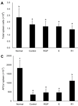
Antibody forming cells 형성능: 면양적혈구로 면역시 킨 마우스의 비장 세포를 면양적혈구와 보체를 혼합하여 배 양하면, 항체 생산세포(B 세포)는 면역글로불린을 방출하게 된다. 방출된 면역글로불린은 적혈구와 결합하고 보체가 작 용하여 항체 생성세포 주위의 적혈구가 용혈되어 plaque가 형성된다. CY에 의해 면역이 저하된 동물에서 RGP, E, R1 분획이 AFCs 형성능에 미치는 영향을 측정하였다. 총 비장 세포수는 정상군과 비교하여 대조군, RGP, E, R1은 각각 24%, 30%, 30%, 40% 수준으로 유의성 있게 감소되었으나 (
P
<0.05), 시료투여에 의한 군 간의 큰 변화는 없었다(Fig.3A). 106 비장 세포당 AFCs(AFCs/106 spleen cells)는 정 상군에 비하여 대조군은 76% 유의성 있게 감소하였으며 (
P
<0.05), 대조군에 비하여 RGP는 44%, E는 48% 증가하 는 경향을 보였고, R1은 321% 수준으로 크게 증가하였다 (P
<0.05, Fig. 3B). 비장당 AFCs(AFCs/spleen)는 정상군 과 비교하여 대조군이 80% 유의성 있게 감소하였으며(P
<b
a a
a a
0.0 0.5 1.0 1.5 2.0 2.5 3.0 3.5
Normal Control RGP E R1
Total spleen cells (×10 8) .
A
ba a
a b
0 2000 4000 6000 8000 10000 12000
Normal Control RGP E R1
AFCs/10 6 spleen cells .
B
b
a a a
c
0 500 1000 1500 2000 2500
Normal Control RGP E R1
AFCs/ spleen (×10 3) .
C
Fig. 3. Antibody forming cells to SRBCs in male Balb/c mice
orally administered with 6-year old red ginseng and its fractions for 10 days. Normal and control groups were administered with distilled water. The three test groups were administered with 100 mg/kg of RGP, E, and R1 for 10 days, respectively. CY was ad- ministrated intraperitoneally as a single dose of 50 mg/kg on day 5 before sacrifice. On the following day of CY injection, mice were immunized with 2.5×10⁸ SRBCs per mouse in 0.5 mL of EBSS i.p.. The results are presented as the mean±SD of 8 animals per group. Values with different letters (a-c) above the bars are significantly different at P<0.05.Table 4. Organ weight in male Balb/c mice orally administered with first residue or its fractions for 10 days
Absolute organ weight Relative organ weightSpleen Liver Thymus Spleen Liver Thymus
Normal Control R1 R2 W
0.110±0.01 0.084±0.01**
0.092±0.01* 0.093±0.01* 0.086±0.01
1.350±0.10 1.312±0.09 1.373±0.05 1.350±0.01 1.349±0.06
0.059±0.01 0.041±0.01**
0.048±0.01 0.045±0.01 0.047±0.01
0.0046±0.0004 0.0036±0.0004**
0.0039±0.0004**
0.0039±0.0006**
0.0036±0.0006*
0.057±0.001 0.056±0.002 0.058±0.002 0.056±0.003 0.057±0.002
0.0025±0.0004 0.0018±0.0003**
0.0020±0.0002 0.0019±0.0003 0.0020±0.0003 Normal and control groups were administered with distilled water.
The three test groups were administered with 100 mg/kg of R1, E, and R2 for 10 days, respectively. CY was administrated intra- peritoneally as a single dose of 50 mg/kg 5 days before sacrifice. On the following day of CY injection, mice were immunized with 2.5×10⁸ SRBCs per mouse in 0.5 mL of EBSS i.p..
The results are presented as the mean±SD of 8 animals per group.
Values are significantly different from normal at *
P<0.05,
**P<0.01.
0.05), 대조군에 비하여 RGP, E는 각각 33%, 32% 증가하 는 경향을 보였으나 R1은 214% 수준으로 유의성 있게 증가 하였다(
P
<0.05, Fig. 3C). R1은 면양적혈구에 대한 AFCs 형성능이 E보다 현저히 높았다. E의 산성 다당체와 진세노 사이드 함량은 각각 0.11%, 6.99%, R1의 산성 다당체와 진세노사이드 함량은 각각 4.93%, 0.40%였다. E는 산성 다 당체보다 사포닌 함량이 높았으나, R1은 산성 다당체의 함 량이 더 높았다(Table 2). 이는 사포닌보다 산성 다당체 함 량이 높은 비사포닌 계열이 면역활성에 더 큰 영향을 주는 것으로 생각된다.2차 분획의 면역활성
장기 무게 변화: 비장 무게는 정상군에 비하여 대조군에 서 23.8% 유의성 있게 감소하였으며(
P
<0.01), 대조군에 비하여 R1, R2, W는 각각 9.6%, 10.6%, 3.1% 증가하는 경향 을 나타내었다(Table 4). 흉선 무게는 정상군에 비해 대조군 에서 29.5% 유의적으로 감소하였으나(
P
<0.01) 대조군에 비하여 R1, R2, W는 각각 16.0%, 8.7%, 12.7% 수준으로 회복되는 경향을 나타내었다. 간 무게는 CY 투여로 큰 변화 가 나타나지 않았다(Table 4). 비장과 흉선의 상대 무게는 CY를 투여한 모든 실험군에서 유의성 있게 감소하였으며 (P
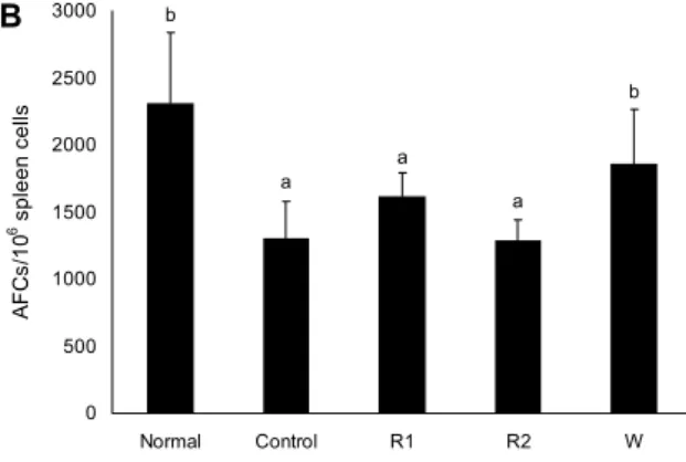
<0.05), 시험물질은 CY 투여에 의해 감소한 비장과 흉선 의 상대 무게에 큰 영향을 미치지 않았다.Antibody forming cells 형성능: 총 비장 세포수는 정 상군에 비하여 대조군, R1, R2, W에서 각각 27%, 21%, 21%, 27% 수준으로 감소되었고, 대조군과 비교하여 시료 투여군에서는 의미 있는 변화가 없었다(Fig. 4A). AFCs/
106 spleen cells은 정상군에 비하여 대조군에서 44% 유의
ab a a ab
b
0.0 0.5 1.0 1.5 2.0 2.5
Normal Control R1 R2 W
Total spleen cells (×108) .
A
b
a a
a b
0 500 1000 1500 2000 2500 3000
Normal Control R1 R2 W
AFCs/106 spleen cells .
B
b
a b
a c
0 50 100 150 200 250 300 350 400 450
Normal Control R1 R2 W
AFCs/ spleen (×103) .
C
Fig. 4. Antibody forming cells to SRBC in male Balb/c mice
orally administered with 100 mg/kg of R1, R2 or W for 10 days, respectively. Normal and control groups were administered with distilled water. CY was administrated intraperitoneally as a single dose of 50 mg/kg on day 5 before sacrifice. On the following day of CY injection, mice were immunized with 2.5×10⁸ SRBCs per mouse in 0.5 mL of EBSS i.p.. Four days later, the number of antibody forming cells (AFCs) was quantified. The results are presented as the mean±SD of 8 animals per group. Values with different letters (a-c) above the bars is significantly different atP<0.05.
적으로 감소하였으며(
P
<0.05), 대조군에 비하여 R1은 24%증가하는 경향을 나타내었고, W는 43% 수준으로 유의성 있게 증가하였으나(
P
<0.05), R2는 영향을 미치지 않았다 (Fig. 4B). AFCs/spleen은 정상군에 비하여 대조군은 57%유의적으로 감소하였다(
P
<0.05). 대조군에 비하여 R1과 W 에서 각각 36%, 42% 유의성 있게 증가하였으나(P
<0.05), R2는 거의 영향을 미치지 않았다(Fig. 4C). 산성 다당체 및 진세노사이드 함량은 W가 각각 7.46%, 0.61%, R1은 각각 4.93%, 0.40%, R2는 각각 0.50%, 0.30%였으며, W가 다른 분획에 비해 산성 다당체 함량이 가장 높았다. 시험물질의 산성 다당체 함량은 W, R1, R2 순으로 높았으며 AFCs 형성 능 결과와 일치하였다. 이는 산성 다당체 함량이 높은 분획 일수록 AFCs 형성이 증가함을 나타내었다.비장 세포 아형 분석
총 비장 세포수, T 및 B 세포수, macrophage 수: AFCs 형성능이 가장 우수한 W 분획이 CY 투여에 의해 감소된 면역세포수에 미치는 영향을 알아보기 위해 총 비장 세포수, T 세포수, B 세포수 및 macrophage 수를 측정하였다. 총 비장 세포수는 정상군에 비하여 대조군에서 62% 유의성 있 게 감소하였으며(
P
<0.05), 대조군에 비하여 W 50, 100, 200 mg/kg은 각각 32%, 42%, 62% 유의성 있게 증가하였 다(P
<0.05, Fig. 5A). T 세포수는 정상군에 비하여 대조군 이 54% 유의성 있게 감소하였으며(P
<0.05), 대조군에 비하 여 W 50, 100 mg/kg은 각각 40%, 57%로 증가하는 경향을 나타내었고, W 200 mg/kg은 54% 유의성 있게 증가하였다(
P
<0.05, Fig. 5B). B 세포수도 정상군에 비하여 대조군에 서 71% 유의적으로 감소하였으며(P
<0.05), 대조군에 비하 여 W 50, 100 mg/kg은 각각 22%, 16% 증가하는 경향을 보였고, W 200 mg/kg은 61% 유의적으로 증가하였다(P
<0.05, Fig. 5C). Macrophage 수는 정상군에 비하여 대조군 이 76% 유의성 있게 감소하였으며(
P
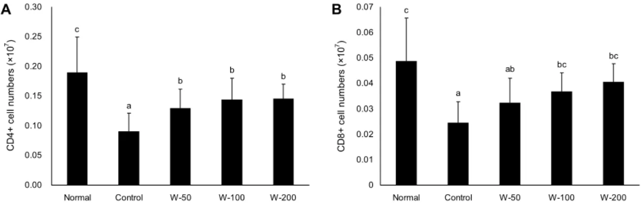
<0.05), 대조군에 비하 여 W 50, 100, 200 mg/kg은 각각 65%, 78%, 69% 증가하 였다(Fig. 5D).CD4+ 및 CD8+ T 세포수: W 분획이 CD4+ T 세포수와 CD8+ T 세포수에 미치는 영향을 측정한 결과 CD4+ T 세포 수는 정상군에 비해 대조군에서 52% 유의성 있게 감소하였 고(
P
<0.05), 대조군에 비하여 50 mg/kg은 43%, 100 mg/kg은 60%, 200 mg/kg은 61% 수준으로 용량 의존적으로 유의성 있게 증가하였다(
P
<0.05, Fig. 6A). CD8+ T 세포수 는 정상군에 비해 대조군에서 50% 수준으로 유의성 있게 감소하였으며(P
<0.05), 대조군에 비하여 50 mg/kg은 32%증가하는 경향을 보였고, 100, 200 mg/kg은 50%, 66% 수 준으로 용량 의존적으로 유의성 있게 증가하였다(
P
<0.05, Fig. 6B). 이상의 결과 산성 다당체 함량이 가장 높은 W 분획은 CY 투여로 인해 감소된 면역세포수를 증가시켰으며 항체형성능 또한 증가시켰다.수삼으로부터 분리된 150,000 Da의 분자량을 갖는다고 보고된 다당체 ginsan은 비장 세포를 IL-2 생성으로 암세포 를 사멸하는 LAK cell(lymphokine activated killer cell)로 유도시켰으며, B16-F10 melanoma의 폐 전이를 억제하였 고(30), macrophage 및 dendritic 세포에서 TNF-α, in-
b
ab b
a c
0.0 0.2 0.4 0.6 0.8 1.0 1.2 1.4 1.6 1.8
Normal Control W-50 W-100 W-200
Total spleen cell numbers (×10 7) .
A
b ab b
a c
0 0.05 0.1 0.15 0.2 0.25 0.3 0.35 0.4
Normal Control W-50 W-100 W-200
T cell numbers (×10 7) .
B
b
ab a a
c
0.0 0.1 0.2 0.3 0.4 0.5 0.6
Normal Control W-50 W-100 W-200
B cell numbers (×10 7) .
C
a a a
a b
0 0.005 0.01 0.015 0.02 0.025 0.03 0.035 0.04 0.045
Normal Control W-50 W-100 W-200
Macrophage cell numbers (×10 7) .
D
Fig. 5. The numbers of splenic cellularity. Male Balb/c mice orally administered with 50, 100, 200 mg/kg of W for 10 days,
respectively. Normal and control groups were administered with distilled water. CY was administrated intraperitoneally as a single dose of 50 mg/kg 3 days before sacrifice. The results are presented as the mean±SD of 8 animals per group. Values with different letters (a-c) above the bars are significantly different at P<0.05. CY: cyclophosphamide, W: water fraction from residue (R1) that red ginseng powder was extracted with 100% ethanol.b b b
a c
0.00 0.05 0.10 0.15 0.20 0.25 0.30
Normal Control W-50 W-100 W-200
CD4+ cell numbers (×107) .
A
bc bc ab
a c
0 0.01 0.02 0.03 0.04 0.05 0.06 0.07
Normal Control W-50 W-100 W-200
CD8+ cell numbers (×107) .
B
Fig. 6. The number of splenic CD4
+ and CD8+ T lymphocytes in male Balb/c mice orally administered with 50, 100, 200 mg/kg of W for 10 days, respectively. Normal and control groups were administered with distilled water. CY was administrated intra- peritoneally as a single dose of 50 mg/kg 3 days before sacrifice. The results are presented as the mean±SD of 8 animals per group. Values with different letters (a-c) above the bars are significantly different at P<0.05. CY: cyclophosphamide, W: water fraction from residue (R1) that red ginseng powder was extracted with 100% ethanol.terferone-γ와 같은 사이토카인을 생성하여 면역 조절작용 을 나타냄이 보고되었으며,
Salmonella
Typhimurium의 경 구 감염에 대하여 항체 생성을 증강시켰다(31). 홍삼은 수삼 을 증숙 후 건조하여 제조한 것으로 제조과정에서 화학성분 의 변화가 일어나 수삼 또는 백삼과는 다른 생리적 활성성분 의 함량 차이를 나타내고 있다(4,5). 다당체의 경우도 백삼 에서 분리되는 다당체와 홍삼에서 분리되는 다당체는 화학적 성상이 다를 것으로 예상되며, 일부 구성당은 분석되었으 나 상세한 이화학적 특성은 아직 밝혀지지 않았다. 인삼 또 는 홍삼에서 분리된 다당체가 항암작용 및 면역조절작용을 나타낸다는 연구는 있으나 사포닌과 비사포닌 중 어떤 성분 이 면역증진 효과에 더 효과를 나타내는지에 대한 비교 연구 는 없었다. 이에 본 실험은 홍삼 분획에 따른 사포닌 분획과 다당체 분획의 면역증진 효과를 비교한 실험으로 사포닌보
다는 비사포닌계의 다당체 성분이 더 큰 면역활성을 갖는 성분임을 나타내었다.
요 약
본 연구에서는 홍삼의 면역력 증진에 기여하는 성분을 체계 적으로 구명하기 위하여 홍삼분말을 에탄올, 물로 분획하면 서 각 분획의 AFCs 형성능 및 Flow cytometery를 이용하 여 비장 T 세포수, B 세포수, macrophage 수, CD4+ 및 CD8+ T 세포수를 측정하였다. 6년근 홍삼분말을 100% 에 탄올로 추출하여 에탄올 분획과 1차 잔사(R1)를 얻었고, 면 역활성이 우수한 1차 잔사(R1)를 물로 추출하여 물분획(W) 과 2차 잔사(R2)를 얻었다. 6년근 홍삼분말의 산성 다당체, 진세노사이드 함량은 각각 4.94%, 1.56%이며, 에탄올 분획 의 산성 다당체, 진세노사이드 함량은 각각 0.11%, 6.99%, 1차 잔사의 산성 다당체, 진세노사이드 함량은 각각 4.93%, 0.40%였다. 6년근 홍삼분말과 1차 분획에 대한 AFCs 형성 능 측정 결과 산성 다당체 함량이 높은 1차 잔사가 가장 우수 하였다. 면역활성이 우수한 1차 잔사를 물 추출하여 2차 분 획을 얻었으며, 2차 분획의 AFCs 형성능을 측정한 결과 물 분획이 가장 우수하였다. 물분획의 산성 다당체, 총 사포닌 함량은 각각 7.46%, 0.61%이며, 다른 분획에 비해 산성 다 당체 함량이 가장 높았다. 면역활성이 가장 우수한 물분획을 용량에 따른 비장 세포아형을 분석한 결과 cyclophos- phamide 투여에 의해 감소된 비장 총 세포수, T 세포수, B 세포수, macrophage 세포수, CD4+ 및 CD8+ T 세포수가 용량 의존적으로 유의성 있게 증가하였다. 이상의 결과로부 터 홍삼의 면역 증진을 나타내는 성분은 사포닌보다는 다당 체 함량이 높은 비사포닌계가 활성을 나타낼 것으로 제시되 었다. 앞으로 단계별로 분획을 제조하여 면역활성 기능 성분 으로서 다당체에 대한 구조를 구명하고 구조에 따른 면역활 성 연구가 필요할 것으로 생각된다.
REFERENCES
1. Goldby Ra, Kindt TJ, Osborne BA, Kudy J. 2003. Immunol-
ogy. 5th ed. WH Freeman and Company, New York, NY,
USA. p 5-11.2. Abo T, Kawamura T, Watanabe H. 2005. Immunologic states of autoimmune diseases. Immunol Res 33: 23-34.
3. Mok SK, Lee IH, Cheon SK. 1996. Korean ginseng (cultiva-
tion). Korea Ginseng & Tobacco Research Institute, Chunill
Press, Daejeon, Korea. p 130-196.4. Nam KY. 1996. Korean ginseng (component and efficacy).
Korea Ginseng & Tobacco Research Institute, Chunill Press, Daejeon, Korea. p 13-43.
5. Shibata S, Tanaka O, Ando T, Sado M, Tsushima S, Ohsawa T. 1966. Chemical studies on oriental plant drugs. ⅩⅣ.
Protopanaxadiol, a genuine sapogenin of ginseng saponins.
Chem Pharm Bull (Tokyo) 14: 595-600.
6. Jung NP, Song SO, Choi SU. 2000. Cytotoxicity of white and red ginseng against cancer cells and their effects on the cell cycle. J Ginseng Res 24: 183-187.
7. Nam KY. 2005. The comparative understanding between red ginseng and white ginseng processed ginseng (Panax
ginseng C.A. Meyer). J Ginseng Res 29: 1-18.
8. Kitagawa I, Taniyama T, Shibuya H, Noda T, Yoshikawa M. 1987. Chemical studies on the crude drug processing.
Ⅴ. On the constituents of white and red ginseng prepared from the same Panax ginseng root. Yakugaku Zassi 107:
495-505.
9. Kitagawa I. 1992. Chemical investigation of naturally occur- ring drug materials. Elucidation of scientific basis for tradi- tional medicines and exploitation of new naturally occurring drugs. Yakugaku Zassi 112: 1-41.
10. Li XG. 1992. Studies on the transforming mechanism of amino acid components in ginseng in the course of ginseng processing. Korean J Ginseng Res 16: 64-67.
11. Keum YS, Park KK, Lee JM, Chun KS, Park JH, Lee SK, Kwon H, Surh YJ. 2000. Antioxidant and anti-tumor pro- moting activities of the methanol extract of heat-processed ginseng. Cancer Lett 150: 41-48.
12. Jeon BH, Kim HS, Chang SJ. 1999. Effect of saponin and non-saponin of on the blood pressure in the renovascular hypertensive rats. J Ginseng Res 23: 81-87.
13. Kang SY, Kim ND. 1992. The antihypertensive effect of red ginseng saponin and the endothelium-derived vascular relaxation. Korean J Ginseng Res 16: 175-182.
14. Kim DH, Kwak KH, Lee KJ, Kim SJ. 2004. Effect of Korea red ginseng total saponin on repeated unpredictable stress induced changes of proliferation of neural progenitor cells and BDNF mRNA expression in adult rat hippocampus. J
Ginseng Res 28: 94-103.
15. Lee JH, Park HJ. 1995. Effects of lipophilic fraction from Korean red ginseng on platelet aggregation and blood coag- ulation in rats fed with corn oil and beeftallow diet. Korean
J Ginseng Res 19: 206-211.
16. Jung KY, Kim DS, Oh SR, Lee IS, Lee JJ, Park JD, Kim SI, Lee HK. 1998. Platelet activating factor antagonist activ- ity of ginsenosides. Biol Pharm Bull 21: 79-80.
17. Bae KC, Kim SH. 1998. Antioxidant effects of Korea gin- seng radix, Korean red ginseng radix and total saponin.
Korean J Oriental Medical Pathology 12: 72-81.
18. Kim JY, Park JY, Kang HJ, Kim OY, Lee JH. 2012. Benefi- cial effects of Korean red ginseng on lymphocyte DNA damage, antioxidant enzyme activity, and LDL oxidation in healthy participants: a randomized, double-blind, place- bo-controlled trial. Nutr J 11: 1-9.
19. Tomoda M, Shimada K, Konno C, Sugiyama K, Hikino H.
1984. Partial structure of panaxan A, a hypoglycaemic gly- can of Panax ginseng roots. Planta Med 50: 436-438.
20. Konno C, Sugiyama K, Kano M, Takahashi M, Hikino H.
1984. Isolation and hypoglycaemic activity of panaxans A, B, C, D and E, glycans of Panax ginseng roots. Planta Med 50: 434-436.
21. Hikino H, Oshima Y, Suzuki Y, Konno C. 1985. Isolation and hypoglycemic activity of panaxans F, G and H, glycans of Panax ginseng roots. Shoyakugaku Zasshi 39: 331-333.
22. Oshima Y, Konno C, Hikino H. 1985. Isolation and hypo- glycemic activity of panaxans I, J, K and L, glycans of
Panax ginseng roots. J Ethnopharmacol 14: 255-259.
23. Konno C, Hikino H. 1987. Isolation and hypoglycemic activ- ity of panaxans M, N, O and P, glycans of Panax ginseng roots. Int J Crude Drug Res 25: 53-56.
24. Konno C, Murakami M, Oshima Y, Hikino H. 1985. Isolation and hypoglycemic activity of panaxans Q, R, S, T and U, glycans of Panax ginseng roots. J Ethnopharmacol 14: 69-
74.
25. Park KM, Kim YS, Jeong TC, Joe CO, Shin HJ, Lee YH, Nam KY, Park JD. 2001. Nitric oxide is involved in the immunomodulating activities of acidic polysaccharide from
Panax ginseng. Planta Med 67: 122-126.
26. Kim YS, Park KM, Shin HJ, Song KS, Nam KY, Park JD.
2002. Anticancer activities of red ginseng acidic poly- saccharide by activation of macrophages and natural killer cells. Yakhak Hoeji 40: 113-119.
27. Byeon SE, Lee J, Kim JH, Yang WS, Kwak YS, Kim SY, Choung ES, Rhee MH, Cho JY. 2012. Molecular mecha- nism of macrophage activation by red ginseng acidic poly- sacchride from Korean red ginseng. Mediators Inflamm 2012: 1-7.
28. In G, Ahn NG, Bae BS, Han ST, Noh KB, Kim CS. 2012.
New method for simultaneous quantification of 12 ginseno- sides in red ginseng powder and extract: In-house method validation. J Ginseng Res 36: 205-210.
29. Kwak YS, Kim EM. 1996. The physicochemical properties of crude polysaccharide fraction isolated from Korean red ginseng (Panax ginseng C.A. Meyer). Korean J Food Sci
Technol 28: 389-392.
30. Kim KH, Lee YS, Jung IS, Park SY, Chung HY, Lee IR, Yun YS. 1998. Acidic polysaccharide from Panax ginseng, ginsan, induces Th1 cell and macrophage cytokines and generates LAK cells in synergy with rIL-2. Planta Med 64:
110-115.
31. Na HS, Lim YJ, Yun YS, Kweon MN, Lee HC. 2012. Ginsan enhances humoral antibody response to orally delivered antigen. Immune Netw 10: 5-14.