홍삼과 발효홍삼의 항염증 작용 및 항알러지 효과 비교
박혜진, 정다혜, 주해미, 강남성, 장선아1, 이재근2, 손은화1*
성균관대학교 약학부, 1강원대학교 생약자원개발학과, 2강원대학교 식물자원응용공학과
The comparative study of anti-allergic and anti-inflammatory effects by fermented red ginseng and red ginseng
Hye-Jin Park, Da-Hye Jung, Haemi Joo, Nam-Sung Kang, Seon-A Jang1, Jae Geun Lee2, and Eun-Hwa Sohn1*
College of Pharmacy, Sungkyunkwan University, Suwon 440-746, Korea
1Department of Herbal Medicine Resource, Kangwon National University, Samcheok 245-711, Korea
2Department of Applied Plant Science, Kangwon National University, Chuncheon 200-701, Korea
Abstract - Red ginseng(RG, steamed and dried root of Panax ginseng C. A. Meyer, family Araliaceae) and fermented red ginseng(FRG, fermented red ginseng by yeast and lactic acid bacteria) are known to show different pharmacological effects by changed composition of saponins through fermentation. We examined the effects of RG and FRG on β-hexosaminidase secretion, ICAM-1 expression, the mitogen-induced proliferation of lymphocyte from mice in ex vivo systems and HaCaT cell(keratinocyte) proliferation to compare the anti-allergic and anti-inflammatory effects between both groups. RG groups showed inhibition of β-hexosaminidase secretion and ICAM-1 expression at 1μg/ml, 10μg/ml and the same effects were observed at all concentrations in FRG groups. In our study, RG increased LPS-induced B cell proliferation at 1μg/ml and ConA-induced B cell proliferation at 100 μg/ml but FRG decreased LPS- and ConA-induced lymphocytes at 100 μg/ml. We showed that FRG increased the proliferation of HaCaT at 1, 10μg/ml but not by RG. These findings suggest that RG and FRG might have anti-inflammatory and anti-allergic effects, which can be needed to proper clinical concentration to applied to various allergic diseases and inflammation.
Key words -red ginseng, fermented red ginseng, ICAM-1, β-hexosaminidase, lymphocyte proliferation
*
교신저자(E-mail) :[email protected]서 언
인삼(Panax ginseng C. A. Meyer)은 오가피 나무 과(Araliaceae)의 인삼속(Panax)에 속하는 다년생 음지 성 숙근초로서 한반도를 중심으로 극동지역에 분포하고 있다. 인삼은 크게 인삼의 원형을 유지하는 1차 가공방법 에 따라 수삼(생인삼), 백삼, 홍삼으로 구분한다. 우리가 흔히 인삼으로 칭하는 것은 수삼(수분 약 75%)을 건조한 백삼(수분 약 12%)을 말하여, 홍삼은 4~6년근 수삼을 엄 격히 선별하여 껍질을 벗기지 않은 상태에서 증기로 쪄서 건조시킨 담황갈색 또는 담적갈색 인삼을 말한다(Cho et al., 2006). 본 원고에서는 수삼 및 백삼을 인삼으로 언급
하여, 홍삼과 구별하여 사용하였다. 인삼의 주요 약리활성 성 분은 사포닌 즉, 진세노사이드(ginsenoside) 성분으로 알려 지고 있으나, 인삼 중에는 산성다당체(acidic polysaccharide), panaxydol 등의 polyacetylene계 성분, 말톨(maltol) 등 의 아미노산 유도체 성분 등을 포함한 비사포닌계의 여러 가지 약리활성 성분도 함유하고 있다. 이러한 성분 중 일부 성분들이 홍삼 제조과정 중 화학구조가 변환되어 새로운 사포닌 성분이 생성되기도 하고 일부 생리활성 성분의 함 량이 증가하기도 한다(Lars et al., 2008).
인삼에 존재하는 사포닌은 주로 MG-Rb1, MG-Rb2, MG- Rc, MG-Rd와 같은 트리테르펜 올리고 배당체(triterpene- oligoglygoside)인 말로닐-진세노사이드(malonyl-ginsenoside;
MG)인데, 홍삼을 제조하는 가열과정 중에 산성사포닌인
MG의 malonic acid가 이탈되면서 홍삼에는 G-Rg3, GRg2, G-Rh2, G-Rs1, G-Rs2, Rh4와 같은 미량 사포닌이 특 이적으로 존재하게 된다. 이러한 약리활성 성분의 변화를 갖는 홍삼은 인삼의 효능과 완전히 다르지는 않으나, 혈액 순환개선효과, 암발생 억제력, 감염방어 작용 등이 인삼보 다 우수하다는 연구가 보고되어 있다(Nam, 2005; Sung et al., 2009). 또한 최근에는 항알러지 작용, 기억력개선 작용, 발기부전효과 등에 대한 홍삼의 새로운 효능이 보고 되고 있는데, 이러한 효능은 인삼과 달리 홍삼에만 존재하 는 G-Rg3, G-Rf와 G-Rh2 등의 성분에서 나타나는 효과 일 수 있다는 가능성이 제기되고 있다(Bae et al., 2005;
Jang et al., 2008).
임상적으로, 홍삼을 복용했을 때 홍삼의 사포닌 성분들 은 체내에서 직접 흡수되는 것이 아니라, Bifidobacterium sp., Lactobacillus sp., Saccharomyces sp. 등의 인체 장내 미생물들에 의해 G-Rh2, compound K 등의 대사물 로 전환된 후 흡수됨으로, 인체에서는 최종적으로 이러한 성분들이 더 높은 약리활성을 나타내게 된다(Trinh et al., 2007; Yang et al., 2007).
따라서 홍삼의 다양하고 우수한 약리활성 작용에도 불구 하고 개인별 장내 미생물의 분포 및 활성화의 차이로 인하 여 실제로 홍삼의 약효에 차이에 나타낼 수 있다. 이를 극 복하기 위하여 인삼을 장내환경과 같은 무산소 상태에서 장내 미생물을 이용하여 발효(fermentation)하는 과정을 거쳐 최종 대사산물의 성분으로 변환 혹은 증가시키는 발 효홍삼 제조에 관한 연구 및 개발이 이루어지고 있다(Kong et al., 2008). 유산균, 효모, 고초균 등 우리에게 유익한 미생물을 이용한 발효기술의 진보로 천연물의 생리활성효 능이 증가된 발효산물을 얻거나 또는 상호간의 상승효과에 의해 생리활성효능이 상승되는 제품들이 개발되고 있다 (Jeon et al., 2005). Bifidobacterium 속 유산균으로 발 효시킨 발효홍삼에서 고지혈증 및 고혈당을 개선시키는 효 과가 보고된 바 있으며(Trinh, et al., 2007), Bacillus lichenifomis 균주로 발효시킨 청국장 담금에 홍삼을 혼합 하여 발효시킨 발효홍삼청국장에서도 고혈당과 고지혈증 을 감소시키는 것으로 나타나(Lee et al., 2005), 발효홍 삼 제품 개발에 많은 주목을 받고 있는 실정이다.
최근 산업화, 도시화의 심화로 변화된 주거환경, 식생활 의 변화, 유전적인 영향, 환경오염에 의해 발생되는 화학 적, 생물학적 유해인자들에 대한 노출은 유년기를 포함한
다양한 연령층에서의 알러지 질환을 크게 증가시키고 있는 실정이다(Heo et al., 2008). 이는 인체가 무해한 외부환 경에 대해서도 민감한 반응을 보이며, 지속적으로 천식이 나 비염과 같은 호흡기계 또는 만성 피부염증, 아토피 질환 등의 피부계 증상 등으로 일상생활은 물론 정서적인 면에 도 악영향을 미치는 것으로 알려져 있다(Lebovidge et al., 2009; Fukamizu et al., 2009). 그러므로 본 연구에 서는 최근 들어 면역증강효과, 항염증 작용 및 항알러지 효 과가 보고되고 있는 홍삼추출물(red ginseng: RG)과 장내 미생물의 발효과정에 의해 최종 대사산물 성분을 높이려는 제조법으로 개발된 발효홍삼(fermented red ginseng:
FRG)을 이용하여 면역증강효과, 항염증, 항알러지 및 피 부세포의 증식효과를 상호 비교함으로써, 홍삼 또는 발효 홍삼을 보다 유효하고 적절하게 선택하여 사용할 수 있도 록 하기 위한 기초 자료를 제공하고자 하였다.
재료 및 방법
세포주 및 재료
RBL-2H3(rat basophilic leukemia) 세포는 American Type Culture Collection(ATCC, Manassas, VA)에서 구입 하였고 15% FBS, 2% penicillin-streptomycin(10,000 U pen/㎖, 10,000 ㎍ strep/㎖)을 첨가한 MEM(GIBCO BRL, Grand Island, NY) 성장배지에 부착 배양하였으며, HaCaT (human keratinocyte) 세포는 10% FBS, 2% penicillin- streptomycin(10,000 U pen/㎖, 10,000 ㎍ strep/㎖)을 첨가한 DMEM(GIBCO BRL, Grand Island, NY) 성장배 지에 부착 배양하였으며, 37℃, 5% CO2 조건을 유지하였다.
홍삼 추출물과 발효홍삼 추출물
홍삼추출물 RG의 제조는 우선 홍삼 원물을 70%의 주정 (발효주정, 대한주정판매주식회사)을 사용하여 8시간 동안 1회, 15% 주정을 사용하여 8시간 동안 2회 추출하였다. 추 출된 시료를 농축(60~64 cm/Hg, 65brix) 하여 주정을 회 수하고 5배의 정제수로 희석하였다. 원심분리(Continuous centrifuge, Kansai, Japan)를 이용하여 여과하고 60℃
이하에서 진공감압농축(60~64 cm/Hg, 67brix)하여 홍 삼농축액을 획득하였다. 발효홍삼 FRG 시료는 얻어진 홍 삼농축액에 6배의 정제수를 가하고 95℃에서 2시간 동안 살균․냉각시킨 후 효모배양액을 1/20 수준으로 첨가하고
26℃를 유지하면서 3일 동안 발효를 수행하였다. 첨가 된 효모배양액은 발효기(KF-500, 한국발효기)를 이용 하여 발효배지(멸균정제수 85%와 홍삼분말 15%)에 1×10⁸ CFU/g까지 배양된 Sacharomyces cerevisise (IFO 0309)와 Kluyveromyces lactis(ATCC 8585)를 1% 수준으로 접종하고 26 ℃에서 5일간 배양하여 1×10⁶ CFU/g이 되도록 하여 조제하였다. 효모에 의해 발효가 완 료된 홍삼추출액에 다시 유산균 배양액을 1/20 수준으로 첨가하고 36℃를 유지하면서 2주 동안 발효를 수행하였다.
첨가된 유산균배양액은 발효기(KF-500, 한국발효기)를 이 용하여 발효배지(멸균정제수 85%와 홍삼분말 15%)에 1×10⁸ CFU/g까지 배양된 유산균 Lactobacillus plantarum (299V Probi AB, Sweden), Lactobacillus casei(Shirota, Yakult, Japna), Bifidobacterium longum(BB536, Morinaga Milk Industry Co., Ltd. Japan)을 1×10⁸ CFU/g까지 배양된 액를 1% 수준으로 접종하고 36℃에서 5일간 배양하여 유산균수가 1×10⁶ CFU/g 이상이 되도록 배양한 액을 사용하였다.
β-Hexosaminidase 분비능 측정
β-Hexosaminidase 분비능은 Dastych 등(Dastych et al., 1999)의 방법에 의해 측정하였다. RBL-2H3을 24-well plate에 2×10⁵ cells/well로 분주하고, anti-DNP IgE 200 ng/㎖을 처리한 다음 37℃, 5% CO2 incubator에 서 16시간 배양하였다. 각 세포들을 TGCM buffer(136 mM NaCl, 2.68 mM KCl, 0.36 mM NaH2PO2· H2O, 1 mM CaCl2, 0.5 mM MgCl2, 11.9 mM NaHCO3, 5 mM Dextrose, 1g/L gelatin, pH 7.4)로 2회 세척한 후 각 well 당 Tyrode’s buffer와 시약을 처리한 후 30분 동안 반응시켰다. 이 후 DNP-BSA 1㎍/㎖를 사용하여 세포를 37℃, 5% CO2
incubator에 30분 반응시키고 ice bath에서 10분 동안 배 양한 후 반응을 종결시켰다. 상층액 15 ㎕를 96-well plate 에 옮기고, pellet은 Triton X-100(0.1%) 200 ㎕를 24 well 에 넣고 녹인 후 96 well에 15 ㎕ 넣는다. 상층액과 pellet 이 담긴 96 well에 substrate buffer(p-nitrophenyl-N- acetyl- β-D-glucosaminide 1 mM, citrate buffer 0.05 M, pH 4.5) 60 ㎕을 넣고 37℃ 에서 1시간 배양시킨 다음 각 well에 stop solution 150 ㎕을 첨가하여 반응을 종결시켰다.
405 nm에서 흡광도를 측정하여, 시료와 대조구의 흡광도
값으로 다음 식에 의해 degranulation(%) 을 산출하였다.
% Degranulation = ODsupernatant/(ODsupernatant+ODtriton x-100) × 100
Western blot analysis
IFN-γ를 이용하여 HaCaT 세포의 염증 환경을 만든 후, RG 또는 FRG를 처리한 실험군과 대조군을 각각 lysis하여 30 ㎍의 단백질을 얻은 후, Bradford’s proetin assay법 으로 단백질을 정량하였다. 동일한 양의 단백질을 sodium dodecyl sulfate-polyacrylamide gel electrophoresis (SDS-PAGE) gel에 전기 영동한 후 PVDF membrane으로 Tank-type transfer unit을 이용하여 transfer 하였다. 단 백질이 옮겨진 membrane은 1시간 동안 상온에서 blocking buffer(5% skim milk와 0.1% Tween 20을 함유한 TBST 용액)를 처리한 후 각 검증 단백질에 대한 1차 항체(anti- ICAM-1)를 가하여 1~2시간 동안 반응시켰다. 이어 0.1%
Tween 20을 함유한 TBST용액으로 10분간 3회 이상 세척 후 2차 항체(HRP-conjugated anti-IgG antibody)를 1시간 동안 상온에서 반응시켰다. 다시 3회 이상 세척 후에 detection reagent(Amersham™)를 가한 후 Fuji Mmedical X-ray film(Biomax Light)에 노출시켜 각 단백질의 발현 량을 확인하였다.
Keratinocyte cell proliferation
HaCaT 각질형성세포주를 96-well plate에 1×10⁵/well 이 되도록 분주하고 RG와 FRG를 농도별로 처리한 후 각각 24시간 동안 배양한 후 MTT assay를 이용하여 세포 증식 능을 측정하였다.
Splenocyte blsatogenesis
8주령이 된 생쥐에서 분리된 비장세포(splenocyte)를 96-well plate에 5×10⁵/well이 되도록 분주하고 mitogen 으로 10 ㎍/㎖ LPS와 2.5 ㎍/㎖ ConA를 첨가하여 각각 B 세포와 T 세포의 세포증식력을 측정하였다. 전체 배양 부 피는 200 ㎕로 하여, 37℃, 5% CO2 배양기에서 48시간 배 양 후 20 ㎕ phenazine methosulphate(PMS; electron- coupling reagent)와 25 ㎕ XTT를 가하고, 3시간 더 배양 한 후 XTT formazan의 생성량을 측정하여 세포증식력을 확인하였다.
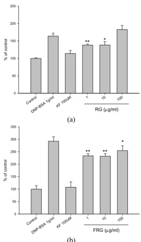
Control DNP-BSA 1g/
ml
KF 100uM 1 10 100
0 50 100 150 200 250
RG (μg/ml)
% of control
** *
(a)
FRG (μg/ml) Control
DNP-BSA 1g/
ml
KF 100uM 1 10 100
% of control
0 50 100 150 200 250 300 350
** **
*
(b)
Fig. 1. (a) The effect of RG and (b) FRG on secretion of β -hexosaminidase from mast cell(RBL-2H3). After sensitization of RBL-2H3 cells with DNP-specific IgE for 16 hrs, cells were reincubated with Tyrode’s buffer and RG, FRG for 30 min, and then stimulated with DNP-BSA for 30 hr. The supernatnats were reacted with an equal volume of substrates and the reaction product was measured at 405 nm. The inhibitory effects of β-hexosaminidase secretion were compared with that of ketotifen fumarate. The results are mean ± S.E.
of quintuplicates from a representative experiment(*p<0.05,
**p<0.01; significantly different from the DNP-BSA antigen- treated groups).
결과 및 고찰
β-Hexosaminidase 분비능에 미치는 영향 초기 알러지 반응을 매개하는 비만세포(mast cell)의 히 스타민(histamine) 유리 작용은 알러지 반응의 병리적 진 행과정에서 필수적이다. 비만세포는 항원 또는 면역학적인 자극에 의해 활성화되었을 때, 세포표면의 FcεRI 수용체가 IgE 및 특이적 알레르겐(allergen)과 결합하여, 세포내 칼 슘농도의 상승으로 탈과립되면서, 다양한 사이토카인을 분 비한다(Church et al., 1997). 이 중 β-hexosaminidase 는 비만세포의 활성화에 의해 히스타민과 함께 분비되기 때문에 일반적으로 in vitro 알러지 반응에 관한 실험적 연 구의 비만세포 탈과립(degranulation) 반응의 주요 표지 자로 사용되고 있다(Matsuda et al., 2002). 본 연구에서 는 RG와 FRG가 항원자극에 의한 비만세포의 탈과립 과정 에 미치는 조절 효과를 알아보기 위하여 RG 또는 FRG를 전처리 하고 항원 자극에 의한 β-hexosaminidase 분비량 을 측정하였다. RG는 1 ㎍/㎖과 10 ㎍/㎖에서 FRG는 1, 10, 100 ㎍/㎖ 모두에서 감소 효과를 나타내었다(Fig. 1).
이는 RG와 FRG가 모두 항원 자극에 의한 비만세포의 탈 과립 효과를 감소시킴으로써 항알러지 효과를 나타낸다고 볼 수 있으며, RG와 FRG는 유효 농도에 차이가 있음을 알 수 있다.
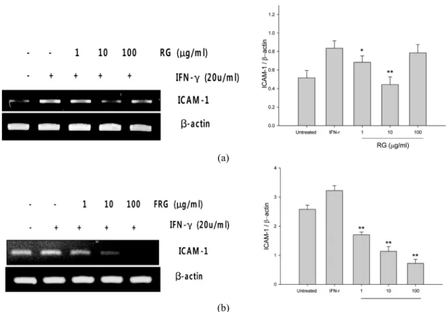
ICAM-1의 발현에 미치는 영향
세포부착분자는 여러 염증성 질환의 발생에 중요한 역할을 하는 세포 표면 당단백질(glycoprotein)로 구조적 차이에 따라 크게 selectins, integrins, immunoglobulin superfamily 등 세 가지로 나눠진다. 세포부착분자의 발현 조절에 의해 발생하는 염증세포의 내피 부착성은 염증 반응 에서 초기에 일어나는 중요한 단계인데, 이 중 immunoglobulin superfamily에 속하는 ICAM-1(intracellular adhesion molecules-1)의 발현은 각종 염증세포의 이동 및 침윤에 관여하는 중요한 과정으로, ICAM-1 세포부착분자의 발현 억제는 염증 반응을 조절하는데 크게 작용한다(Koyama et al., 1992). 본 연구에서는 RG 또는 FRG가 IFN-γ 염증성 사이토카인에 의해 유발된 HaCaT 세포의 ICAM-1의 발현 증가에 대하여 각각 어떤 영향을 미치는지를 확인하였다.
실험결과 RG는 저농도 1 ㎍/㎖과 10 ㎍/㎖에서 IFN-γ에 의해 유발된 ICAM-1의 발현을 감소시켰으며, FRG는 1,
10, 100 ㎍/㎖ 모두에서 ICAM-1의 발현을 농도 의존적으로 감소시켰다(Fig. 2). 이러한 결과는 RG와 FRG가 모두 ICAM-1의 발현 억제 조절을 통해 항염증효과를 나타낼 수 있다는 것을 보여주며, β-hexosaminidase 분비량 조절효과 에서와 같이 ICAM-1의 발현 조절에도 RG와 FRG의 유효 농도에서 차이가 있음을 알 수 있다. RG 및 FRG가 일부 약리 활성에 있어 농도 의존적으로 나타나지 않고 오히려 고농도에서 작용을 나타내지 않는 이유는 과잉의 유효 성
(a)
(b)
Fig. 2. (a) The effect of RG and (b) FRG on ICAM-1 expression in IFN-γ-treated keratinocyte. HaCaT cells were treated with RG or FRG in the absence or presence of IFN-γ (20u/㎖) for 12 hrs. Total protein was extracted and subjected to Western blot anaysis for ICAM-1 expression in protein level. Densitometric analysis of the immunoreactive bands. The control value was set as 100 % and presented as a bar graph. Similar observations were obtained in two other experiments.
분에 의한 상경적 작용이 수용체와의 결합을 오히려 방해 할 수 있기 때문으로 사료된다.
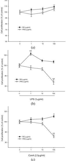
Mitogen에 의한 림프구(lymphocyte)의 세포증식효과 Resting lymphocyte는 외부 자극에 의하여 활성화되는 단계에서 proliferation이 일어난다. 이러한 활성화 정도는 mitogen-stimulated response에 의하여 쉽게 확인할 수 있으며, 이는 mitogen이 특이적으로 감작된 어떤 lymphocyte subpopulation에 결합하여 cell cycle을 G0 phase에서 G2 phase로 자극함으로써 mitosis를 일으켜 lymphocyte population을 유용하여 측정할 수 있게 하는 방법이다 (Burleson et al., 1995). RG 또는 FRG가 lymphocyte의 세포증식효과에 영향을 미치는지 알아보기 위하여 생쥐의 비 장(spleen)으로부터 비장세포(splenocyte)를 분리하여 B세 포의 mitogen LPS와 T세포의 mitogen ConA (concanavalin A)를 사용하여 세포증식능을 측정하였다. 실험에 사용된 비
장세포는 LPS 처리에 의해 각각 182.83%로 세포증식을 나 타내었고, ConA 처리에 의해 151.85%로 세포증식이 일어 남을 확인함으로써 LPS와 ConA가 본 실험에 mitogen으 로써 적절히 활용되었음을 확인하였다(data not shown).
Fig. 3(a)에 의하면 mitogen을 처리하지 않은 RG 또는 FRG를 단독 처리한 실험군에서는 유의성 있는 세포증식변 화를 나타내지 않았다. 그러나, LPS를 처리한 B 림프구 증 식에서 RG는 1 ㎍/㎖ 농도에서 유의적인 증가 효과를 나타 내었며, FRG에서는 100 ㎍/㎖에서 오히려 B 림프구 증식 을 감소시켰다(Fig. 3(b)). ConA를 처리한 T 림프구 증식 에 있어서도 RG는 100 ㎍/㎖ 농도에서 유의적인 증가 효 과를 나타내었으나, FRG 100 ㎍/㎖ 농도에서는 오히려 T 림프구 증식을 감소시켰다(Fig. 3(c)). 이러한 결과는 FRG 의 항염증 효과와 같이 과도한 면역반응을 억제한다는 의 미에서 긍정적으로 평가될 수도 있으나, FRG의 100 ㎍/㎖
농도에서 림프구의 면역능을 오히려 감소시킬 수 있다는
0 1 10 100 50
60 70 80 90 100 110 120
Cell proliferation (% of control)
RG (μg/ml) FRG (μg/ml)
(a)
0 1 10 100
60 80 100 120
RG (μg/ml) FRG (μg/ml)
Cell proliferation (% of control)
**
**
LPS (1μg/ml)
(b)
0 1 10 100
60 80 100 120
Cell proliferation (% of control)
**
ConA (2.5μg/ml)
RG (μg/ml) FRG (μg/ml)
*
(c)
Fig. 3. (a) The effect of RG and FRG on proliferation of splenocytes from mice. (b) The effect of RG and RRF on proliferation of LPS-treated splenocytes from mice. (c) The effect of RG and RRF on proliferation of ConA-treated splenocytes from mice. Splenocytes from mice were treated with FRG and RG respectively in the absence or presence of LPS or ConA for 68hrs. The proliferation of splenocytes was assessed by XTT assay. Cell density was measured at a test wavelength of 450 nm and a reference wavelength of 690 nm. Cell proliferations were represented by the percentage of control. The results are mean ± S.E. of quintuplicates from a representative experiment(*p<0.05, **p<0.01;
significantly different from the LPS alone or ConA alone treated group).
Control 1 10 100
0.1 0.2 0.3 0.4
RG (μg/ml) FRG (μg/ml)
Cell proliferation (O.D. 540nm) **
*
Fig. 4. The effects of FRG and RG on proliferation of keratinocyte. HaCaT cells were treated with RG or FRG for 24 hrs. The proliferation of keratinocytes(HaCaT) was assessed by MTT assay. Cell density was measured at 540 nm. Cell proliferations were represented by the percentage of control. The results are mean ± S.E. of quintuplicates from a representative experiment(*p<0.05, **p<0.01; significantly different from the control).
양면성을 지니기도 한다. 따라서 FRG의 사용에 있어서는 100 ㎍/㎖ 고농도 보다는 저농도의 사용이 건강기능성 식 품 사용에 대한 안전한 유효농도라고 평가된다.
Keratinocyte 세포 증식 효과
각질형성세포(keratinocyte)는 피부의 각질층 구성에 필 수적인 세포로 피부에서 발생하는 면역반응의 1차적인 방 어체로 작용한다. 인체와 외부환경 사이의 투과막인 피부 각질장벽이 손상되면 경표피수분손실(transepidermal water loss)이 증가되고 수분결합력(water-bindng capacity)이 감소되어 소양증이 심해지고 피부가 건조해진다(Schaefer et al., 1991). 그러므로 이러한 각질형성세포의 증식은 알 러지성 염증 질환을 완화시킬 수 있다. 실험 결과 RG는 각 질형성세포의 세포 증식능에 영향을 주지 않았으나, FRG는 1 ㎍/㎖과 10 ㎍/㎖ 농도에서 유의성 있는 세포 증식력을 나 타내었다(Fig. 4).
적 요
홍삼은 인삼을 증숙하는 과정에서 생겨나는 화학적으로 변화된 생리활성물질들에 의해 인삼과 차별화된 효과를 보 여준다. 또한 홍삼은 장내미생물의 생물학적 전환에 의해 변화되어 생체내 약리활성을 보이는데, 이는 개인의 장내 미 생물의 분포 차이에 의해 약리작용의 차이를 보일 수 있다.
최근 이 같은 차이를 극복하기 위해 인위적으로 장내미생 물에 의해 사전 발효시킨 발효홍삼은 홍삼과 다른 성분량 의 차이를 보임으로써 독특한 약리작용을 보인다고 알려져 있다. 이에 본 연구는 홍삼과 발효홍삼의 추출물이 알러지 및 염증반응에 끼치는 영향에 대해 알아보기 위해 비만세 포의 탈과립 과정에서 분비되는 β-hexosaminidase 분비 량 비교, 염증매개 세포부착물질인 ICAM-1 발현비교, 생쥐 림프구의 증식능, 각질형성세포 증식능을 상호 비교하였다.
실험결과 홍삼은 1, 10 ㎍/㎖에서 β-hexosaminidase 분 비량 억제 및 ICAM-1 발현 억제 조절 효과를 나타내었으 며, 발효 홍삼은 모든 농도에서 유의적인 억제 효과를 나타 내었다. 그러므로 홍삼과 발효홍삼에서는 모두 항알러지 및 항염증 효과가 있다고 사료된다. 그러나 LPS에 의한 B 세포의 증식능에서 홍삼 1 ㎍/㎖은 증가효과를 발효홍삼에 서는 100 ㎍/㎖에서 억제효과를 나타내었고, ConA에 의한 T세포의 증식능에서는 홍삼 100 ㎍/㎖에서 증가효과를 발 효홍삼 100 ㎍/㎖에서는 억제효과를 나타냄으로써 림프구 증식능에 관한 실험에서는 두 추출물간에 상반된 효과를 나타내었다. HaCaT세포를 이용한 각질형성세포 증식능에 서는 발효홍삼 1, 10 ㎍/㎖에서만 효과를 나타내었다. 이러 한 결과를 종합해 볼 때, 홍삼과 발효홍삼에 존재하는 성분 량의 변화에 따라 그 효능에서 차이를 나타내고 있는 것으 로 사료되며, 홍삼 및 발효홍삼의 고농도 100 ㎍/㎖에서는 저농도와는 다른 효과를 보이는 경향을 나타내고 있으므 로 홍삼 및 발효홍삼의 사용에 있어 적절한 적응증과 주성 분의 검증 및 유효 농도에 대한 연구가 더욱 필요하다고 판단된다.
사 사
본 연구는 중소기업청에서 지원하는 2009년도 산학연공 동기술개발사업(No. 00038453)의 연구수행으로 인한 결과 물임을 밝힙니다.
인용문헌
Bae, E.A., M.J. Han, Y.W. Shin and D.H. Kim. 2005.
antiallergic and antipsoriatic effects of korean red ginseng.
J. Ginseng Res. 29(2):80-85.
Burleson, G.R., J.H. Dean, and A.E. Munson, 1995. Methods
in immunotoxicology. Wiley-Liss, New York, USA. Vol 1.
197-210.
Church, M.K., and F. Levi-Schaffer. 1997. The human mast cell. J. Allergy Clin. Immunol. 99:155.
Cho, W.C., W.S. Chung, S.K. Lee, A.W. Leung, C.H. Cheng and K.K. Yue. 2006. Ginsenoside Re of Panax ginseng possesses significant antioxidant and antihyperlipidemic efficacies in streptozotocin-induced diabetic rats. Eur. J.
Pharmacol. 550:173-179.
Dastych, J., A. Walczak-Drzewiecka, J. Wyczolkowska and D.D. Metcalfe. 1999. Murine mast cells exposed to mercuric chloride release granule-associated N-acetyl-beta- dhexosaminidase and secrete IL-4 and TNFalpha. J Allergy Clin Immunol 103:1108-1114.
Fukamizu R, M.H. Jang and M. Miyasaka. 2009. The role of eosinophils in allergic inflammation and intestinal immunity.
Nippon Rinsho. 67(11):2088-2093.
Heo, Y. and H.A. Kim. 2008. Correlation between skin prick test and enzyme-linked immunoabsorbent assay using serum for identification of subjects positive to major respiratory allergens. J. Env. Hlth. sci., 24(5):369-373.
Jang, D.J., M.S. Lee, B.C., Shin, Y.C. Lee and E. Ernst.
2008. Red ginseng for treating erectile dysfunction: a systematic review. Br. J. Clin. Pharmacol. 66(4):444-450.
Jeon, B.S., J.W. Park, B.K. Kim, H.K. Kim, T.S. Jung, J.R.
Hahm, D.R. Kim, Y.S. Cho and J.Y. Cha. 2005. Fermented mushroom milk supplemented dietary fiber prevents the onset of obesity and hypertriglyceridemia in Otsuka Long- Evans Tokushima Fatty (OLETF) rats. Diabetes Obes.
Metab. 7: 709-715.
Kong B.M., M.J. Park, J.W. Min, H.B. Kim, S.H. Kim, S.Y.
Kim and D.C. Yang. 2008. Physico-Chemical Characteristics of White, Fermented and Red Ginseng Extracts. J. Ginseng Res. 32:238-243.
Koyama S., T. Ebihara and K. Fukao. 1992. Expression of intercellular adhesion molecule-1 during the development of invasion and/or metastasis of gastric carcinoma. J.
Cancer Res. Clin. Oncol. 118:619-614.
Lars, P.C. 2008. Ginsenosides: Chemistry, Biosynthesis, Analysis, and Potential Health Effects. Advances in Food and Nutrition Research. 55:1-99.
Lebovidge, J.S., H. Strauch, L.A. Kalish and L.C. Schneider.
2009. Assessment of psychological distress among children and adolescents with food allergy. J. Allergy Clin. Immunol.
124(6):1282-1288.
Lee, S.I., J.G. Shin and S.D. Kim. 2005. Effect of ginseng- chungkukjang effects on lipid profiles of serum in alcohol administered diabetic-induced rats. J. Korean Soc. Food Sci. Nutr. 34: 1362-1366.
Matsuda, H., T. Morikawa, K. Ueda, H. Managi and M.
Yoshikawa. 2002. Structural requirement of flavonoids for inhibition of antigen-induced degranulation, TNF-α and IL-4 production from RBL-2H3 cells. Bioorg. Med.
Chem. Lett. 10:3123-3128.
Nam, K. Y. 2005. The comparative understanding between red ginseng and white ginsengs, processed ginsengs(panax ginseng C. A. Meyer). J. Ginseng Res. 29:1-18.
Trinh HT, S.J. Han, S.W. Kim, Y.C. Lee and D.H. Kim.
2007. Bifidus fermentation increases hypolipidemic and hypoglycemic effects of red ginseng. J. Microbiol Biotechnol.
17(7):1127-1133.
Schaefer, L. and K. Kragballe. 1991. Abnormalities in epidermal lipid metabolism in patients with atopic dermatitis.
J Invest Dermatol 96:10-5.
Sung, H., Y.S. Jung and Y.K. Cho. 2009. Beneficial effects of a combination of Korean red ginseng and highly active antiretroviral therapy in human immunodeficiency virus type 1-infected patients. Clin. Vaccine Immunol. 16(8):1127-1131.
Yang, C.S., S.R. Ko, B.G. Cho, J.Y. Lee, K.H. Kim, D.M.
Shin, J.M. Yuk, H.J. Sohn, Y.S. Kim, J.J. Wee, J.H. Do and E.K. Jo. 2007. Compound K (CK) Rich Fractions from Korean Red Ginseng Inhibit Toll-like Receptor(TLR) 4- or TLR 9- mediated Mitogen activated Protein Kinases Activation and Pro-inflammatory Responses in Murine Macrophages. J. Ginseng Res. 31:181-190.
(접수일 2010.3.4; 수락일 2010.9.1)