Received February 1, 2011; Revised February 17, 2011;
Accepted March 7, 2011
Corresponding author: Ki-Young Lee
Tel: +82 (63) 469-1832 e-mail: [email protected] 1225-3480/24376
Gametogenic Cycle and the Spawning Season by Quantitative Statistical Analysis and the Biological Minimum Size of Cyclina sinensis in
Western Korea
Ee-Yung Chung1, Chang Hoon Lee2, Young Je Park3, Moon Sul Choi4, Ki-Young Lee4 and Dong-Ki Ryu5
1Korea Marine Environment & Ecosystem Institute, Dive Korea, Bucheon 420-857, Korea;
2Neoenbiz Co., Bucheon 420-806, Korea;
3Sea Green Industry Institute, Bucheon 420-851
4Department of Marine Biotechnology, Kunsan National University, Gunsan 573-701, Korea
5Department of Aquaculture and Aquatic Sciences, Kunsan National University, Gunsan 573-701, Korea
ABSTRACT
The gametogenic cycle and the spawning season in female and male Cyclina sinensis were investigated by quantitative statistical analysis using an image analyzer system, and the biological minimum size (the size at 50%
of sexual maturity) was calculated by combination of quantitative data by size and von Bertalanffy's equation.
Compared the gametogenic cycle by quantitative statistical analysis with the previous qualitative results in female and male C. sinensis, monthly changes in female and male gametogenic cycles calculated by quantitative statistical analysis showed similar patterns to the gonadal stages in female and male reproductive cycles by qualitative histological analysis. Comparisons of monthly changes in the portions (%) of each area to eight kinds of areas by quantitative statistical analysis in the gonads in female and male C. sinensis are as follows. Monthly changes in the portions (%) of the ovary areas to total tissue areas in females and also monthly changes in the portions of the testis areas to total tissue areas in males increased in March and reached the maximum in May, and then showed a rapid decrease from June to October. Monthly changes in the portions (%) of oocyte areas to ovarian tissue areas in females and also monthly changes in the portions of the areas of the spermatogenic stages to testis areas in males began to increase in March and reached the maximum in June in females and males, and then rapidly dropped from July to October in females and males when spawnig occurred. From these data, it is apparent that the number of spawning seasons in female and male C. sinensis occurred once per year, from July to October. Monthly changes in the number of the oocytes per mm2 and in the mean diameter of the oocyte in captured image which were calculated for each female slide showed a maximum in May and reached the minimum from December to February. Therefore, C. sinensis in both sexes showed a unimodal gametogenic cycle during the year. The percentage of sexual maturity of female and male clams ranging from 25.1 to 30.0 mm in length was over 50% and 100% for clams over 40.1 mm length. In this study, the biological minimum size (sexually mature shell lengths at 50% of sexual maturity) in females and males were 26.85 and 26.28 mm, respectively.
Key words: Cyclina sinensis, gametogenic cycle, spawning season, quantitative statistical analysis
INTRODUCTION
The Chinese cyclina, Cyclina sinensis (Bivalvia:
Veneridae), is one of the commercially important edible clams in East Asian countries, including Korea, China, and Japan (Kwon et al., 1993; Min et al., 2004). The species is mainly found intertidally along the coast of Simpo, Jollabuk-do, Korea, inhabiting silty bottoms. As a consequence of reckless over-harvesting, the standing stock of this clam has
dramatically declined in recent years (Kim et al., 2000b) and the species has been denoted as a target organism and fisheries resource that should be managed using a more reasonable fishing regimen (Kim et al., 2000b). For propagation and management, it is important to understand the population characteristics with regard to the gametogenic cycle and the biological minimum size (the size at 50% of sexual maturity).
To date, regarding Cyclina sinensis, several studies have focused on reproductive ecology, including early embryonic development and growth (Choi, 1971, 1975), artificial fertilization and development (Choi and Song, 1973), reproductive cycle (Chung et al., 2003), and sexual maturity (Chung et al., 1991; Kim et al., 2000b) by qualitative histological analysis, and on the aspect of physiology, including parasite infection by Trematoda (Kim et al., 2000a).
Some authors (Ropes and Stickney, 1965;
Brousseau, 1978) reported that the gametogenic cycle can be classified into the unimodal or bimodal gametogenic cycles by quantitative statistical analysis using an image analyzer system. It is known that in particular, Mya arenaria and Mercennaria mercennaria in bivalve mollusc exhibited a change from a unimodal to a bimodal cycle with decrease in latitude (Ropes and Stickney, 1965; Brousseau, 1978).
In particular, in case of different local populations of the same species (e.g., Ruditapes philippinarum), there are some differences in the number of spawning seasons per year in other areas of the world: In Canada, USA, northern Japan, Vostok Bay, Korea, and the northwestern part of the Sea of Japan, there is one spawning season during the year (Quayle and Bourne, 1972; Holland and Chew, 1974; Pourovsky and Yakovlev, 1992; Choi et al., 2005), while there are two spawning seasons during the year in southern parts of Tokyo Bay (Tanaka, 1954; Ohba, 1959). Thus, the number of spawning seasons of R. philippinarum varied with latitudinal gradients (locations) of the world.
As mentioned above, it is well-known that the number of spawning seasons per year vary with latitudinal gradients (locations) of the world. In
particular, in case of Korea, the number of spawning seasons of C. sinensis were once a year by qualitative histological analysis (Chung et al., 1991; Kim et al., 2000b; Chung et al., 2003, 2004). However, in fact, the spawning season and the number of spawning seasons per year by qualitative histological analysis is not correct because two or more stages often occurred simultaneously within each tissue section. In this case, therefore, the staging criteria decisions were made according to conditions of majority of tissue sections by researcher’s individual subjectivity. Thus, sometimes the qualitative analysis of gonad developmental stages by individual subjectivity is not correct. Therefore, the period of spawning season and the number of spawning seasons needs to be studied by quantitative statistical analysis for the confirmation of the unimodel or bimodel gametogenic cycles of gonads per year.
Thus, despite the studies referred above, little information is available on the gametogenic cycle and the spawning season by quantitative statistical analysis, and the biological minimum size (the size at 50% of sexual maturity) of this species. Understanding the gametogenic cycle and the number of spawning seasons of this species will provide necessary information for the determination of age and recruitment period and for the estimation of the spawning frequency during the spawning season.
Additional information on the biological minimum size (the size at 50% of sexual maturity) of this species would be very useful for propagation, aquaculture, and resource management. In particular, information on the size at which individuals reach 50% of sexual maturity could be useful in determining a prohibitory measure for adequate natural resource management. In the present study the gametogenic cycle and the number of spawning seasons by quantitative statistical analysis, and biological minimum size of C. sinensis were studied because we could not find mentioned above studies.
MATERIALS AND METHODS 1. Sampling
Specimens of the Chinese cyclina, C. sinensis, were collected monthly at the intertidal and subtidal zones
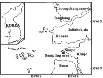
Fig. 1. Map showing the sampling station.
of Simpo, Jollabuk-do, Korea (Fig. 1) from January to December 2007. The clams ranging from 17.4 to 54.6 mm in shell length were used for the present study.
After the clams were transported alive to the laboratory, shell length and total weight were immediately measured.
2. Production of histological tissue section slides of ovarian tissues
For light microscopic examination of histological tissue section slides by quantitative analysis, a total of 245 individuals were used for the production of histological tissue section slides of ovarian tissues.
Ovarian tissues were removed from shells and preserved in Bouin’s fixative for 24h. They were then washed with running tap water for 24h. then dehydrated in alcohol and embedded in paraffin molds. Embedded tissues were sectioned at 5-7 μm thickness using a rotary microtome. Sections were mounted on glass slides, stained with Hansen’s hematoxylin-0.5% eosin. After production of histological tissue section slides of ovarian tissues, they were examined using a light microscope (Zeiss Axiovert 10 microscope) for quantitative statistical analysis.
3. Quantitative statistical analysis using an image analyzer system
Tissue slides were observed for quantitative analysis by an image analyzer system. Slides were viewed on a stereozoom microscope (Nikon, SMZ-U)
from where the images were captured by a TOSHIBA Model IK-642K CCD camera and were then viewed on a SAMSUNG color video monitor. The image analyzer (BMI plus, WINATech Co.) is capable of automatic measurement of the area and diameter of polygons encircled by the operator, counting objects that are contrasted by background color (in black and white mode), and statistical analysis of numerous characteristics of objects in the captured images.
Measurements for female were carried out for areas of total tissue, the ovary, the follicle, and the oocyte, the number of the oocyte per unit area, and the diameter of each oocyte. Measurements for male were carried out for areas of total tissue, the testis, and the spermatogenic stages. Measurements on the areas of total tissue, the ovary and the testis were conducted at magnification of 7.5 at which the field area of captured image was 60 mm2, while the other measurements were done at magnification of 75 (field area: 0.524 mm2). 15-20 individuals per month and two fields per slides were analyzed. Areas of total tissue, the ovary, the testis, the follicle, and the oocyte were measured by manually tracking the margins of objects one by one with a pointer on the captured image. Before the measurements of the areas of the oocytes, and the diameter of the oocytes and the spermatogenic stages, captured color image was converted to a gray scale image and then to a black-and-white image with an appropriate threshold at which only the spermatogenic stages or oocytes were contrasted with black, while the other parts of the tissue were in white. Measurements on the areas of the spermatogenic stages and the oocytes, and the diameter of oocytes were carried out by the automatic procedure provided by the BMI plus software.
Number of oocyte was also recorded during this automatic procedure.
From the measured values of image analyses, (A) percent of field occupied by the ovary to total tissue, (B) percent of field occupied by the follicle to total tissue, (C) percent of field occupied by the follicle to the ovary, (D) percent of field occupied by the oocyte to the ovary, (E) the number of the oocyte per mm2 of the ovary, and (F) mean diameter of the oocyte in
captured image were calculated for each slide of female clams.
From the measured values of image analyses, (A) percent of field occupied by the testis to total tissue, and (B) percent of field occupied by the spermatogenic stages to the testis were calculated for each slides of male clams.
4. Statistical analysis
A one-way ANOVA (multiple comparisons by Duncan's procedure, a=0.05) was applied to compare the means of monthly data. One-way t-tests were used to determine significant differences in the data of two adjacent months. All statistical analyses were done using the SPSS package.
5. Percentages (%) of sexual maturities by sizes using qualitative analysis of histological section slides
For determination of the percentages (%) of sexual maturity, a total of 280 (143 females and 137 males) clams of histological preparations (17.4-54.6 mm in shell length) were examined % of sexual maturity by shell length through histological observations, from May to October, 2007. The formular of the percentage (%) of sexual maturity is as follows: The percentage (%) of sexual maturity = No. of mature individuals x 100 / No. of total individuals investigated.
6. Calculation of the size at rate (50%) of group sexual maturity (RM50) by quantitative analysis.
For the investigation of age composition by size, von Bertalanffy's equation used by Son et al. (1996) is as follows: SLt = SL∞(1- e-k (t-t0)). Where, SLt: shell length (mm) at age t, SL∞: theoretical maximum shell length, k: constant expressing rate of approach to SL∞, t: age t, to: theoretical age at which SL = 0 mm, TW: total weight (g) at age t.
To calculate the size at the rate (50%) of sexual maturity after fitting the rate of sexual maturity to an exponential equation, the size equivalent to the size at 50% of sexual maturity was estimated to be the sexually mature length of the population (Chung and Ryou, 2000). The exponential equation of the rate of sexual maturity is as follows: RM = 100/1+exp (a-bx), where, RM: rate of sexual maturity; a, b:
constants, x: shell length.
RESULTS
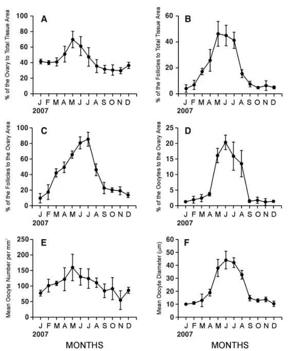
1. Gametogenic cycle by quantitative statistical analysis Female C. sinensis showed a unimodal gametogenic cycle (Fig. 2). The percent of field occupied by the ovary area to total tissue area in female began to increase in April, and reached a maximum in May (69.8%), and then gradually decreased from June to October (61.3%-30.7%, p=0.002). During the periods of January-March and September-December, the ovarian tissue area occupied about 30%-40% of total tissue. There were no significant differences during the periods of January-April, May-June, July-August, and August-December (one-way ANOVA, p=0.121, 0.178, 0.101, and 0.110, respectively, Fig. 2A).
The percent of field occupied by the follicle area to total tissue area (Fig. 2B) and the percent of field occupied by the follicle area to the ovarian tissue area (Fig. 2C) in females showed similar patterns each other. The peak of the follicle area to total tissue were found in May (Fig. 2B), while that of the follicle area to the ovary area were in July (Fig. 2C). The follicle area in the ovary increased gradually from January to July (9.6%-85.5%, p < 0.001), and then decreased rapidly until October. From September to December, it fluctuated with lower value less than 20% (Fig. 2C). The percent of ovarian tissue occupied by oocytes also showed a distinct seasonal pattern. During the periods of June-August, the percents of oocyte areas showed higher values (13.5%-20.3%, p = 0.114), whereas during January-April and September-December, it showed lower values (1.2%-3.7%, p = 0.163). The variations among individuals were great especially during May-August. Monthly changes the portions (%) of oocyte areas to ovarian tissue areas in females began to increase in March and reached the maximum in June, and then rapidly dropped from July to October when spawnig occurred (Fig. 2D). The number of the oocytes increased from January to May (77.3-159.7 eggs·mm-2, p = 0.055), after reaching a peak in May, and then decreased from June to November (129.8-54.6 eggs·mm-2, p = 0.020) with slight fluctuations. However, the variations of the
Fig. 2. Monthly changes in quantitative reproductive traits in Cyclina sinensis. A: Percent of the area occupied by the ovary to total tissue area. B: Percent of the area occupied by follicles to total tissue area. C: Percent of the area occupied by follicles to the ovary area. D: Percent of the area occupied by oocytes to the ovary area. E: Percent of mean oocyte number per mm2. F: Mean Oocyte Diameter (μm).
number of oocytes among individuals were greater than between variations of all combinations of adjacent months (t-test, a = 0.05), the differences of means of the number of oocytes between adjacent months were not statistically significant (Fig. 2E).
The seasonal pattern was well reflected also in the mean diameter of oocytes. As shown in Fig. 2F, the mean oocyte diameter increased from April to June (18.9-44.0 μm, p = 0.002), reached a maximum in June, and then declined from July to October
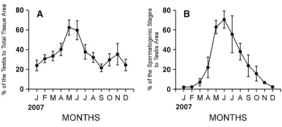
Fig. 3. Monthly changes in quantitative reproductive traits in Cyclina sinensis. A: Percent of area occupied by testis to total tissue area. B: Percent of area occupied by spermatogenic stages to testis area.
(42.1-12.7 μm, p < 0.001). There were good coincidences of patterns among the area of spermatogenic stages in male, the area of follicles, area of oocytes, and the mean diameter of oocytes in female.
Male C. sinensis showed a unimodal gametogenic cycle (Fig. 3). The percent of field occupied by the testis to total tissue in male began to increase in February (Fig. 3A). The testis area gradually increased from February to March (30.9%-33.5%, p = 0.423), and greatly increased from April to May (40.2%-62.4%, p=0.004), and then reached its maximum in May, and then decreased from June to September (59.7%-21.5%, p=0.002). However, it showed somewhat irregular changes. From October to December, the testis area fluctuated within the range of 25%-35%. Variations of the testis area among individuals were so high that there were nosignificant differences during January-March, February-April, May-Jun, July-August, and October-December (one way ANOVA, p=0.091, 0.069, 0.574, 0.069, and 0.057, respectively).
The percent of field occupied by the spermatogenic stages to the testis area in males changed from 2.0% in January to 70.5% in June (Fig. 3B). Seasonal cycle was found evidently from the area of the spermatogenic stages. It greatly increased from March to May (7.0%-62.9%, p < 0.001), reached maximum in June, and
then decreased from July to October, and continuously to December (55.6%-2.2%, p =0.007). There were no significant differences during January-March, March-April, May-July, August-September, and October-December (one way ANOVA, p=0.172, 0.082, 0.108, 0.084, and 0.172, respectively).
Monthly changes in the portions of the areas of the spermatogenic stages to testis areas in males began to increase in March and reached the maximum in June, and then rapidly dropped from July to October in males when spawnig occurred (Fig. 3B).
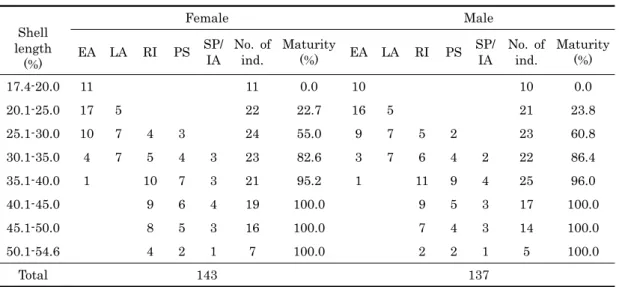
2. Sizes at sexual maturity by histological observations A total of 280 (143 females and 137 males) individuals of C. sinensis were investigated histologically to determine the shell lengths of clams that reach maturation and participate in reproduction from May (maturation before spawning) to late October (after spawning). As shown in Table 1, percentages of smaller female and male individuals ranging from 17.4−20.0 mm in shell length were 0%, and those individuals were in the early active stage, which is characterized by a small number of oogonia and early developing oocytes in oogenic follicles in the ovary, and the appearance of a small number of spermatogonia and spermatocytes in the acini of the testis. It is supposed that their sizes at sexual maturities can not be reached until October when
Shell length
(%)
Female Male
EA LA RI PS SP/
IA
No. of ind.
Maturity
(%) EA LA RI PS SP/
IA
No. of ind.
Maturity (%)
17.4-20.0 11 11 0.0 10 10 0.0
20.1-25.0 17 5 22 22.7 16 5 21 23.8
25.1-30.0 10 7 4 3 24 55.0 9 7 5 2 23 60.8
30.1-35.0 4 7 5 4 3 23 82.6 3 7 6 4 2 22 86.4
35.1-40.0 1 10 7 3 21 95.2 1 11 9 4 25 96.0
40.1-45.0 9 6 4 19 100.0 9 5 3 17 100.0
45.1-50.0 8 5 3 16 100.0 7 4 3 14 100.0
50.1-54.6 4 2 1 7 100.0 2 2 1 5 100.0
Total 143 137
EA, early active stage; LA, late active stage; RI, ripe stagel; PS, partially spawned stage;
SP/LA, spent/inactive stage.
Table 1. Shell lengths of sexual maturity in female and male Cyclina sinensis during the breeding season
spawning was completed. Percentages of sexual maturities of female and male clams ranging from 20.1-25.0 mm shell length were 22.7% and 23.8%, respectively, and those individuals were in the early active stage and late active stages. At this time, younger animals had a small number of oogonia and developing oocytes in the late active stage in the follicles of the ovary, and a number of spermatocytes, spermatids, and a small number of spermatozoa appeared in acini of the testis. It is supposed that sexual sizes of most individuals can not be reached until October September when spawning of a few mature individuals were completed. However, percentages of sexual maturities of female and male clams ranging from 25.1-30.0 mm in shell length were 55.0% and 60.8%, but those individuals were in the early active, late active, ripe, and partially spawned stages during the breeding season. Percentages of sexual maturities of all individuals of shell length greater than 40.1 mm are 100%, and those individuals were in the late, ripe, partially spawned, and spent/inactive stages. Therefore, it is assumed that most individuals can reach full maturity until September if they are larger 25.1 mm in shell length at that time. In this study, percentages of sexual maturities of female and male clams ranging from 25.1 to 30.0 mm were over 50.0%. So, we can not
understand accurate sizes at 50% sexual maturities of female and male individuals. Therefore, we have to calculate their accurate shell lengths at 50% of sexual maturities by quantitative analysis. Percentages were 100% for female and male clams over 40.1 mm length.
3. Size at the rate (50%) of sexual maturity (RM50)
As shown in Fig. 4, shell lengths at 50% sexually mature clams (the biological minimum size = sizes at the rate (50%) of sexual maturity, RM50) that were fitted to an exponential equation were 26.85 mm in females and 26.28 mm in males, respectively.
DISCUSSION
1. Comparisons of the ovarian gametogenic cycle by qualitative and quatitative analyses
To compare the results of the gametogenic cycle by qualitative and quantitative analyses of C. sinensis in this study, we quoted the results on the reproductive cycle in female C. sinensis already reported by Kim et al. (2000b). They reported that the reproductive cycle in female C. sinensis in Komso Bay, Jeollabuk-do, Korea by qualitative analysis (histological observation) could be classified into 5 successive stages: the early acive stage (February to April), late active stage (March to June), ripe stage (April to August), partially spawned stage (July to October), with a peak
Fig. 4. Relationships between the rates of sexual maturity (%) and shell lengths (mm) in female and male Cyclina sinensis.
spawnig between July and August), and spent/inactive stage (September to February).
In this study, according to the results of gametogenic cycle by quantitative analysis, the results of monthly changes in proportions (%) of the oocyte areas to the ovary areas began to rapid increase in March, and reached a maximum in June, thereafter, their proportions (%) gradually decreased from July to October when spawning occurred, and the main spawning occurred between July and August. In particular, peak mature oocyte level occurred in June followed by a significant decrease from July to October which indicated spawning (the main spawning occurred between July and August).
In females, the results of monthly changes in the number of the oocytes per mm2, and mean diameter of the oocytes showed the same or similar patterns:
the maximum in May-June, and then rapidly dropped from July to October which indicated spawning.
Therefore, compared female gametogenic cycle by quantitative statistical analysis with those by qualitative histological analysis (Kim et al., 2000b;
Chung et al., 2004), the results the gametogenic cycle studied by quantitative statistical analysis in this study coincided with gonadal maturation and the spawning season studied by qualitative histological analysis (Kim et al., 2000b; Chung et al., 2004). In females, spawning of this species was clarified from
July to October by qualitative analysis (Kim et al., 2000b; Chung et al., 2004),
According to the results of qualitative histological analysis by Kim et al. (2000b), the reproductive cycle in male C. sinensis in Gomso Bay, Jeollabuk-do, Korea could be classified into five successive stages: early acive stage (February to April), late active stage (March to June), ripe stage (April to August), partially spawned stage (July to October), and spent/inactive stage (September to February).
In this study, according to the results of gametogenic cycle in male C. sinensis by quantitative analysis, monthly changes in the percent of field occupied by the spermatogenic stages to the testis area increased from March to May reached maximum in June, and then decreased continuously from July to October which indicated spawning. Compared the results of the gametogenic cycle by quantitative statistical analysis with qualitative histological analysis already reported by Kim et al. (2000b), in this study, the spawning period of this species by quantitative statistical analysis using an Image Analyzer System was from July to October. Therefore, on the whole, the results of gametogenic cycle in female and male C. sinensis by quantitative analysis coincided with the reproductive cycle, gonadal maturation and the spawning season by qualitative histological analysis.
2. Number of the spawning season
From the results of the number of spawning seasons investigated by quantitative statistical analysis using an Image Analyzer System, the gametogenic cycle in both sexes was clarified to be a unimodal gametogenic cycles showing a maximum maturity in June and one spawning season per year, from July to October with peak spawning between July and August. Regarding the spawning season of different local populations of R. philippinarum, it is well-known that the number of spawning seasons by qualitative analysis (histological observations) varied with latitudinal gradients (Rand, 1973).
Giese (1959) and Sastry (1979) reported that in general, latitudinal differences in timing of the reproductive cycles of marine molluscs. In particular, some authors (Ropes and Stickney, 1963; Brousseau, 1978; Heffernan et al., 1989a) reported that Mya arenaria and Mercennaria mercennaria in Bivalve mollusc exhibited a change from a unimodal to a bimodal cycle with decrease in latitude. However, another authors (Heffernan and Walker, 1989;
Heffernan et al., 1989b) reported that several other bivalves (i.e, Geukensia demisa, Crassostrea virginica and Spisular solidissima similis) showed unimodal gametogenic cycle in the southeastern U.S. waters (Kanti et al., 1993). In this study, the gametogenic cycle in female and male C. sinensis by quantitative statistical analysis showed a unimodal gametogenic cycle.
Cyclina sinensis showed a unimodal gametogenic cycle as found in other clams in Korean coasts.
Quantitative results by image analyses of C. sinensis showed the peaks of maturity in May-June (Figs. 1, 2). Compared to qualitative results (frequencies of gonadal phases) of other clams in previous works, the period of maturity of C. sinensis is quite similar to May-June of Ruditapes philippinarum (Chung et al., 1994), Mactra veneriformis (Chung and Ryou, 2000), and Meretrix lusoria (Chung, 2007), In this study, peaks of the oocyte area in females and the spermatogenic stages in males in June (Fig. 2D, 3B) implies the readiness of gonadal maturation.
Spawning seemed to initiate in July by decrease of both the oocyte area and the area the spermatogenic
stages. Significant decreases in the oocyte area and the area of the spermatogenic stages and were found during July-October, indicating the spawning period.
The spawning period of C. sinensis was also similar to other clams mentioned above (June-September for R.
philippinarum, M. veneriformis, and M. lusoria May-October for S. purpuratus).
3. Size at sexual maturity
The percentage of first sexual maturity of female and male clams ranging from 25.1 to 30.0 mm length were over 50%, and this was 100% for clams over 40.1 mm length. According to the growth curves for the mean shell length fitted to von Bertalanffy’s equation by Kim et al. (1986), individuals ranging from 25.1 to 30.0 mm in shell length were considered to be two years old because Kim et al.(1986) reported that 31.2 mm in shell length was considered to be two years old.
We assume that both sexes of this population begin reproduction about two years of age. To clarify the biological minimum size, we should calculate shell lengths at 50% of sexual maturity to clarify acurate shell lengths which were fitted to an exponential equation by von Bertalanffy's equation already used by Kim et al (1986) in female and male clams.
4. Biological minimum size (the size at the rate (50%) of sexual maturity (RM50))
Biological minimum size (sizes at the rate (50%) of sexual maturity, RM50) that fitted to an exponential equation were 26.85 mm in females and 26.28 mm in males.
In terms of natural resource management, the present study suggests that harvesting clams less than 26.28 mm in shell length (< 2 years old) can potentially lead to a drastic reduction in recruitment.
A prohibitory measure should be taken for adequate natural resources management.
ACKNOWLEDGEMENTS
The authors are grateful to Dr. Tae Hwan Lee, the University Of Michigan for helpful comments on the manuscript. This research was supported in part by
the fund from the Research Projects of Costal Research Center, Kunsan National University.
REFERENCES
Brousseau, D.J. (1978) Spawniong cycle. fecundity and recruitment in a pupulation of soft-shell clam. Mya arenaria from Cape ann, Massachusetts. Fisheries Buletin. 76: 155-166.
Choi, K.C. (1971) Ecological studies of the clams, Meretrix lusoria and Cyclina sinensis for increasing seed clam yield. Korean Journal of Limnology, 4:
9-19.
Choi, S.S. aand Song, Y.K. (1973) Studies on the artificial fertilization and development of Cyclina sinensis. Bulletin of Korean Fisheries Society, 6:
76-80.
Choi, S.S. (1975) Comparative studies on the early embryonic development of Cyclina sinensis. Bulletin of Korean Fisheries Society, 8: 185-195.
Choi, K.H., Park, G.M. and Chung, E.Y. (2005) Ovarian maturation in female Ruditapes philippinarum on the west coast of Korea. Development and Reproduction, 9: 123-134.
Chung, E.Y, Lee, T.Y and An C.M. (1991) Sexual maturation of the venus clam, Cyclina sinensis, on the west coast of Korea. Journal of Medical &
Applied Malacology, 3: 125-136.
Chung, E.Y, Ryou D.K, and Lee J.H. (1994) Gonadal development, age and growth of the shortnecked clam, Ruditapes philippinarum (Pelecypoda:
Veneridae), on the coast of Kimje. Korean Journal of Malacology, 19: 38-54.
Chung, E.Y and Ryou D.K. (2000) Gametogenesis and sexual maturation of the surf clam, Mactra veneriformis on the west coast of Korea.
Malacologia, 42: 149-163.
Chung, E.Y., Hur, Y.B., Kwak O.Y. and Choi, K.H.
(2003) Ovarian maturation, artificial spawning and spawning frequency of the venus clam, Cyclina sinesis, in the Gimje Coastal Waters of Korea.
Korean Journal of Malacology, 19: 153-159.
Chung, E.Y., Park, K.H., Kim, J.B. and Lee, C.H. (2004) Seasonal changes in biochemical components of the adductor muscle and visceral mass tissues in female Cyclina sinensis, in relation to gonad developmental phases. Korean Journal of Malacology, 20: 85-92.
Chung, E.Y. (2007) Oogenesis and sexual maturation in Meretrix lusoria (Roding 1978, Bivalvia: Veneridae) in western Korea. Journal of Shellfish Research, 26:
71-80.
Giese, A.C. 1959. Compartative Physiology: Annual reproductive cycles of marine invertebrates. Review of Physiology, 21: 547-576.
Heffernan, P.B., Walker, R.L. and Carr, J.L. (1989a) Gametogenic cycles of three bivalves in Wassaw Sound, Georgia Ⅰ: Mercenaria mercenaria
(Linnaeus, 1758). Journal of Shellfish Research, 8:
51-60.
Heffernan, P.B., Walker, R.L. and Carr, J.L. (1989b) Gametogenic cycles of three bivalves in Wassaw Sound, Georgia II: Crassostrea virginica (Gmelin, 1971). Journal of Shellfish Research, 8: 61-70.
Heffernan, P.B. and Walker, R.L. (1989) Gametogenic cycles of three bivalves in Sassaw Sound, Georgia
Ⅲ: Geukensia demissa (Dillwyn) Journal of Shellfish Research, 8: 327-334.
Holland, D.A. and Chew K.K. (1974). Reproductive cycle of the manila clam Washington. Proceedings of National Shellfish Research Association, 64: 53-58.
Kanti, A, Heffernan, PB, and Walker, R.L. (1993) Gametogenic cycle of the southern surfclam, Spisula solidissimasimilis (Say, 1822), from St. Catherine Sound, Georgia. Journal of Shellfish Research, 12:
255-261.
Kim, J.R., Chung, E.Y., Choi, M.S. and Ryou, M.H.
(1986) Environment in Busa Bay and Marine Resource Biological Studies. Bulletin of the Institute of Natural Sciences, Kunsan National University, I:
151-197.
Kim, Y.G., Chung, E.Y. and Kim, Y.H. (2000a) Studies on reproductive ecology and parasite of the venus clam, Cyclina sinensis on the west coast of Korea.
2. On the Metacercaria of Himasthia kussigi Yamaguchi, 1939 (Trematoda) found in the venus clam, Cyclina sinensis. Korean journal of Malacology, 16: 43-48.
Kim, Y.H., Chung, E.Y. and Kim, Y.K. (2000b) Reproductive ecology and parasite of the venus clam, Cyclina sinensis (Gmelin), on the west coast of Korea 1. Reproductive ecology. Korean journal of Malacology, 16: 35-41.
Kwon, O..K, Park, G.M, and Lee, J.S. (1993) Coloured shells of Korea. Academy Publication Co. Seoul 288 pp.
Min, D.K., Lee, J.S, Ko, D.B, Je, J.G. (2004) Mollusks in Korea. Hanguel Graphics, Busan, Korea 566 pp.
Ohba, S. (1959) Ecological studies in the natural population of a clam, Tapes japonica, with special reference to seasonal variations in the size and structure of population and to individuals growth.
Biological Journal of Okayama University, 5: 13-42.
Ponurovsky, S.J. and Yakovlev, Y.M. (1992) The reproductive biology of the Japanese littleneck.
Tapes philippinarum (A. Adams & Reeve, 1850) (Bivalvia: Venerida). Journal of Shellfish Research, 11: 265-277.
Quayle, D.B.. and Bourne, N. (1972) The clam fisheries of British Columbia. Fisheries Research Board Canadian Bulletin, 179: 70.
Rand, W.M. (1973) A stochastic model of the temporal aspect of breeding strategies. Journal of Theoretical Biology, 40: 337-351.
Ropes, J.W. and Stickney, A..P. (1965) Reproductive cycle of Mya arenaria in New England. Bioogical
Bulletin, 128: 315-327.
Sastry. A.N. (1979) Pelecypoda (Excluding Ostreidae). pp 113-292. In: Reproduction in marine invertebrates.
Vol. 5: Pelecypods and lesser classes. A. C. Giese and J.S. Pearse (eds.) Academic Press, New York.
Tanaka, Y. (1954) Spawning season of important bivalves in Ariake Bay Ⅲ. Tapes philippinarum.
Bulletin of Japanese Society Science Fisheries, 19:
1165-1167.