Received: December 14, 2012 ; Accepted: December 21, 2012
Corresponding author: Ee-Yung Chung
Tel: +82 (32) 328-5145 e-mail: [email protected] 1225-3480/24461
Gametogenic Cycle and the Number of Spawning Seasons by Quantitative Statistical Analysis, and the Size at 50% of Group Sexual Maturity
in Atrina ( Servatrina ) pectinata (Bivalvia:
Pinnidae) in Western Korea
Jae Seung Chung
1, Ee-Yung Chung
2and Chang-Hoon Lee
31Department of Urology, College of medicine, Inje University, Busan
2Korea Marine Environment and Ecosystem Institute, Bucheon 420-857, Korea
3Institute of Environmental Protection and Safety, Neoenbiz Co., Bucheon 420-806, Korea
ABSTRACT
The gametogenic cycle, the number of spawning seasons per year and first sexual maturiity of the pen shell,
Atrina(Servatrina) pectinata, were investigated by quantitative statistical analysis using an Image Analyzer System. Compared two previous results (the spawning periods in the reproductive cycles in 1998 and 2006) by qualitative histological analysis with the present results by quantitative statistical analysis, there are some differences in the spawning periods: the spawning period (June to September) by quantitative statistical analysis was one month longer than those of two previous reports (June to July or June to August) by qualitative histological analysis. However, the number of spawning seasons studied by the qualitative and quatitative analyses occurred once per year. In quantitative statistical analysis using an image analyzer system, the patterns of monthly changes in the percent (%) of the areas occupied by follicles to the ovary area in females (or that of the areas occupied by spermatogenic stages to the testis area in males) showed a maximum in May, and then sharply droped from June to September, 2006. From these data, it is apparent that the spawning season of A. (S.)
pectinata occurred once a year from June to September, indicating a unimodal gametogenic cycle during the year.Shell heights of sexually mature pen shells (size at 50% of group sexual maturity, GM
50) that were fitted to an exponential equation were 15.81 cm in females and 15.72 cm in males (considered to be one year old).
Key words: Atrina
(Servatrina) pectinata, gametogenic cycle, spawning season, first sexual maturity, quantitative statistical analysis
INTRODUCTION
The pen shell, A. (S.) pectinata (Pinnidae), is widely distributed along the coasts of Korea, China, Japan (Kwon et al., 1993). In Korea, it is distributed in the south and west coasts of Korea (Min et al., 2004). More specifically, it is mainly found in the subtidal zones of Nokdo along the Boryeong coastal
waters of Korea, inhabiting silty sans bottom upto 15-20 m in depth. Recently, it is one of the most commercially important edible fisheries products.
In particular, for the propagation of a living natural resource, it has been noted as a target organism for the development of aquaculture techniques. As a consequence of reckless over-harvesting, the standing stock of this species has dramatically reduced in recent years.
To date, regarding the study of A. (S.) pectinata, there have been many studies on aspects of reproduction, including reproduction and growth (Baik, 1998, 2002), reproductive biology (Chung et al., 2006) and biochemical components of two organs
related to reproduction (Baik et al., 2001) and reproductive ecology (Yoo and Yoo, 1984), on aspect of ecology, including age and growth (Ryu et al., 2001) on aspect of aquaculture (Yoo et al., 1998). However, although there have been many studies on reproduction of this species, there are still gaps in our knowledge on reproductive ecology.
In particular, understanding the gametogenic cycle and spawning period of this species will provide information needed for the determination of age and recruitment period (Chung, 2002; Chung et al., 2005, 2010). However, exceptionally, in the case of A. (S.) pectinata, the spawning seasons of this species vary with local populations throughout the world.
Regarding the studies on the unimodal or bimodal gametogenic cycle of bivalves for the confirmation of the number of spawning seasons during the year, particularly, the unimodal cycle of R. philippinarum in Korea has been concluded by quantitative statistical analysis using an Image Analyzer System (Chung et al., 2010). According to latitudinal gradient, it is known that particularly, Mya arenaria and Mercennaria mercennaria in Bivalve mollusc exhibited a change from a unimodal to a bimodal cycle with decrease in latitude (Ropes and Stickney, 1963;
Heffernan et al., 1989a; Kanti et al., 1993). To date, regarding this species, the periods from the beginning month of spawning to the final month of spawning for determination of age and the beginning of recruitment month were different and have not yet been determined clearly, therefore, it is hard to perform age determination and clarify the recruitment period or assess population dynamics of bivalve species.
Baik (2002) and Chung et al. (2006) reported that the number of spawning season of A. (S.) pectinata was once a year by qualitative histological analysis.
However, in fact, the beginning and final spawning months by qualitative histological observations were different or not correct according to each researcher because two or more stages often occurred simultaneously within each tissue section. In this case, therefore, the staging criteria decisions were made according to conditions of majority of tissue
sections by researcher’s individual subjectivity. Thus, sometimes the qualitative analysis of gonad developmental stages by individual subjectivity is not correct. Therefore, the number of spawning seasons per year needs to be studied by quantitative reproductive analysis (statistical analysis) for the confirmation of the unimodal or bimodal cycle of gonads.
Although there have been a few studies on the reproductive cycle and the number of spawning seasons per year by qualitative hisological analysis for aquacuclture and management of natural living resource of A. (S.) pectinata, we could not find any results associated with first sexual maturity and the size at 50% of group sexual maturity and the number of spawning seasons per year by quantitative statistical analysis of this species in other areas of the world. Therefore, the purpose of this study is to describe the gametogenic cycle of this species, and clarify the number of spawning seasons per year by quantitative statistical analyses using an image analyzer system. And our additional aim is to clarify the size at 50% of group sexual maturity of A. (S.) pectinata for aquaculture and natural living resources management.
MATERIALS AND METHODS
1. Sampling Methodology
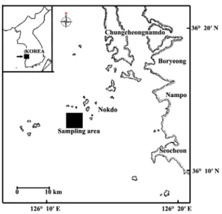
Specimens of A. (S.) pectinata were collected monthly at the subtidal zones of Nokdo, Boryeong-si in Chungcheongnam-do, Korea (Fig. 1) from January to December, 2008. The pen shell ranging from 8.2 cm to 31.3 cm in shell height were used for the present study. After the specimens were transported alive to the laboratory, shell length and total weight were immediately measured.
2. Production of Histological Tissue Section Slides of the Ovarian Tissues
For light microscopic examination of histological tissue section slides by quantitative analysis, a total of 398 individuals were used for the production of histological tissue section slides of ovarian tissues.
Ovarian tissues were removed from shells and
Fig. 1. Map showing the sampling area.
preserved in Bouin’s fixative for 24h. They were then washed with running tap water for 24h. then dehydrated in alcohol and embedded in paraffin molds. Embedded tissues were sectioned at 5-7 μm thickness using a rotary microtome. Sections were mounted on glass slides, stained with Hansen’s hematoxylin-0.5% eosin, and examined using a light microscope (Zeiss Axiovert 10 microscope).
3. Quantitative Statistical Analysis using An Image Analyzer System
Histological Tissue slides were observed for quantitative analysis by an image analyzer system.
Slides were viewed on a stereo-zoom microscope (Nikon, SMZ-U) from where the images were captured by a TOSHIBA Model IK-642K CCD camera and were then viewed on a SAMSUNG color video monitor. The image analyzer (BMI plus, WINATech Co.) is capable of automatic measurement of area and diameter of polygons encircled by the operator, counting objects that are contrasted by background color (in black and white mode), and statistical analysis of numerous characteristics of objects in the captured images. Measurements for male were carried out for areas of total tissue, the testis, and the spermatogenic stages. Measurements for female were carried out for areas of total tissue, the ovary, the
follicles, and the oocytes, the number of the oocytes per unit area, and the diameter of each oocyte.
Measurements on the areas of total tissue, the testis, and the ovary were conducted at magnification of 7.5, at which the field area of captured image was 60 mm2, while the other measurements were done at magnification of 75 (field area: 0.524 mm2). 15-20 individuals per month and two fields per slides were analyzed. Areas of total tissue, the testis, the ovary, the follicle, and the oocyte were measured by manually tracking the margins of objects one by one with a pointer on the captured image. Before the measurements of the areas of the oocytes, and the diameter of the oocytes and the spermatogenic stages captured color image was converted to a gray scale image and then to a black-and-white image with an appropriate threshold at which only the spermatogenic stages or oocytes were contrasted with black, while the other parts of the tissue were in white. Measurements on the areas of the spermatogenic stages and the oocytes, and the diameter of oocytes were carried out by the automatic procedure provided by the BMI plus software.
Number of oocyte was also recorded during this automatic procedure. From the measured values of image analyses, and (A) percent of field occupied by the ovary to total tissue, (B) percent of field occupied by the follicle to total tissue, (C) percent of field occupied by the follicle to the ovary, (D) percent of field occupied by the oocyte to the ovary, (E) the number of the oocyte per mm2 of the ovary, and (F) mean diameter of the oocyte in captured image were calculated for each slide of female. For each male slide, (A) percent of field occupied by the testis to total tissue and (B) percent of field occupied by the spermatogenic stages to the testis were calculated.
One-way ANOVA (multiple comparison by Duncan's procedure, α = 0.05) was applied to compare the means of monthly data. One-way t-tests were used to compare the means of two adjacent months. All statistical analyses were done using SPSS package.
4. Size at First Sexual Maturity by Light Microscopical Observation
For determination of the size at 50% of first sexual maturity, a total of 398 individuals (201 females and 197 males) of A. (S.) pectinata ranging from 8.2 to 31.3 cm in shell height, were examined in order to certify the size at 50% first sexual maturity (=
biological minimum size) by histological observations from April to September, 2008. The percentage (%) of first sexual maturity = No. of mature individuals x 100 / No. of total individuals investigated.
5. Size at the Rate (50%) of Group Sexual Maturity (RM50) (= Biological Minimum Size)
To calculate the size at the rate (50%) of sexual maturity after fitting the rate of group sexual maturity to an exponential equation, the size equivalent to the size at 50% of sexual maturity was estimated to be the sexually mature length of the population (method used by Chung and Ryou, (2000)).
The exponential equation of the rate of sexual maturity is as follows: GM = 100/[1+exp(a-bx)], where, GM: rate of sexual maturity; a, b: constants, x: shell height.
RESULTS
A detailed insight into the gametogenic cycle of A.
(S.) pectinata population was ascertained from a combination of quantitative data gathered during the study period from January to December 2008.
1. Quantitative Results
Baik (2001) and Chung et al. (2006) reported the reproductive cycle (or gametogenic cycle) of A. (S.) pectinata by qualitative histological analysis.
Therefore, in this study, we did not described them by the same method. We have to check the gametogenic cycle of A. (S.) pectinata by quantitative statistical analysis because the spawning period of the reproductive cycle (or gametogenic cycle) of A. (S.) pectinata by qualitative histological analysis varied with author's observations of histological tissue section slides. The quantitative statistical results of slide sections are as follows:
Females: A. (S.) pectinata in females showed a unimodal gametogenic cycle in the results by
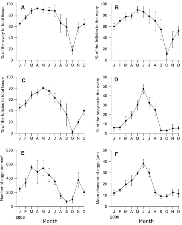
quantitative analysis (Fig. 2). Monthly changes in the percent of field occupied by the ovary to total tissue area in female began to increase in March and reached a maximum in April-May (91.9% - 89.9%, p=
0.70), and then gradually decreased from May to October (89.93%-17.3.7%, p<0.05), thereafter, grad- ually increased from November to January (58.0%- 65.0%, p=0.590) (Fig. 2A). During the winter period gametogenic cycle (December, January and February), the ovarian tissue was found to have relatively lower proportion of the ovary to total tissue.
Over all, according to the time, variations of the ovary area to total tissues among individuals were so high that there were significant differences (one-way ANOVA, P<0.05), however, variations of the ovary area to total tissue area among individuals were no significant differences during February-March and March-June (Duncan's Multiple range test, p=0.059, p=0.574, respectively) (Fig. 2A).
As shown in Fig. 2B, monthly changes in the percent of field occupied by the follicle area to the ovary area began to increase in March and reached a maximum in May (89.7%), and then gradually decreased from June to October (85.3%-11.1%, p<
0.05), thereafter, gradually increased from November to January (36.7%-60.0%, p>0.05) (Fig. 2B). The follicle area in the ovary increased rapidly from near 11.1% in October to 77.3% in March (p<0.05). Over all, according to the time, there were significant differences in follicle area to the ovary area during the investigated period (one-way ANOVA, P<0.05), however, there was no significant differences in follicle area to the ovary area during February-April and March-June (Duncan's multiple range test, p=
0.113, 0.196, respectively) (Fig. 2B).
In Fig. 2C, monthly changes in the percent of field occupied by follicle to total tissues area showed similar patterns to follicle area to the ovary area mentioned above. Monthly changes in the percent of field occupied by the follicle area to total tissue began to increase in March and reached a maximum in May (80.5%), and then gradually decreased from June to October (75.3%-2.2%, p<0.05), thereafter, gradually increased from November to January (20.5.% - 45.0%,
Fig. 2. Monthly changes in quantitative reproductive traits in female Atriana (Servatrina) pectinata. A: percent of area occupied by ovary to total tissue area, B: Percent of area occupied by follicles to total tissue area, C: Percent of area occupied by follicles to ovary area, D: Percent of area occupied by oocytes to ovary area, E: Mean number of oocytes per mm2, F: Mean diameter of oocytes (μm).
p<0.05). Over all, according to the time, there were some significant differences in follicle area to the ovary area during the investigated period (one-way ANOVA, P<0.05) (Fig. 2C).
The percent of field occupied by oocytes to the ovary area showed similar patterns to follicle area to the ovary area mentioned above. Monthly changes in the percent of field occupied by the oocytes to ovary
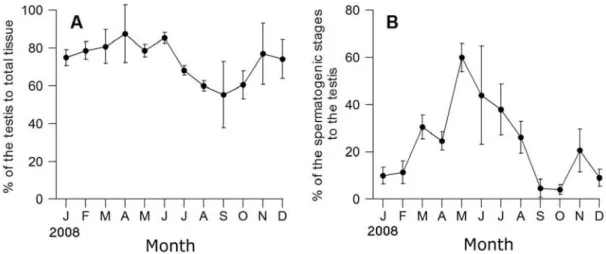
Fig. 3. Monthly changes in quantitative reproductive traits in male Atriana (Servatrina) pectinata. A: Percent of area occupied by testis to total tissue area, B: Percent of area occupied by spermatogenic stages to testis area.
area began to increase in March and reached a maximum in June (50.0%), and then gradually decreased from July to September (35.3%-3.0%, p<
0.05), thereafter, gradually increased from October to January (4.2.%-7.0%, p>0.05) (Fig. 2D).
As shown in Fig. 2E, monthly changes in the number of eggs per mm2 of the ovary began to increase in February and reached a maximum in March (570 eggs/mm2), and then gradually decreased from April (500 eggs/mm2) to September-Octover (25-50 eggs/mm2, p<0.05) thereafter, temporary rapidly increased from October (25 eggs/ mm2) to November (380 eggs/mm). Over all, according to the time, there were some significant differences in the number of eggs per mm2 during the investigated period (one-way ANOVA, P<0.05).
In Fig, 2F, monthly changes in the mean diameter of eggs began to increase in February (12 μm) and reached a maximum in June (mean 38 μm), and then gradually decreased from August to October (12.3 μ m-9.0 μm, p > 0.05). Over all, according to the time, there were some significant differences in the mean diameter of eggs during the investigated period (one-way ANOVA, P<0.05).
Males: A. (S.) pectinata in males showed a unimodal gametogenic cycle in the results by quantitative analysis (Fig. 3). The percent of field occupied by the testis to total tissue in male began to increase in
March (80.8%) and reached a maximum in April-May (87.6%-78.6%, p = 0.460), and then decreased from June to September (85.4%-55.3%, p<0.05), thereafter, gradually increased from October to December (Fig.
3A). Over all, according to the time, there were significant differences; (one-way ANOVA, P<0.05), however, variations of the testis area to total tissue area among individuals were no significant differences during February-June and September-October, and October-November (Duncan's Multiple range test, p=
0.312, p=0.577, p=0.059, respectively) (Fig. 3A).
As shown in Fig. 3B, the percent of field occupied by the spermatogenic stages to the testis area in males begin to increase in March (30.6%) and reached a maximum in May (60.0%), and then sharply droped from June to October (44.0%-4.1%, p=0.05), thereafter, slightly increased from November to January (20.6%-11.4%, p>0.05). Over all, according to the time, there were significant differences in the spermatogenic stages to the testis area during during the investigated period (one-way ANOVA, P<0.05), however, variations of the spermatogenic stages to the testis area among individuals were no significant differences during March-April, September-November (Duncan's Multiple range test, p=0.210, p=0.052, respectively) (Fig. 3A).
2. Size at first sexual maturity
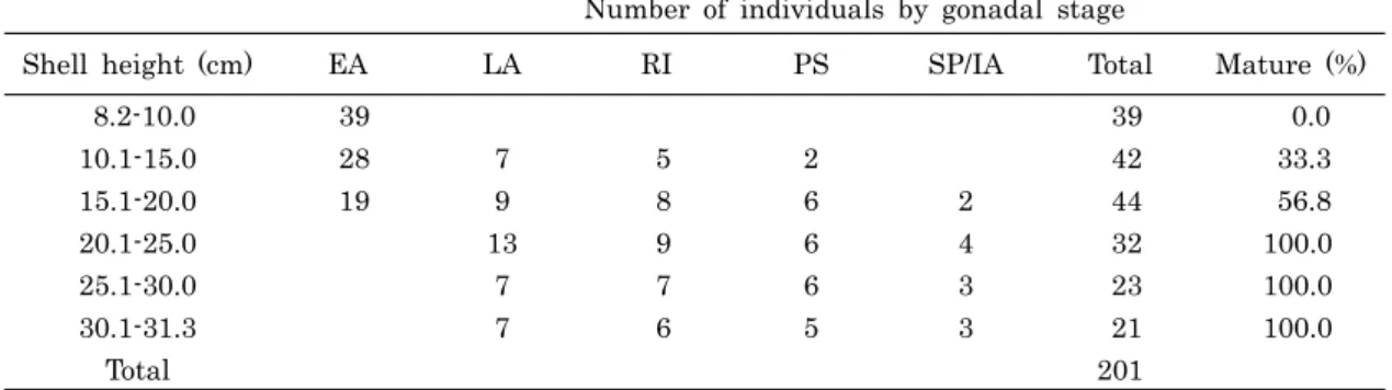
Number of individuals by gonadal stage
Shell height (cm) EA LA RI PS SP/IA Total Mature (%)
8.2-10.0 39 39 0.0
10.1-15.0 28 7 5 2 42 33.3
15.1-20.0 19 9 8 6 2 44 56.8
20.1-25.0 13 9 6 4 32 100.0
25.1-30.0 7 7 6 3 23 100.0
30.1-31.3 7 6 5 3 21 100.0
Total 201
*Abbreviations: EA, early active stage; LA, late active stage; RI, ripe stagel; PS, partially spawned stage;
SP/IA, spent/inactive stage.
Table 1. Shell height of first sexual maturity in female Atrina (Servatrina) pectinata from April to September, 2008
A total of 398 (201 females and 197 males) individuals of A. (S.) pectinata were investigated histologically to determine the shell heights of the pen shell that reach maturation and participate in reproduction from May (before spawning) to Septem- ber (after spawning).
Females: As shown in Table 1, it was found that the percentage of first sexual maturity of smaller individuals ranging from 8.2-10.0 cm in shell height was 0%, and that those individuals were in the early active stage, characterized by a small number of oogonia and the appearance of previtellogenic oocytes.
It is supposed that their sizes at sexual maturity could have not been reached until September when spawning was completed. In addition, the percentage of first sexual maturity of female pen shell ranging from 10.1-15.0 cm in shell height was 33.3%, and those individuals were in the early active, late active and ripe stages during the period between June and August, when spawning was observed among older individuals. However, younger animals had a small number of oogonia and a number of previtellogenic oocytes. A number of vitellogenic oocytes and a small number of mature oocytes were present in the follicles of the ovary. It is supposed that their sizes at sexual maturity could not be reached until late August when the spawning of a few mature individuals was completed. In addition, the percentage of first sexual maturity of female scallops ranging from 15.1-20.0 cm in shell height was 56.8%, and
those individuals were in the early active, late active, ripe, and partially spawned, spent/inactive stages during the breeding season. In contrast, the percentage of first sexual maturity of all individuals of shell height greater than 20.1 cm was 100%, and that those individuals were in the late, ripe, partially spawned, and spent/inactive stages. Accordingly, it is assumed that most individuals can reach full maturity by late September if they are larger than 20.1 cm in shell height.
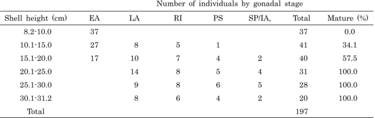
Males: As shown in Table 2, the percentage of first sexual maturity of smaller individuals ranging from 8.2-10.0 cm in shell height was 0%, and those individuals were in the early active stage, which is characterized by a small number of spermatogonia and the appearance of a number of spermatocytes in the acini of the testis. It is supposed that their sizes at first sexual maturity could not be reached until late August when discharging of spermatozoa were completed. The percentage of first sexual maturity of male clams ranging from 10.1-15.0 cm shell height was 34.1%, and those individuals were in the early active, late active and ripe stages during the period between June and August, when spawning was observed among older individuals. However, younger animals had a small number of spermatogonia, spermatocytes, a number of spermatids, and spermatozoa in the acini of the testis. It is supposed that their sizes at sexual maturity of most individuals could have not been reached until late September
Number of individuals by gonadal stage
Shell height (cm) EA LA RI PS SP/IA, Total Mature (%)
8.2-10.0 37 37 0.0
10.1-15.0 27 8 5 1 41 34.1
15.1-20.0 17 10 7 4 2 40 57.5
20.1-25.0 14 8 5 4 31 100.0
25.1-30.0 9 8 6 5 28 100.0
30.1-31.2 8 6 4 2 20 100.0
Total 197
*Abbreviations: EA, early active stage; LA, late active stage; RI, ripe stagel; PS, partially spawned stage;
SP/IA, spent/inactive stage
Table 2. Shell height of first sexual maturity in male Atrina (Servatrina) pectinata from April to September, 2008
Fig. 4. Relationship between the rate of group sexual maturity (%) and shell height (cm) in Atriana (Servatrina) pectinata. A, Female; B, Male.
when sperm discharging of a few mature individuals were completed. In addition, the percentage of first sexual maturity of male pen shells ranging from 15.1-20.0 cm in shell height was 57.5%, but those individuals were in the early active, late active, ripe, and partially spawned stages during the breeding season. The percentage of first sexual maturity of all individuals of shell height greater than 20.1 cm was 100%, and those individuals were in the late, ripe, partially spawned, and spent/inactive stages.
Therefore, it is assumed that most individuals can reach full maturity by late August if they are larger than 20.1 cm in shell height at that time.
In this study, the percentage of first sexual maturity of female and male pen shells ranging from 15.1 to 20.0 mm was over 50.0%. The percentage was
100% for female and male pen shells over 20.1 mm in shell height.
3. Size at the Rate (50%) of Group Sexual Maturity (GM50) (= Biological Minimum Size)
As shown in Figs. 4A, 4B, shell heights of sexually mature pen shell (sizes at 50% of group sexual maturity, GM50) that were fitted to an exponential equation were 15.81 cm in females and 15.72 cm in males.
DISCUSSION
1. Comparisons of the ovarian gametogenic cycle by qualitative and quatitative analyses
To compare the results of the gametogenic (reproductive) cycle by qualitative and quantitative
analyses in female A. (S.) pectinata, in this study, we quoted two results on gametogenic cycle by qualitative histological analysis already reported by Baik (1988) and Chung et al. (2006). They reported that the reproductive cycle in female A. (S.) pectinata can be classified into 5 successive stages: the early acive stage (November to march), late active stage (February to May), ripe stage (April-July), partially spawned stage (June to July or June to August), and spent/inactive stage (August to October). In this study, according to the results of ovarian gametogenic cycle by quantitative analysis, the results of monthly changes in portions (%) of ovary areas to total tissue areas began to rapid increase in March, and reached a maximum in May, thereafter, their proportions (%) are gradually decreased from June-September when spawning occur. And monthly changes in proportions (%) of follicle areas to ovary areas also began to rapid increase in March, and reached a maximum in May, thereafter, their proportions (%) are gradually decreased from June- September when spawning occur, and the main spawning occurred between July and August. In particular, peak mature oocyte level occurred in May followed by a significant decrease from June to September which indicated spawning, and the main spawning occurred between July and August.
In addition, monthly changes in proportions (%) of the oocyte areas to the ovary areas, the number of the oocytes per mm2, and mean diameter of the oocytes showed the same or similar patterns: the maximum in May, and then rapidly dropped from June to September which indicated spawning.
Therefore, compared female gametogenic cycle by qualitative analysis with those by quantitative statistical analysis, the results of female gametogenic cycle and gonadal maturation by qualitative histological analysis coincided with those studied by quantitative statistical analysis. Judging from the results confirmed by quatitative statistical analyses using an Image Analyzer System, testicular gametogenic cycle was confirmed to be a unimodal gametogenic cycles showing a maximum maturity and one spawning season per year from June to
September. Giese (1959) and Sastry (1979) reported that in general, latitudinal differences in timing of the reproductive cycles of marine molluscs. In particular, some authors (Ropes and Stickney, 1963;
Brousseau, 1978; Heffernan et al., 1989a) reported that Mya arenaria and Mercennaria mercennaria in Bivalve mollusc exhibited a change from a unimodal to a bimodal cycle with decrease in latitude. However, some authors (Heffernan and Walker, 1989; Heffernan et al., 1989b) reported that several other bivalves (i.e, Geukensia demisa, Crassostrea virginica and Spisular solidissima similis) showed unimodal gametogenic cycle in the southeastern U.S. waters (Kanti et al., 1993). In this study, the gametogenic cycle in A. (S.) pectinata by quantitative statistical analysis showed a unimodal gametogenic cycle.
2. Number of spawning seasons per year by quantitative analysis
Exceptionally, in case of the spawning seasons of R.
philippinarum, there have been many studies on the number of spawning seasons studied by the qualitative histological analysis in other areas of the world: there are one spawning season in northern districts of Tokyo Bay (Kurashige, H. (1943; Yoshida, 1953; Ponurovsky and Yakovlev, 1992; Quayle and Bourne, 1972; Holland and Chew, 1974; Chung et al., 1994, 2010), two spawning seasons in southern parts of Tokyo Bay (Tanaka, 1954; Ohba, 1959; Ko, 1957), and three spawning season during the year in Spain (Sarasquaete et al. 1991). Perhaps, it is assumed that the number of spawning seasons during the year of bivalve species vary with latitudinal gradients (locations) of the world. If two or three spawning seasons were investigated by qualitative histological analysis in some areas of the world, the investigation of the number of spawning seasons (periods) should be clarified by quantitative statistical analysis in order to confirm the initial and final spawning months.
Korean latitude is similar to the location of the northern district of Tokyo Bay, Japan, Accordingly, it is assumed that the number of spawning seasons during the year of most Korean bivalves may be once
a year.
Thererfore, it is assumed that some local variations and timing of spawning of this clam might be related to the geographical differences in the water temperatures, time of the food production (phytoplanktons), and some other environmental factors (Ko, 1957;Momoyama and Iwamoto, 1979).
3. Quantitative analysis
The pen shell, A. (S.) pectinata, showed a unimodal gametogenic cycle as found in other scallops on the Korean coastal waters. Quantitative reproductive results by an image analysis of this species showed the peaks of maturity in May. Compared to qualitative results (frequencies of gonadal phases) of other clams in previous works, the period of maturation of A. (S.) pectinata, is quite similar to the May-July results of Cyclina sinensis (Chung et al., 1991), Mactra veneriformis (Chung and Ryou, 2000), Meretrix lusoria (Chung, 2007), and Saxidomus purpuratus (Chung at al., 1999). The peak of oocyte area to ovarian tissue in June implies the readiness of maturation. The spawning seemed to be initiated in June with a decrease in oocyte area. A significant decrease in oocyte area was found during June-September, indicating the major period of spawning. The spawning period of this species was also similar to other clams mentioned above (June-September for C.
sinensis, M. veneriformis, and M. lusoria, May-October for S. purpuratus).
The follicle area to the ovarian tissue rapidly increased in March but the increase in the oocyte area to the ovarian tissue at this period was relatively low. During and after spawning, the follicle area did not decrease in proportion to the oocyte area; rather it remained high until November when the oocyte area decreased to less than 10%. The follicular tissue seemed to be formed earlier (in March) than the period of maturation (April-May) and to degenerate later (in November) than the period of spawning (June-September). In observation of slides after the spawning period (September-November), a large portion of empty space was found in most of the follicles, which indicated the post-release of oocytes.
However, during this period, follicles were not entirely empty.
They seemed to be remnant eggs undischarged during the spawning period and to be reabsorbed to the body. From this, it can be explained that the number of eggs and mean diameter of oocytes during September-November was not zero (Figs. 6E, F). In conclusion, A. (S.) pectinata did not seem to release eggs entirely during the spawning period. In case of C. sinensis the size of follicle tended to increase toward the spawning period and slightly variable among individuals. So, variability in the number of follicles per field was great and heterogeneous even within an individual. In addition, the size of eggs also was different between months and variation in egg size in a field was not homogeneous among months, especially during the spawning period. Therefore, the number of eggs per unit area of ovary was applied instead of the number of eggs per follicle in this study.
4. Size at First Sexual Maturity and Size at 50% of Group Sexual Maturity
Ryu et al. (2001) investigated the determination of age of the pen shell, A. (S.) pectin. According to the growth curves for the mean shell height fitted to the von Bertalanffy equation by Park (2002), ages (year) and mean shell lengths (mm) were estimated as follows: 1 year (14.51 cm), 2 years (19.47 cm), 3 years (22.24 cm), 4 years (24.47 cm), 5 years (26.09 cm), 6 years (27.33 cm), 7 years (28.31 cm), 8 years (29.11 cm), 9 years (29.76 cm), and 10 years (30.35 cm).
In this study, the percentages of first sexual maturity of female and male individuals of 15.1-20.0 mm in shell height were 56.8% in females and 57.5%
in males, respectively. It is assumed that female and male pen shells ranging from 15.1-20.0 cm in shell height are one year old. And the percentages of first sexual maturity of female and male individuals of over 20.1 cm in shell height were 100% in both sexes.
Accordingly, it is supposed that female and male pen shells ranging over 20.1 cm in shell height were approximately three years old.
According to the growth curve for shell height
fitted to von Bertalanffy equation, shell heights at 50% of group sexual maturities (GM50) were 15.81 cm in females and 15.72 cm in males (Figs. 4A, 4B).
In this study, these shell heights (GM50) in both sexes were considered to be one year old (Ryu et al., 2001).
Therefore, we assumed that these female and male populations achieve maturity and begin reproduction at one year of age.
In the aspect of natural resource management, the present study suggests that harvesting pen shells less than 15.72 cm in shell height for GM50 (one year old) can potentially lead to a drastic reduction in recruitment. Accordingly, a measure indicating a prohibitory fishing size should be taken for adequate fisheries management.
5. Ovarian development and maturation
A wide range of exogenous factors has recently been suggested as controls for gonadal development and maturation in marine bivalves. Of various factors, water temperature and food availability seem to be particularly important. Sastry (1966, 1968; Chung and Ryou, 2000) stated that these and other factors (salinity, day length, etc) probably interact with endogenous factors (neuroendocrine activity) in a complex manner to control the initiation of gametogenesis. Sastry (1968) stated that sea water temperature acts as a triggering stimulus for the initiation of the germ cell growth phase. The water temperatures required for activating the growth of germ cells at the beginning of oogenesis and spermatogenesis and for attaining maturity ultimately limit the annual period of gonad activity and gametogenesis in the natural environment. In this study, gamete differentiation of A. (S.) pectinata began in the winter - early spring seasons, and reached maturity in the population from April to July when water temperatures were increased. After basic metabolic requirements are satisfied, gonad activity and gametogenesis of this species occur under temperature conditions that allow nutrients mobilization to the gonads (Sastry, 1966). The periods of food abundance and of gonad development of R.
philippinarum are nearly coincident gonad growth
and gametogenesis in spring coincided with peak food levels, although food concentrations remained high throughout the summer months (Kim, 2005).
Therefore, it is assume that if food and temperature criteria are met, growth of germ cells is initiated in conjunction with the transfer of nutrients from digestive diverticular to the gonad. However, it is assumed that the amount of nutrients mobilized for the gonad maturation depends not only on the food level, but also on the water temperature and the basic metabolic requirements of the clams. In Korean coastal waters, growth and production of bivalves is relatively high from spring to early summer seasons (Chung et al., 1994; Kim, 2005; Chung, 2008) due to the abundance in phytoplankton. Thus, abundant food supply (e.g., bivalves) is available to R. philippinarum during the period of gonadal development and maturation. Therefore, it is suggested that gonadal development and maturation of the Korean R.
philippinarum is closely related to temperature change and food availability. Fretter (1984) observed that in temperate zones, the seasonal temperature fluctation associated with changing illumination is a controlling factor in gametogenesis. In consequence, gonadal development and maturation of this species may be retarded under low illumination, due to the decrease in food availability caused by diminished primary production of phytoplankton.
ACKNOWLEDGEMENTS
The authors are grateful to Dr. William Heard, the Florida State University for helpful comments on the manuscript, and also correction the manuscript. This research was supported in part by the funds (2008) from the Research Projects of the Korea Marine Environment & Ecosystem Research Institute, Korea.
REFERENCES
Baik, S.H., Kim, K.J., Chung, E.Y. Choo, J.J. and Park, K.H. (2001) Seasonal variations in biochemical components of the visceral mass and adductor muscle in the pen shell, Atriana pectinata. The Korean Journal of Malacology, 17: 71-78. [in Korean]
Baik, S.H. (2002) Reproduction, age and growth of the pen shell Atrina pectinata on the west coast of Korea. Ph. D. Thesis, Kunsan National University, 98 pp.
Chung, E.Y, Lee, T.Y. and An C.M. (1991) Sexual maturation of the venus clam, Cyclina sinensis, on the west coast of Korea. Journal of Medical &
Applied Malacology, 3: 125-136.
Chung, E.Y, Ryou D.K, and Lee J.H. (1994) Gonadal development, age and growth of the shortnecked clam, Ruditapes philippinarum (Pelecypoda:
Veneridae), on the coast of Kimje. Korean Journal of Malacology, 19: 38-54.
Chung, E.Y and Ryou D.K. (2000) Gametogenesis and sexual maturation of the surf clam, Mactra veneriformis on the west coast of Korea.
Malacologia, 42: 149-163.
Chung, E.Y., Park, YJ, Lee, J.Y. and Ryu, D.K. (2005) Germ cell differentiation and sexual maturation of the hanging Cultured female scallop Pationpecten yessoensis on the East coast of Korea. Journal of Shellfish Research, 24: 913-921.
Chung, E.Y. (2007) Oogenesis and sexual maturation in Meretrix lusoria (Roding 1978), (Bivalvia: Veneridae) in western Korea. Journal of Shellfish Research, 26:
71-80.
Chung, E.Y., Lee, C.H., Choi, K.H., Choi, M.S. and Lee, K.Y. (2010) Gametogenic cycle and the number of spawning seasons by quantitative reproductive analysis in female Ruditapes philippinarum in Western Korea. The Korean Journal of Malacology, 26: 245-254.
Chung, E.Y. (2008) Ultrastructural studies of oogenesis and sexual maturation in female Chalmys (Azumapecten) farreri farreri (Jones & Preston, 1904, Pteriomorphia: Pectinidae) on the western coast of Korea. Malacologia, 50: 279-292.
Fretter, V. (1984) Prosobranchs. pp.1-45. In: Laar, eds., The Mollusca, Vol. 7, Academic Press, New York 352 pp.
Giese, A.C. 1959. Compartative Physiology: Annual reproductive cycles of marine invertebrates. Review of. Physiology, 21:547-576.
Heffernan, P.B., Walker, R.L. and Carr, J.L. (1989a) Gametogenic cycles of three bivalves in Wassau Sound, Georgia I: Mercenaria mercenaria (Linnaeus, 1758). Journal of Shellfish Research. 8: 51-60.
Heffernan, P.B., Walker, R.L. and Carr, J.L. (1989b) Gametogenic cycles of theree bivalves in Wassaw Sound, Georgia II: Crassostrea virginica (Gmelin, 1971). Journal of Shellfish Research, 8: 61-70.
Heffernan, P.B., Walker, R.L.and Carr, J.L. (1989c) Gametogenic cycles of three bivalves in Sassaw Sound, Georgia Ⅲ: Geukensia demissa (Dillwyn) Journal of Shellfish Research, 8: 327-334.
Holland, D.A. and Chew KK (1974). Reproductive cycle of the manila clam Washington. Proceedings of National Shellfish Research Association, 64: 53-58.
Kanti, A, Heffernan PB, and Walker RL (1993) Gametogenic cycle of the southern surfclam, Spisula solidissimasimilis (Say, 1822), from St. Catherine Sound, Georgia. Journal of Shellfish Research, 12:
255-261.
Kim, Y.M. (2005) A Study on reproductive ecology of the hard clam, Meretrix lusoria, on the west coast of Korea. Ph. D. Thesis, Kunsan National University 123 pp.
Ko, Y. (1957) Some histological note on the gonads of Tapes japonica Deshayes. Bulletin of Japanese Society Fisheries, 23: 394-399.
Kurashige, H. (1943) Seasonal variation in the weight and volume as well as the chemical composition of the soft body of Tapes philippinarum with special reference to its spawning. Bulletin of Korean Fisheries Experimental Station, 8: 115-140.
Kwon, O..K, Park, G.M, and Lee, J.S. (1993) Coloured shells of Korea. Academy Pub Co. Seoul 288 pp.
Loosanoff, V.L, and Davis, H.C. (1963) Rearing of bivalve mollusks. Advance Marine Biology, Academic press, 1: 110-112.
Min, D.K., Lee, J.S, Ko, D.B, Je, J.G. (2004) Mollusks in Korea. Hanguel Graphics, Busan, Korea 566 pp.
National Fisheries Research and Development Institute (1999) Development of optimal technology for sustaining production in a shellfish farm. NFRDI 208 pp.
Momoyana, G. and Iwamoto, T. (1979) On the spawning season of the short necked clam in Yamaguch and Okai Bay. Bulletin of yamaguchi Prefecture Fisheries Experimental Sttation, 7: 19-28.
Ohba, S. (1959) Ecological studies in the natural population of a clam, Tapes japonica, with special reference to seasonal variations in the size and structure of population and to individuals growth.
Biological Journal of Okayama University, 5: 13-42.
Ponurovsky, S.J. and Yakovlev, Y.M. (1992) The reproductive biology of the Japanese littleneck.
Tapes philippinarum (A. Adams & Reeve, 1850) (Bivalvia: Venerida). Journal of Shellfish Research, 11: 265-277.
Quayle, D.B.. and Bourne, N. (1972) The clam fisheries of British Columbia. Fisheries Research Board Canadian Bulletin, 179: 70.
Rand, W.M. (1973) A stochastic model of the temporal aspect of breeding strategies. Journal of Theoretical Biology, 40: 337-351.
Ropes, J.W. and Stickney, A..P. (1965) Reproductive cycle of Mya arenaria in New England. Biological Bulletin, 128: 315-327.
Ryu, D.K., Baik, S.H., Park , K.H. and Chung, E.Y.
(2001) Age and growth of the pen shell, Atrina (Servatrina) pectinata japonica (Reeve), on the west Coast of Korea. The Korean Journal of Malacology, 17: 71-78. [in Korean]
Sarasquete, M.C. Gimeno, S., and Gonzalez de Canales, M.L. (1990) Cycle reproducteur de la palourude
Ruditapes philippinarum (Adams and Reeve, 1850) de la cote sud ouest atilantique (Espagne). Review of International Oceanography. Med IXXXXVII:
90-99.
Sastry, A.N. (1966) Temperature effects in reproduction of the bay scallop, Aquipecten irradians Lamarck.
Biological Buletin, 130: 118-134.
Sastry, A.N. (1968) Relationship among food, temperature and gonad development of the bay scallop, Aquipecten irradians Lamarck. Physiol Zool, 41: 44-53.
Sastry. A.N. (1979) Pelecypoda (Excluding Ostreidae). pp 113-292. In: Reproduction in marine invertebrates.
Vol. 5: Pelecypods and lesser classes. A. C. Giese and J. S. Pearse (eds.) Academic Press, New York.
Tanaka, Y. (1954) Spawning season of important bivalves in Ariake Bay Ⅲ. Tapes philippinarum.
Bull Jap Soc Sci Fish, 19: 1165-1167.
Yoshida, H. (1953) Studies on larvae and young shells of industrial bivalves in Japan. J Shimonoseki Fisheries College, 3: 1-106.
Yoo, S.K. and Yoo, M.S. (1984) Studies on the pen shell culture development (1) Reproductive ecology of pen shell in Yeoja Bay. Bulletin of the Korean Fisheries Society, 17: 529-535. [in Korean]
Yoo, S.K., Lim, H.S., Ryu H.Y. and Kang, K.H. (1988) Improvement of the seed production method of the pen shell. The occurrence of larvae and the early growth of the spat. Bulletin of the Korean Fisheries Society, 21: 206-216, [in Korean]