한국표면공학회지 J. Kor. Inst. Surf. Eng.
Vol. 41, No. 6, 2008.
<연구논문>
Adhesion of Human Osteoblasts Cell on TiN Thin Film Deposited by Cathodic Arc Plasma Deposition
Vuong Hung Pham
a, Sun Kyu Kim
b*, Vinh Van Le
b, Byoung Se Kwon
a,c,da
Department of Immunology and Biomedicine, University of Ulsan, Ulsan 680-749, Korea
b
School of Materials Science and Engineering, University of Ulsan, Ulsan 680-749, Korea
c
National Cancer Center, Seoul 410-769, Korea
d
LSU Eye Center, Louisiana State University Health Sciences Center, New Orleans, Louisiana 70112. U.S.A.
(Received November 6, 2008; revised November 20, 2008; accepted December 30, 2008)
Abstract
Interaction between human osteoblast and TiN films was conducted in vitro. TiN films were produced by cathodic arc plasma deposition. The surface was characterized by atomic force microscopy (AFM). TiN films, glass substrates and Ti films were cultured with human osteoblasts for 48 and 72 h hours. Actin stress fiber patterns and microtubules of osteoblasts were found slightly more organized and distributed on TiN films compared to those on the Ti films and the glass substrates. Human osteoblasts also showed slightly higher cell attachment, proliferation, and focal contact adhesion on TiN films compared to those on Ti films and glass substrates. Our results demonstrated that TiN films showed slightly better cellular adhesion of osteo- blasts than Ti films and glass substrates in a short-time culture period.
Keywords: Human osteoblast, TiN film, Cell adhesion, Surface modification, Cytoskeleton
1. Introduction
Pure titanium is widely used as an implant material because of its suitable mechanical properties and biocompatibility
1-4). Biocompatibility of pure titanium is due to the TiO
2layer formed on its surface
5). However, Ti is a soft material with low shear resistance on the surface, which is mainly due to the naturally formed oxide
6). Therefore, there is a need for wear resistant coatings for these materials and investigation of their biocompatibility. Biocompatibility of biomaterials is closely related to the behavior of the contacting cells, in particular their adhesion
7). Cell adhesion to the surface is affected by surface chemistry, topography, surface energy
8,9)and method of surface modification
10).
Cell adhesion to TiN films has been studied previously. Groessner-Schreiber et al. used sputtering to modify the surface of titanium to enhance fibroblast cell adhesion
11), while Czarnowska
et al.used ion
plating to enhance fibroblast cell growth
12). Cyter
etal.
used sputtering to deposit TiN films to enhance neuron cell adhesion
13). Yeung
et al.reported that TiNi substrates with TiN film exhibited the largest degree of osteoblast proliferation compared to untreated substrates and stainless steel substrates
14). Many researchers used fibroblast and neuron cells and not much attention has been directed toward the formation of cytoskeleton organization and adhesion behavior of osteoblast cells when subjected to TiN films.
In this work, we evaluated the osteoblast cell adhesion to TiN films deposited by cathodic arc plasma deposition. The choice of cathodic arc plasma deposition is from the fact that there is a good adhesion of coated layer with the substrate and a simple wear resistance coating method. Cathodic arc plasma deposition process is widely used in the industry because it offers wide range of deposition and the adhesion of films deposited by this process is superior to other methods. For comparative purpose, Ti film, which is known as a biocompatible surface, was also tested.
*
Corresponding author. E
-mai
l : [email protected]2. Experimental
Standard round glass coverslips of 12mm (Marienfeld, Germany) were used as substrates. Prior to deposition, the substrates were ultrasonically cleaned with ethanol (95%) for 20 min and then dried by Ar gas. Finally, they were loaded into the deposition chamber. The detailed experimental procedures were described elsewhere
15). Ti cathode was used for depositions of both TiN and Ti films. The arc current was kept in constant at 45 A for deposition of TiN films, while it was 90 A for deposition of Ti films. Deposition was done without applying bias potential at 200
oC.
Surface roughness was measured by AFM. The apparatus used was thermomicroscope (CP-research system) with a cantilever(ARROW-CONTRO50, Nanoworld).
Human osteoblasts (hFOB 1.19) were used as a model for studying the interaction between the cell and the surface. The cells were maintained in Dulbecco’s modified Eagles Medium (DMEM) containing 10% fetal bovine serum and 1%
antibiotics at 37
oC in humidified air and 5% CO
2. For actin staining, attached cells were fixed by 4%
paraformaldehyde and pearmeabilized with 0.1%
Triton X-100. Cells were stained with Alexa-flour phallodin 488. For tubulin staining, anti-tubulin monoclonal antibody was used as the primary antibody. Alexa 488 goat anti-mouse was used as the secondary antibody. Cells were rinsed twice with phosphate buffer saline (PBS), and fixed with 4%
paraformaldehyde for 10 min. Then they were permeabilized with 0.1% Triton X-100 in PBS after PBS washing, blocked with 1% bovine serum albumin (BSA) and stained with the primary antibody. They were rinsed again with PBS and then stained with the secondary antibody.
Adherence of the cells on the thin films was determined by counting in seven randomly chosen fields of view at magnification 100 under an optical microscope (Axiovert 135).
Cell proliferation was observed by visualization of the immunofluorescent cells after 48 and 72 h of incubation.
For focal contact adhesion analysis, anti-vinculin (hVIN-1) was used as the primary antibody, and Alexa 488 goat anti-mouse as the secondary antibody.
Cells were rinsed twice with PBS, and fixed with 4%
paraformaldehyde for 10 min. Then the cells were permeabilized with 0.1% Triton X-100 in PBS, blocked with 1% BSA and stained with the primary
antibody for 30 min. The cells were then rinsed with PBS and stained with the secondary antibody. All visualization of the stained cell was done by a confocal microscope (Olympus 1 X 81).
For the cell adherence analysis, the
t-test was used to assess the statistical significance of results between surfaces. The statistical analysis was performed with the software GraphPad Prism 4 at the confidence level of 95%. A probability value of p<0.05 was considered significant.
3. Results and Discussion
Using AFM, the average surface roughness of Ti film, TiN film and glass substrate were inspected.
The surface roughness of Ti and TiN film was 0.268 nm and 0.480 nm, respectively while the value on glass substrate was only 0.161 nm.
Examination of actin cytoskeleton organization of the cells focused on the analyzing the cells cultured on the films on which stress fibers were formed.
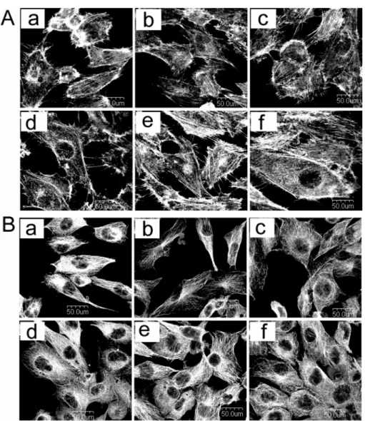
Actin stress fibers were oriented in parallel direction with the main cellular axis on all the surfaces after 48 and 72 h of incubation (Fig. 1A). At 48 h, the pattern of actin stress fibers seemed to be thicker on TiN films than those on the Ti films and the glass substrates. Interestingly, the more evidence of a thicker actin stress fibers on TiN films were observed after 72 h of incubation. Microtubules of osteoblasts cell were expanded on the TiN films, whereas cells on the glass substrates were narrower and had a lower expression of tubulin represented by less microtubles in the cells (Fig. 1B). We found slight differences in the development of microtubules between the TiN and Ti films after 48 and 72 h of incubation.
Fig. 2 shows the cell attachment on three tested surfaces after 48 h of incubation. The number of attached cells was significantly greater on the TiN films than on the Ti films (p<0.05) or the glass substrates (p<0.05).
The cell proliferation on the films and the glass substrates after 48 and 72 h of incubation is shown in Fig. 3. Cell proliferation increased with the incubation time on the three tested surfaces. Cells on the TiN films slightly more proliferated than on the Ti films and the glass substrates. It is also clear from the micrographs that the cells nearly reached the confluence after 72 h of incubation on all of the three tested surfaces.
Fig. 4 shows the focal contact adhesion of
Fig. 1. Cytoskeleton analysis of osteoblast on tested specimens. (a and d: glass substrate, b and e: Ti film, c and f:
TiN film. a-c: 48 h of incubation. d-f: 72 h of incubation). (A: cytoskeleton of actin stress fiber on surfaces tested, B: cytoskeleton of microtubule on surfaces tested).
Fig. 2. Cell attachment on the surfaces tested (upper panel is immunofluoresence actin visualization of the cell on the
surface, lower panel is quantification of cell density by counting. a: glass substrate, b: Ti film, c: TiN film).
osteoblasts on the glass substrates and the films.
Focal contact adhesions were found on all the three tested surfaces. However, focal contact adhesion was more abundant on TiN films than on Ti films and glass substrates.
Cathodic arc plasma deposition produced many particles thus cause the surface of the TiN and Ti films to be rougher than that of glass substrates.
Mukherjee
et al.
16)reported that TiN films revealed a higher surface roughness compared with Ti films when the films were produced by plasma immersion ion implantation-assisted deposition without bias voltage. In our study, the surface roughness of TiN film was also higher than Ti film when the films were deposited without bias voltage. Huang et al.
studied influence of TiN films prepared by cathodic arc plasma deposition and ion-nitrided titanium films, and polished Ti substrates on osteoblast cell adhesion
in vitro17).