https://doi.org/10.22889/KJP.2020.51.3.151
151
콜라겐-유도의 사람 혈소판에서 PI3K/Akt 및 MAPK 조절을 통한 Isoscopoletin의 항혈소판 효과
이동하*
남서울대학교 임상병리학과·분자진단연구소
Anti-platelet Effects of Isoscopoletin through Regulation of PI3K/Akt and MAPK on Collagen-induced Human Platelets
Dong-Ha Lee*
Department of Biomedical Laboratory Science, Molecular Diagnostics Research Institute, Namseoul University
Abstract When blood vessels are damaged, a rapid hemostatic reaction occurs to minimize blood loss and maintain normal circulation. Platelet activation and aggregation is essential in this process. However, excessive platelet aggregation or abnormal platelet aggregation may be the cause of cardiovascular disease, such as thrombosis, stroke and atherosclerosis. Therefore, it is important to prevent and treat cardiovascular disease by finding substances that can regulate platelet activation and suppress aggregation reactions. Isoscopoletin, which is mainly found in the roots of plants Artemisia or Scopolia, has been reported to have potential pharmacological effects on anticancer and Alzheimer’s disease, but its role and mechanisms for platelet aggre- gation and thrombus formation are unknown. This study confirmed the effect of isoscopoletin on major regulation of collagen- induced human platelet aggregation, TXA2 production and intracellular granular secretion (ATP and serotonin release). In addi- tion, the effects of isoscopoletin on phosphorylation of phosphorylated proteins PI3K/Akt and MAPK involved in signal trans- duction in platelet aggregation was studied. As a result, isoscopoletin significantly inhibited the phosphorylation of PI3K/Akt and MAPK, significantly inhibiting platelet aggregation through TXA2 production and intracellular granular secretion (ATP and serotonin release). Therefore, we suggest that isoscopoletin is an anti-platelet substance that regulates phosphorylation of phos- phorus proteins such as PI3K/Akt and MAPK and is valuable as a preventive and therapeutic agent for platelet-derived car- diovascular disease.
Keywords Isoscopoletin, PI3K/Akt, MAPK, TXA2, Granule secretion
혈액은 각 장기와 조직에 산소와 영양분을 공급함과 동시 에 폐기물 제거를 지원함으로써 생존에 절대적인 역할을 하 며, 이 작업을 정상적으로 수행하려면 혈액 순환이 원활해 야 한다. 따라서, 혈관이 손상되면 혈액 손실을 최소화하고 정상적인 순환을 원활하게 유지하기 위해 빠른 지혈 반응 이 일어나며, 이 과정은 혈소판의 활성화 및 응집으로 시작 된다. 그러나, 혈소판의 과다 또는 비정상적인 응집은 혈전 증, 뇌졸중 및 죽상 동맥 경화증과 같은 심혈관 질환의 원 인이 될 수 있다. 따라서, 혈소판의 활성화를 조절하고 응 집을 억제 할 수 있는 물질을 찾는 것이 심혈관 질환의 예 방 및 치료에 중요하다.1,2) 혈관이 손상되면 혈소판이 부상
부위로 모집되어 혈관 활성화제(collagen, ADP 및 thrombin 등)에 의해 활성화된다. 이때, 혈소판 막에서 활성화 된 phospholipase C는 phosphatidylinositol 4,5-bisphosphate를 inositol 1,4,5-triphosphate(IP3) 및 diacylglycerol(DG)로 가 수 분해하고, 생성 된 I P3는 혈소판 내 dense tubular system 에 위치한 Ca2+ 채널을 열어 세포질 내 Ca2+ 농도를 증가시 킨다.3) 혈소판이 활성화 될 때, 세포질 막의 phospholipase A2(cPLA2)에 의해 가수 분해된 arachidonic acid은 TXA2 synthase(TXAS)와 cyclooxygenase-1(COX-1)의 효소 작용 으로 인해 TXA2로 전환되고 혈소판으로부터 분비된다.4,5) 방출 된 TXA2는 다른 혈소판 막 수용체에 결합하여 지속 적으로 혈소판 응집을 촉진하는 작용제로서 기능한다고 알 려져 있다.6) 또한, TXA2의 유사 체인 U46619(9,11-dideoxy- 9a.la-methanoepoxy prostaglandin F2a)는 세포질에서 Ca2+
*교신저자(E-mail):[email protected] (Tel): +82-41-580-2148
수준을 증가시켜 myosin light chains와 pleckstrin을 인산화 하여 혈소판 응집을 유도하는 것으로 알려져 있다.7,8)
Mitogen-activated protein kinases(MAPK)는 세포 내 신호 전달에 작용하는 것으로 알려진 인산화 효소로서, c-Jun N- terminal kinase(JNK), extracellular signal-regulated kinase (ERK) 및 p38 MAPK로 분류되며, 이들의 역할은 꾸준히 연구되어 왔다.9) JNK, ERK 및 p38 MAPK는 사람 혈소판 에서 검출되고 여러 작용제가 유발하는 단백질 인산화에 의 해 활성을 갖는 것으로 보고되어 있다.10-12) 이 신호 전달 분 자의 인산화는 혈소판 과립 분비를 유도하는 데 중요한 역 할을 하는 것으로 잘 알려져 있다.13,14) 또한 MAPK는 세포 막에 위치한 cPLA2를 인산화하여 혈소판 막의 인지질에서 arachidonic acid을 생성하여 TXA2로 변환되어 다른 혈소판 의 활성화 및 응집을 유발하는 것으로 알려져 있다.15,16) 또 한, PI3K/Akt 경로는 혈소판에서 dense 과립의 분비 및 혈 소판 응집을 포함하여 혈소판 기능의 조절에 중요한 역할 을 보여주었다.17)
Scopolia 또는 Artemisia 속 식물의 뿌리에서 주로 발견되 는 isoscopoletin은 항암 및 Alzheimer’s disease에 도움이 될 수 있는 잠재적인 약리학적 특성에 대해 연구된 바 있다.18,19) 이전의 연구에 따르면, isoscopoletin와 유사한 구조를 가진 물질인 scoparone은 PI3K/Akt/NF-κB 경로를 조절함으로써 IL-1β 매개의 염증 반응을 억제한다고 보고된 바 있다.20-22) 그러나, 혈소판 활성화에서 isoscopoletin의 역할 및 사람 혈 소판 응집에서 isoscopoletin의 작용기전은 알려져 있지 않 다. 이 연구에서는 collagen이 유도한 혈소판 응집 및 이 과 정에 중요한 인자들로 알려진 TXA2 생성, 혈소판에서의 과 립 분비, PI3K/Akt 및 MAPK의 인산화 단백질에 대한 isoscopoletin의 효과를 확인하였다.
재료 및 방법
시약 Isoscopoletin(6-hydroxy-7-methoxy-2H-chromen-2- one)은 Avention Corporation(Seoul, Korea)에서 구입했다(Fig.
1). Collagen은 Chrono-Log Corporation(PA, Havertown, USA)로부터 제공 받았다. TXB2 enzyme immunoassay(EIA) kit, ATP assay kit 및 serotonin EIA kit는 Cayman
Chemical Co.(Ann Arbor, MI, USA)에서 구입하였다. 다른 시약은 Sigma Chemical Corporation(St. Louis, MO, USA) 에서 구입 하였다. Western blotting에 사용된 antibodies 및 lysis buffers는 Cell Signaling(Beverly, MA, USA)로부터 입 수하였다. Enhanced chemiluminescence solution(ECL) 및 polyvinylidene difluoride(PVDF) membrane은 General Electric Healthcare(Chalfont St. Giles, Buckinghamshire, UK)로부터 구입 하였다.
세척된 사람 혈소판의 제조 대한 적십자사 혈액원(수원) 에서 사람 혈소판이 풍부한 혈장(Platelet Rich Plasma, PRP) 을 제공받았다. 세척된 혈소판은 이전에 수행한 방법에 따 라 준비되었다.23) PRP를 1,300 ×g에서 10분 동안 원심 분리 하여 혈소판을 모은 후, 세척 완충액(138 mM NaCl, 12 mM NaHCO3, 0.36 mM NaH2PO4, 2.7 mM KCl, 5.5 mM glucose 및 1 mM Na2EDTA, pH 6.9으로 2회 세척하였다. 세척한 혈소판을 현탁액 완충액(138 mM NaCl, 12 mM NaHCO3, 0.36 mM NaH2PO4, 2.7 mM KCl, 0.49 mM MgCl2, 5.5 mM glucose, 0.25% gelatin, pH 7.4)으로 현탁시켜 108cells/mL 의 최종 농도로 준비하였다. 저온에서 혈소판 응집을 피하 기 위하여 모든 과정을 25°C에서 수행하였으며, 이 실험은 남서울대학교의 생명윤리 기관 검토위원회(IRB)의 승인을 받아 수행되었다(1041479-HR-201803-003).
혈소판 응집능 측정 세척된 혈소판(108cells/mL)에 isoscopoletin을 전처리 하거나 하지 않고 2 mM CaCl2을 첨 가하여 37°C에서 총 3분 동안 예비 배양한 다음, collagen (2.5 μg/mL)을 첨가하여 자극하였다. 혈소판 응집 분석을 수 행하기 위해, aggregometer에서 5분 동안 반응을 확인하였 다. 혈소판 응집률(%)은 광 투과율의 증가에 의해 결정되었 다. isoscopoletin을 0.1% dimethyl sulfoxide(DMSO)의 용 액을 사용하여 용해시켰고, isoscopoletin을 전처리 하지 않 은 대조군에도 동량의 DMSO을 첨가하여 실험하였다.
세포 독성 측정 세포 독성은 세포질로부터 누출되는 lactate dehydrogenase(LDH)를 통해 측정되었다. 사람 세척 혈소판(108cells/mL)을 다양한 농도의 isoscopoletin과 함께 실온에서 2시간 동안 배양한 다음, 12,000 xg에서 2분 동안 실온에서 원심분리 하여 상층을 분리하였고, 상층액을 490 nm 의 광학 파장에서 LDH assay kit(Cayman Chemical Co., Ann Arbor, MI, USA)를 사용하여 측정하였다.
TXB2의 측정 세척된 혈소판(108cells/mL)의 배양을 다 양한 농도의 isoscopoletin을 첨가 한 후 37oC에서 3분 동안 수행 한 후, 2 mM CaCl2를 첨가하고 5분 동안 collagen (2.5 μg/mL)으로 자극하였다. TXB2은 TXA2의 안정적인 대 사 산물이기 때문에 TXB2을 측정하는 것으로 TXA2의 생 성량을 확인할 수 있으며, TXB2 EI A kit를 사용하여 synergy HT multi-reader(BioTek Instruments, Winooski, VT, USA)로 측정하였다.
Fig. 1. The structure of isoscopoletin. PIN: 6-hydroxy-7- methoxy-2H-chromen-2-one, Chemical formula: C10H8O4, Molar mass: 192.17 g/moL.
ATP 및 Serotonin 방출량 측정 세척된 혈소판(108 cells/mL)을 다양한 농도의 isoscopoletin을 첨가하여 37oC에 서 3분 동안 배양한 후, 2 mM CaCl2를 첨가하고 collagen (2.5 μg/mL)으로 5분 동안 자극 하였다. Ice-cold 2 mM EDTA를 처리하여 반응을 정지시킨 후, 원심 분리에 의해 상층을 분리시키고, 상층액에 방출된 ATP 및 serotonin을 ATP assay kit 및 serotonin EIA kit를 사용하여 synergy HT multi-reader(BioTek Instruments, Winooski, VT, USA) 로 측정하였다.
Western Immunoblotting 혈소판 응집반응을 1x lysis buffer를 첨가하여 종결하였다. 혈소판 용 해물의 단백질 농 도를 측정하고 평가하기 위해 BCA protein assay kit(Pierce Biotechnology, IL, USA)를 사용하였다. 단백질(20 μg)을 4
~20% SDS-PAGE를 통해 분리 한 후, PVDF 막으로 옮겼 다. 1차 항체에 대해 1:1,000의 희석이 설정되었고, 2차 항 체에 대해 1:2,000의 희석이 설정되었다. ECL 시약(General Electric Healthcare, Buckinghamshire, UK)의 사용으로 단 백질 밴드를 시각화하였다.
통계 분석 실험 결과는 평균±표준편차를 사용하여 표현 되었다. 통계 분석은 ANOVA 또는 Student’s t-test를 사용하 여 완료하였다. 분산 분석에 따라 그룹 평균간에 유의 한 차 이가 있는 경우 Scheffe의 방법을 사용하여 그룹을 비교하였다.
결 과
Collagen 유도의 사람 혈소판 응집에 대한 Isoscopoletin 의 효과 혈소판을 2.5 μg/mL collagen으로 처리하여 응집 을 유도할 때, 응집 률은 98.0±2.0%였다. Isoscopoletin을 다 양한 농도(10~500 μM)로 첨가하였을 때, collagen에 의한 증가된 응집이 isoscopoletin의 100 μM 이상에서 세포 독성 없이 용량 의존적으로 억제되었고, 양성대조군으로 항혈소 판 물질인 ticagrelor 10 μM의 혈소판 응집억제 효과와 비 교하여 나타내었다(Fig. 2A, C). 이 때, isoscopoletin의 half- maximal inhibitory concentration(IC50)는 240.93 μM로 산출 되었다(Fig. 2B). 이는 isoscopoletin이 collagen-유도 혈소판 응집에 강력한 억제 효과를 갖는다는 것을 보여준다.
TXA2 생성에 대한 Isoscopoletin의 효과 혈소판 응집 을 증폭시키는 autacoid인 TXA2의 생성에 대한 isoscopoletin 의 효과를 확인하였다. Fig. 3에 나타낸 바와 같이, 유도되 지 않은 세포에서 14.02±0.62 ng/108cells인 TXA2 생산은 collagen에 의해 92.43±9.46 ng/108 cells로 증가되었다. 그러 나, isoscopoletin(100, 200 및 300 μM)은 TXA2 생성을 각 각 47.52±2.71, 25.24±4.94, 및 15.40±1.86 ng/108 cells로 크게 감소시켰다(Fig. 3).
ATP 및 Serotonin 방출에 대한 Isoscopoletin의 효과 혈소판에서의 과립 방출의 지표로서 혈소판 응집과 관련된
ATP 및 serotonin의 방출에 대한 isoscopoletin의 효과를 확 인하였다. 결과적으로 collagen(2.5 μg/mL)는 손상되지 않은 세포에서 0.21±0.02 μM의 ATP 방출을 8.86±0.12 μM로 ATP 방출을 강하게 증가시켰지만, 증가 된 ATP 방출은 isoscopoletin(100, 200 및 300 μM)에 의해 크게 억제되었다 Fig. 2. Effects of isoscopoletin on collagen-induced platelet aggregation. (A) Effects of isoscopoletin pretreatment on col- lagen-stimulated platelet aggregation. (B) IC50 value of isosco- poletin on collagen-induced platelet aggregation. (C) Cytotoxicity of isoscopoletin on human platelets. Data are expressed as mean±SD (n=4). *p<0.05, **p<0.001 compared with the col- lagen-stimulated platelets.
(Fig. 4A). 또한, collagen(2.5 μg/mL)은 손상되지 않은 세포 의 7.38±1.99 ng/108 cells의 serotonin 방출을 173.90±10.77 ng/108 cells로 증가시켰다. 그러나, isoscopoletin(100, 200 및
300 μM)은 collagen에 의해 증가된 serotonin 방출을 96.33
±11.82, 70.10±2.15 및 46.38±1.01 ng/108 cells로 각각 감소 시켰다(Fig. 4B). 이 결과들은 isoscopoletin이 혈소판 내 과 립 분비를 유의하게 억제한다는 것을 보여준다.
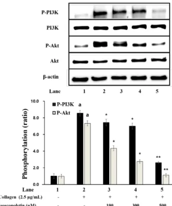
PI3K 및 Akt 인산화에 대한 Isoscopoletin의 효과 혈 소판으로부터 과립의 방출에 관여하는 단백질인 PI 3K 및 Akt의 인산화에 미치는 isoscopoletin의 효과를 확인하였다.
Fig. 5에 나타낸 바와 같이, collagen은 손상받지 않은 세포 와 비교하여 PI 3K의 인산화를 유의하게 증가시켰다. 그러 나, collagen에 의해 증가된 PI 3K의 인산화는 100 μM 이상 의 isoscopoletin에 의해 유의하게 감소되었다(Fig. 5). 또한, collagen은 PI 3K 표적분자로 알려진 Akt의 인산화를 유의 적으로 증가시켰지만, 100 μM 이상의 isoscopoletin에 의해 유의하게 감소되었다(Fig. 5). 이는 isoscopoletin이 collagen 에 의해 증가된 PI 3K 및 Akt의 인산화를 억제한다는 것을 보여준다.
MAPK 인산화에 대한 Isoscopoletin의 효과 혈소판 과 립 방출 및 TXA2 생성에 관여하는 단백질 MAPK(ERK, JNK 및 p38)의 인산화에 미치는 isoscopoletin의 효과를 확 인하였다. Fig. 6에 나타낸 바와 같이, collagen은 손상받지 Fig. 3. Effects of isoscopoletin on TXB2 production. Measure-
ment of TXB2 production was described in “Materials and Methods” section. Data are expressed as mean±SD (n=4).
ap<0.05 compared with no-stimulated platelets, *p<0.05,
**p<0.001 compared with the collagen-stimulated platelets.
Fig. 4. Effects of isoscopoletin on granule secretion. (A) Effects of isoscopoletin on ATP release. (B) Effects of isosco- poletin on serotonin release. Measurement of ATP and sero- tonin release was described in “Materials and Methods”
section. Data are expressed as mean±SD (n=4). ap<0.05 com- pared with no-stimulated platelets, *p<0.05, **p<0.001 com- pared with the collagen-stimulated platelets.
Fig. 5. Effects of isoscopoletin on PI3K and Akt phosphory- lation. Western blotting was determined as described in “Mate- rials and Methods” section. Data are expressed as mean±SD (n=4). ap<0.05 compared with no-stimulated platelets, *p<0.05,
**p<0.001 compared with the collagen-stimulated platelets.
않은 세포와 비교하여 JNK 및 p38의 인산화를 유의하게 증 가시켰지만 ERK의 인산화에는 유의미한 영향을 미치지 않 았다. 또한, collagen에 의해 증가 된 JNK 및 p38의 인산화 는 각각 100 μM 이상의 isoscopoletin에 의해 유의하게 억 제되었다(Fig. 6). 또한, isoscopoletin은 500 μM에서 ERK 인산화를 유의하게 억제했다(Fig. 6). 이는 isoscopoletin이 MAPK로 알려진 ERK, JNK 및 p38의 인산화를 억제하여 혈소판 응집의 신호 과정을 조절한다는 것을 보여준다.
고 찰
혈소판 응집에 대한 isoscopoletin의 효과 및 다양한 신호 전달 물질, 특히, phosphoprotein에 관련한 연구는 알려진 바 가 없기에 본 연구에서는 collagen 유도의 혈소판 응집에 대 한 isoscopoletin의 효과를 명확히 하고자 하였다.
PI3K/Akt 경로는 혈소판 신호 전달에서 기능하는 단백질 로 잘 알려져 있으며, 이들의 인산화는 혈소판 내 dense 과 립의 분비 및 혈소판 응집을 포함하여 혈소판 기능의 조절 에 중요한 역할을 하는 것으로 보고되었다.17) 또한, mitogen-
activated protein kinases(MAPK)는 extracellular signal- regulated kinase(ERK), c-Jun N-terminal kinase(JNK) 및 p38 MAPK를 포함하는 인산화 효소로서 혈소판 활성화 및 응집에 관련한다고 알려져 있다.9) MAPK는 사람 혈소판에 서 검출되며 혈소판이 여러 작용제에 의해 활성화 될 때 인 산화를 통해 활성화되는 것으로 보고되었다.10-12) Mei-Chi 등의 연구에 따르면 p38과 같은 MAPK의 인산화는 TXA2 의 전구체인 arachidonic acid의 방출 및 TXA2의 생성에 중 요하며 혈소판 응집과 관련한다고 보고되었다. 혈소판 억제 활성에 대한 성분 또는 물질을 평가할 때 중요한 지표로서 TXA2의 생성인데, 이는 TXA2가 다른 혈소판을 추가로 활 성화하고 응집시키는 강력한 autacoid로 역할을 하기 때문 이다.24) 따라서, TXA2의 생성을 억제하는 물질이 항 혈소 판 물질로서 유용하게 사용되며, 그 예로 aspirin 및 ozagrel 과 같은 물질이 알려져 있다.25,26)
본 연구에서 isoscopoletin은 collagen에 의한 혈소판 응집 에 대해 농도 의존적으로 억제 효과를 가지며, IC50은 240.93 μM로 나타났다(Fig. 2). 이는 isoscopoletin과 유사하게 Artemisia 및 Scopolia 속 식물의 뿌리에서 추출할 수 있는 물질로 알려진 scoparone이 가지는 collagen 유도의 혈소판 응집억제 효과(IC50은 46.52 μM)에 비해서는 약한 것으로 보인다.27)
혈소판 응집에서 중요한 지표인 TXA2의 생성 및 혈소판 과립 분비(ATP 및 serotonin 방출)에 대한 isoscopoletin의 효과를 측정 하였다. 또한, 본 연구에서는 isoscopoletin과 PI3K/Akt 및 MAPK의 인산화 사이의 관계를 밝히고자 하 였다. 그 결과, isoscopoletin은 collagen에 의한 TXA2의 증 가를 억제하였고, 세포 내 과립 분비의 지표인 ATP와 serotonin의 방출을 크게 감소시켰다(Fig. 3, 4) 또한, 신호 분자로서 혈소판 활성화를 조절하는 것으로 알려진 단백질 인 PI3K/Akt 및 MAPK의 인산화가 isoscopoletin에 의해 현 저히 억제됨을 확인하였다. 특히, MAPK에서 ERK의 경우, collagen에 의해 인산화가 강하게 유발되지 않았으며, isoscopoletin에 의한 억제 효과도 약한 편이었다(Fig. 6). 혈 소판 응집을 억제하는 물질에서 ERK의 인산화 억제 정도 가 JNK 나 p38보다 약하다는 결과가 있으며,28) ERK 경로 의 억제는 작용제-유도의 사람 혈소판 응집에 영향을 미치 지 않는 것으로 보고된 바가 있다.29) 이와 관련하여, MAPK 의 ERK, JNK 및 p38의 인산화가 사람 혈소판의 응집 억 제에 어떻게 관여하는지에 대한 추가 연구가 필요한 것으 로 보인다. 어쨌든, 본 연구의 결과를 통해 isoscopoletin이 PI3K/Akt 및 MAPK와 같은 단백질의 인산화를 억제함으로 써 TXA2 생성 및 세포 내 과립 분비(ATP 및 serotonin 방 출)를 감소시켜 혈소판 응집을 억제한다는 것을 확인하였다.
결론적으로, 우리는 isoscopoletin이 PI3K/Akt 및 MAPK 와 같은 신호전달 과정에 관여하는 phosphoproteins의 인산 Fig. 6. Effects of isoscopoletin on MAPK phosphorylation.
Western blotting was determined as described in “Materials and Methods” section. Data are expressed as mean±SD (n=4).
ap<0.05 compared with no-stimulated platelets, *p<0.05,
**p<0.001 compared with the collagen-stimulated platelets.
화를 조절하는 항혈소판제로서 가치가 있음을 확인하였다.
따라서, isoscopoletin은 혈소판 응집에 의해 야기되는 심혈 관 질환에서 효과적인 치료제 및 예방제로서 가치가 있음 을 제안한다.
사 사
Funding for this paper was provided by Namseoul University.
Conflict of interest
The authors declare no conflict of interest.
인용문헌
1. Jackson, S. P. (2011) Arterial thrombosis?insidious, unpre- dictable and deadly. Nat. Med. 17: 1423-1436.
2. Schwartz, S. M., Heimark, R. L. and Majesky, M. W. (1990) Developmental mechanisms underlying pathology of arteries.
Physiol. Rev. 70: 1177-1209.
3. Payrastre, B., Missy, K., Trumel, C., Bodin, S., Plantavid, M.
and Chap, H. (2000) The integrin alpha IIb/beta 3 in human platelet signal transduction. Biochem. Pharmacol. 60: 1069- 1074.
4. Morello, F., Perino, A. and Hirsch, E. (2009) Phosphoinos- itide 3-kinase signalling in the vascular system. Cardiovasc.
Res. 82: 261-271.
5. Jennings, L. K. (2009) Role of platelets in atherothrombosis.
Am. J. Cardiol. 103: 4A-10A.
6. Sabatine, M. S. and Jang, I. K. (2000) The use of glyco- protein IIb/IIIa inhibitors in patients with coronary artery dis- ease. Am. J. Med. 109: 224-237.
7. Cattanco, M., Tenconi, P. M., Lecchi, A. and Mannucci, P. M.
(1991) In vitro effects of picotamide on human platelet aggre- gation, the release re- action and thromboxane B2 production.
Thromb. Res. 62: 717-724.
8. Su, C. Y., Shiao, M. S. and Wang, C. T. (1999) Differential effects of ganodermic acid S on the thromboxane A2-sig- naling pathways in human platelets. Biochem. Pharmacol.
58: 587-595.
9. Adam, F., Kauskot, A., Rosa, J. P. and Bryckaert, M. (2008) Mitogenactivated protein kinases in hemostasis and throm- bosis. J. Thromb. Haemost. 6: 2007-2016.
10. Bugaud, F., Nadal-Wollbold, F., Lévy-Toledano, S., Rosa, J.
P. and Bryckaert, M. (1990) Regulation of c-jun-NH2 ter- minal kinase and extracellular-signal regulated kinase in human platelets. Blood 94: 3800-3805.
11. Kramer, R. M., Roberts, E. F., Strifler, B. A. and Johnstone, E. M. (1995) Thrombin induces activation of p38 MAP kinase in human platelets. J. Biol. Chem. 270: 27395-27398.
12. Nadal-Wollbold, F., Pawlowski, M., Lévy-Toledano, S., Ber- rou, E., Rosa, J. P. and Bryckaert, M. (2002) Platelet ERK2 activation by thrombin is dependent on calcium and con- ventional protein kinases C but not Raf-1 or B-Raf. FEBS Lett. 531: 475-482.
13. Patrono, C. (1994) Aspirin as an antiplatelet drug. N. Engl. J.
Med. 330: 1287-1294.
14. Flevaris, P., Li, Z., Zhang, G., Zheng, Y., Liu, J. and Du, X.
(2009) Two distinct roles of mitogen-activated protein kinases in platelets and a novel Rac1-MAPK-dependent inte- grin outside-in retractile signaling pathway. Blood 113: 893- 901.
15. Kramer, R. M., Roberts, E. F., Um, S. L., Börsch-Haubold, A.
G., Watson, S. P., Fisher, M. J. and Jakubowski, J. A. (1996) p38 mitogenactivated protein kinase phosphorylates cytosolic phospholipase A2 (cPLA2) in thrombin-stimulated platelets.
Evidence that proline-directed phosphorylation is not required for mobilization of arachidonic acid by cPLA2. J.
Biol. Chem. 271: 27723-27729.
16. McNicol, A. and Shibou, T. S. (1998) Translocation and phosphorylation of cytosolic phospholipase A2 in activated platelets. Thromb. Res. 92: 19-26.
17. Chuang, W. Y., Kung, P. H., Kuo, C. Y. and Wu, C. C. (2013) Sulforaphane prevents human platelet aggregation through inhibiting the phosphatidylinositol 3-kinase/Akt pathway.
Thromb. Haemost. 109: 1120-1130.
18. Ali, M. Y., Jannat, S., Jung, H. A., Choi, R. J., Roy, A. and Choi, J. S. (2016) Anti-Alzheimer's disease potential of cou- marins from Angelica decursiva and Artemisia capillaris and structure-activity analysis. Asian. Pac. J. Trop. Med. 9: 103- 111.
19. Dong, J., Yuan, J., Wang, J. L., Ji, R. F., Quan, Q. H., Guo, X. Y., Gao, J. and Liu, Y. G. (2017) Study on screening anti- tumor active fractions and chemical components in active fractions from root of Anaycclus pyrethrum. Zhongguo Zhong Yao Za Zhi 42: 3932-3937.
20. Huang, H. C., Chu, S. H. and Chao, P. D. (1991) Vaso- relaxants from Chinese herbs, emodin and scoparone, possess immunosuppressive properties. Eur. J. Pharmacol. 198: 211- 213.
21. Huang, H. C., Lee, C. R., Weng, Y. I., Lee, M. C. and Lee, Y. T. (1992) Vasodilator effect of scoparone (6,7-dimethoxy- coumarin) from a Chinese herb. Eur. J. Pharmacol. 218: 123- 128.
22. Lu, C., Li, Y., Hu, S., Cai, Y., Yang, Z. and Peng, K. (2018) Scoparone prevents IL-1β-induced inflammatory response in human osteoarthritis chondrocytes through the PI3K/Akt/NF- κB pathway. Biomed. Pharmacother. 106: 1169-1174.
23. Shin, J. H., Kwon, H. W. and Lee, D. H. (2019) Ginsenoside F4 inhibits platelet aggregation and thrombus formation by dephosphorylation of IP3RI and VASP. J. Appl. Biol. Chem.
62: 93-100.
24. Chang, M. C., Wang, T. M., Yeung, S. Y., Jeng, P. Y., Liao, C. H., Lin, C. C., Lin, B. R. and Jeng, J. H. (2011) Anti- platelet effect by p-cresol, a uremic and environmental tox- icant, is related to inhibition of reactive oxygen species, ERK/
p38 signaling and thromboxane A2 production. Atheroscle- rosis 219: 559-565.
25. Cipollone, F., Patrignani, P., Greco, A., Panara, M. R., Pado- vano, R., Cuccurullo, F., Patrono, C., Rebuzzi, A. G., Liuzzo, G., Quaranta, G. and Maseri, A. (1997) Differential sup- pression of thromboxane biosynthesis by indobufen and aspi- rin in patients with unstable angina. Circulation 96: 1109- 1116.
26. Patrono, C. (2001) Aspirin: new cardiovascular uses for an old drug. Am. J. Med. 110: 62S-65S.
27. Lee, D. H. (2020) Inhibitory effects of scoparone through regulation of PI3K/Akt and MAPK on collagen-induced human platelets. J. Appl. Biol. Chem. 63: 131-136.
28. Irfan, M., Jeong, D., Saba, E., Kwon, H. W., Shin, J. H., Jeon, B. R., Kim, S., Kim, S. D., Lee, D. H., Nah, S. Y. and Rhee, M. H. (2019) Gintonin modulates platelet function and inhib- its thrombus formation via impaired glycoprotein VI sig- naling. Platelets 30: 589-598.
29. McNicol, A. and Jackson, E. C. (2003) Inhibition of the MEK/ERK pathway has no effect on agonist-induced aggre- gation of human platelets. Biochem. Pharmacol. 65: 1243- 1250.
(2020. 6. 6 접수; 2020. 6. 23 심사; 2020. 7. 28 게재확정)