ISSN 1225-6552, eISSN 2287-7630 https://doi.org/10.7853/kjvs.2018.41.1.51
< Case Report >
Veterinary Service
Available online at http://kjves.org
*Corresponding author: Eun-Jin Choi, Tel. +82-54-912-0460, Fax. +82-54-912-0465, E-mail. choiej@korea.kr
Pathological findings of the mixed infection with canine distemper virus and Streptococcus canis on farmed badger
Ji-hyeon Kim, Kyunghyun Lee, Ji-Youl Jung, Eun-Jin Choi*, Ha-Young Kim, ByungJae So
Animal Disease Diagnostic Division, Animal and Plant Quarantine Agency, Gimcheon 39660, Korea (Received 23 February 2018; revised 14 March 2018; accepted 19 March 2018)
Abstract
Herein, we report a case of badgers showing high morbidity and mortality rate due to the mixed in- fection of canine distemper virus (CDV) and Streptococcus canis (S. canis) in a farm where wild ani- mal, badger, is being reared for herbal medicine. During the period of about one month, 120 out of 320 badgers showed severe respiratory symptoms and died, and 3 bodies were submitted to the Animal and Plant Quarantine Agency for disease diagnosis. The lung with the most severe necropsy findings failed to collapse and showed dark reddening and had yellowish nodules on the cut surface. The charac- teristic and common histopathologic findings include multifocal necrosis with hemorrhage of the lung, severe lymphoid depletion of the spleen and intracytoplasmic or intranuclear inclusion bodies in almost all organs. Finally, CDV and S. canis were identified by immunohistochemistry and bacterial isolation, respectively. This is the first mixed infection case of CDV and S. canis in badgers being raised on the farm.
Key words : Badger, Canine distemper virus (CDV), Mixed infection, Pathology, Streptococcus canis (S.
canis)
INTRODUCTION
Canine distemper virus (CDV), a relatively large sin- gle-stranded RNA virus, is a member of the genus Mobilivirus of the family Paramyxoviridae. CDV causes canine distemper (CD), a highly contagious systemic disease affecting terrestrial carnivores including Canidae, Mustelidae, Ailuridae, Mephitidae, and Ursidae (Deem et al, 2000). CDV spreads by aerosol droplet secretions from infected animals and enters the respiratory tract (Ford, 2012). Virus initially replicates in the tonsils and lymphatic tissue, which is followed by infection of res- piratory, gastro-intestinal tract, urinary tract, and central nervous system (Dungworth, 1993). In the early stage of typical infection, a transient fever usually occurs and there may be accompanied by anorexia, nasal discharge,
and lethargy (Ford, 2012). In addition, multisystemic disease with severe neurological signs can occur (Van- develde et al, 2005; Moretti et al, 2006). Affected ani- mals with CDV are prone to opportunistic infections as a consequence of generalized lymphoid depletion and profound immunosuppression (Rudd et al, 2006; Chvala et al, 2007). In previous cases, double infection with the Toxoplama gondii and CDV and triple infection with canine adenovirus (CAV) type 2, Mycoplasma cynos and CDV in a dogs were reported (Chvala et al, 2007;
Aguiar et al, 2012).
Streptococcus canis (S. canis) is generally an oppor- tunist pathogen and a group G beta-hemolytic species of Streptococcus (Facklam, 2002). S. canis is important to the skin and mucosal health of cats and dogs, but under certain circumstances, the bacteria have been reported to cause diseases in a variety of mammals (Facklam, 2002).
The lungs have been considered the organ seriously af-
Fig. 1. Gross and microscopic findings in the lung of three badgers. The mottled dark red spots and yellowish nodules on the surface (A). Bar=2 cm. Severe suppurative bronchopneumonia (B). HE. Bar=200 m. Acidophilic intracytoplasmic inclusion bodies in the bronchial epithelial cells (arrows) (C). HE. Bar=50 m. CDV antigens in bronchial epithelial cells (D). IHC. Bar=50 m.
fected by S. canis infection, and acute infection ap- peared to be superimposed on chronic preexisting pul- monary infection (Murase et al, 2003).
Mustelids are the most susceptible to CDV disease among the species. The badger is a carnivore belonging to the family Mustelidae (Phillippa et al, 2008). In Korea, CD case has been reported in wild badgers with single infection in 1997 (Kim et al, 1997). This is the first report of the mixed infection with CDV and S.
canis in reared badgers through pathological findings and etiological examination.
CASE
Three hundred twenty badgers raised for herbal medi- cine were kept on a farm located in Gyeongbuk prov- ince, republic of Korea. All of them did not have any
vaccination history. About one hundred twenty badgers showed by dyspnea, reduced appetite, weight loss, half-closed eyes with lacrimation, and death over a month period. Three 5-year-old female badgers among them were submitted to the Animal and Plant and Quarantine Agency for laboratory diagnosis.
At necropsy, the lungs failed to collapse and were rubbery and mottled dark red appearances on the surface.
On the cut surface of the lungs, yellowish nodules were multifocally observed (Fig. 1A). The tissue samples in- cluding brain, lung, spleen, liver, kidney, stomach, and urinary bladder were fixed in 10% neutral buffered for- malin, processed routinely, and embedded in paraffin wax. Tissue sections (4 m) were cut and stained with hematoxylin and eosin. Immnunohistochemistry (IHC) was performed using mouse monoclonal antibody against canine distemper virus (MCA1893, AbD Serotec, Dues- seldorf, Germany).
Table 1. Immunohistochemical results for antigen detection of canine distemper virus in three badgers
Organ Immunohistochemical reactivity*
Badger 1 Badger 2 Badger 3
Brain + - +
Kidney ++ +++ +++
Liver ++ +++ -
Lung +++ ++ +++
Spleen +++ +++ +++
Stomach + + +++
Urinary bladder NT +++ ++
*The immunohistochemical reactivity was measured under microscopy (×400). and classified according to the number of CDV antigen-con- taining cells observed per slide as follows; -, no antigen detected; +, mild (<10%); ++, moderate (10∼50%); +++, severe (>50%), NT, not tested.
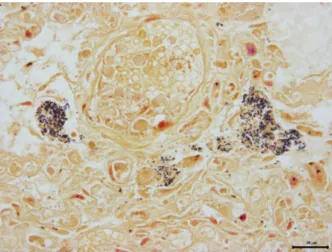
Fig. 2.Gram staining of the lung. Dense clusters of gram-positive bacteria was observed in parenchyma of lung. Gram stain. Bar=20
m.
Histopathologic examination revealed a variety of le- sions related to CDV infection. Significant histological changes were observed in the lung. The lung showed suppurative bronchopneumonia with multifocal abscesses (Fig. 1B). Additional findings were multifocal necrosis with cell debris and hemorrhages in the alveolar lumen.
Many intracytoplasmic inclusion bodies in the bronchial epithelial cells (Fig. 1C) and alveolar epithelial cells were observed. Those inclusion bodies were also found in the epithelial cells of the brain, spleen, liver, kidney, stomach, and urinary bladder. Further, splenic alteration was manifested as severe lymphocytic depletion and liv- er was observed fatty change and lymphohistiocytic hep- atitis with multifocal necrosis.
Immunohistochemically, CDV antigen was strongly detected in the bronchial epithelial cells, which fre- quently also contained acidophilic intranuclear or intra- cytoplasmic bodies (Fig. 1D) and macrophages in the al- veolar lumen. In addition to the lung, CDV antigens were easily identified in the ependymal cells of the brain, epithelial cells of white and red pulp of the spleen, biliary tract of the liver, biliary tract of the liver, collecting tubule of the kidney, mucosa of the stomach, and the transitional cells of the urinary bladder with various of levels (Table 1). The other organs showed negative reaction in three badgers.
Dense clusters of Gram-positive bacteria were ob- served in selected sections of the lung tissues of 3 badgers (Fig. 2). For the identification of the bacteria,
lung tissue samples from all badgers were collected aseptically, inoculated on blood agar and incubated 24 hours at 37°C in aerobic condition. The -hemolytic colonies in all samples were uniformly cultured on blood agar and these isolates were identified as S. canis by VITEK II (BioMerieux, Hazelwood, MO, France).
DISCUSSION
Based on the histopathological features and bacterial examination, this case was diagnosed as a mixed in- fection with CDV and S. canis. Although CDV in- fection have commonly reported in Mustelids (Deem et al, 2000), there are only two case of complex infection as a consequence of CDV-induced immunosuppression in Yellow-throated Marten (Martes flavigula koreana) and badger (Meles meles) (Hammer et al, 2004; Park et al, 2016).
In the present case, significant features of three badg- ers were severe bronchopneumonia with multifocal ab- scess and the presence of both intracytoplasmic and in- tranuclear inclusion bodies in the epithelial cells of many organs. This findings were consistent with pre- vious reports of CDV infection in other species (Headley and Sukura, 2009; Cho et al, 2015) and badgers (Kim et al, 1997). None of three badgers showed the character- istic hyperkeratosis of the footpad or nose often asso-
ciated with CDV infection.
CDV have been commonly reported to infection of the central nervous system (CNS) leading to a variety of neuological signs (Meertens et al, 2003; Amude et al, 2006; Aguiar et al, 2012). In a previous cases, CDV in- fected carnivores showed neuological signs such as seiz- ure, tremor and histopathological alteration including de- myelination and encephalitis with inclusion bodies in the CNS (Liang et al, 2007; Chen et al, 2008; Headley et al, 2009). Fischer (1965) described lesions of the central nervous system in four free-ranging badgers from Switzer- land. In the present case, CDV three badgers had no neurological clinical signs before death and neuro- pathologic lesions. In addition, unlike the strong im- munopositivitiy in other organs such as lungs, liver, and kidney, there were negative or mild reaction in the brain of three badgers.
Canine distemper is a highly contagious disease that can be easily spread between unvaccinated animals (Perrone et al, 2010). In this case, badgers were im- munologically naive, so it may have been easily infected from stray dogs around the farm. Further, the 120 badg- ers that had the similar clinical signs on the farm almost died, suggesting that CDV inflow from outside may have affected high morbidity rated in populated area such as that farm.
In the present case, S. canis was also isolated in the lungs of three badgers. The observed lymhopenia and lymphoid depletion in spleen is the evidence of im- munosuppressive effects of CDV, suggesting that badg- ers infected with CDV were more susceptible to develop the secondary infection by S. canis. On the basis of the severe necrotic lesions and histopathologic features, there was strong viral replication in the lungs and S.
canis seem to have been affected to severity of bron- chopneumonia with necrosis and abscesses. Aguiar et al.
(2012) and Munson et al. (2008) reported that bacterial coinfections impaired immune function and worsen clin- ical symptoms in CDV-infected animals. Coinfections may affect morbidity and mortality (Goller et al, 2010).
Recently, it has been reported that CDV infection af- fects not only wildlife but also aquatic mammals and the seriousness of the disease is also shown in farm-reared wildlife (Deem et al, 2002; Sliki et al,
2002; Perrone et al, 2010). Therefore, it is important to investigate the prevalence of CDV in free-ranging carni- vore species and surveillance to susceptible infectious diseases that can have a large impact on the animal of interest. Further, early diagnosis of infection is important to quarantine the suspectible CDV infection animals. The immunohistochemical demonstration of CDV antigen proved to be a reliable diagnostic methods (An et al, 2008). In agreement with present reports, IHC examina- tion of biopsy samples from the lungs, liver, and kidney could be recommended for diagnosis of CDV infection.
However, compared to IHC, reverse transcriptase poly- merase chain reaction (RT-PCR) is the preferred method to early diagnosis for CDV infection because of its sen- sitivity and specificity (Cho and Park, 2005; An et al, 2008). Therefore, molecular methods are additionally needed for diagnosis of CD cases. We also emphasize to take measures to prevent or reduce secondary pro- liferation of bacteria in reared farm.
ACKNOWLEDGEMENTS
This project was supported by a grant (Project code no. N-1543069-2015-99-01) from the Animal and Plants Quarantine Agency (APQA), the Ministry of Agriculture, Food and Rural Affairs (MAFRA), Republic of Korea.
REFERENCES
Aguiar DM, Amude AM, Santos LGF, Ribeiro MG, Ueno TEH, Megid J, Paes AC, Alfieri AF, Alfieri AA, Gennari SM.
2012. Canine distemper virus and Toxoplasma gondii co-infection in dogs with neurological signs. Arq Bras Med Vet Zootec 64: 221-224.
Amude AM, Alfieri AA, Alfieri AF. 2006. The nervous form of canine distemper. Vet. e Zootec 13: 125-136.
An DJ, Kim TY, Song DS, Kang BK, Park BK. 2008. An im- munochromatography assay for rapid antemortem diag- nosis of dogs suspected to have canine distemper. J Virol Methods 147: 244-249.
Chen CC, Pei JC, Liao MH, Mortenson JA. 2008. Canine dis- temper virus in wild Ferret-Badgers of Taiwan. J Wildl Dis 44: 440-445.
Cho AR, Roh YS, Cho HS, Lim CW, Kang SJ, Kim HY, Kim JW, Kim BS. 2015. Case report : Canine distemper virus
infection in a wild Korean raccoon dog. J Prev Vet Med 39: 29-32.
Cho HS, Park NY. 2005. Detection of canine distemper virus in blood samples by reverse transcription loop-mediated isothermal amplification. J Vet Med B Infect Dis Vet Public Health 52: 410-413.
Chvala S, Benetka V, Möstl K, Zeugswetter F, Spergser J, Weissenböck H. 2007. Simultaneous canine distemper virus, canine adenovirus type 2, and Mycoplasma cynos infection in a dog with pneumonia. Vet Pathol 44:
508-12.
Deem SL, Spelman LH, Yates RA, Montali RJ. 2000. Canine dis- temper in terrestrial carnivores: a review. J Zoo Wildl Med 31: 441-451.
Dungworth DL. 1993. The respiratory system. In: Pathology of Domestic Animals, ed. Jubb KVF, Kennedy PC, and Palmer N, 4th ed. pp.539-698. Academic Press, London.
Facklam R. 2002. What happened to the streptococci: overview of taxonomic and nomenclature changes. Clin Microbiol Rev 15: 613-630.
Fischer K. 1965. Staupe-Encephalitis bei Dachsen. Schweizer Archiv fur Tierheilkunde 107: 87-91.
Ford RB. 2012. Canine infectious repiratory disease. In:
Infectious diseases of the dog and cat, ed. Greence CE.
4th ed., pp. 55-65. Elsevier, St Louis, MO.
Goller KV, Fyumagwa RD, Nikolin V, East ML, Kilewo M, Speck S, Muller T, Matzke M, Wibbelt G. 2010. Fatal canine distemper infection in a pack of African wild dogs in the Serengeti ecosystem, Tanzania. Vet Microbiol 14: 245-252.
Hammer AS, Dietz H, Andersen T, Nielsen L, Blixenkrone- Møller M. 2004. Distemper virus as a cause of central nervous disease and death in badgers (Meles meles) in Denmark. Vet Rec 154: 527-530.
Headley SA, Sukura A. 2009. Naturally occurring systemic ca- nine distemper virus infection in a pup. Braz J Vet Pathol 2: 95-101.
Kim JH, Roh IS, Bae EJ, Jean YH, Hwang EK, Sohn JH, Choi SH. 1997. Canine distemper virus infection in badgers.
Korean J Vet Pathol 1: 145-148.
Liang CT, Chueh LL, Pang VF, Zhuo YX, Liang SC, Yu CK, Chiang H, Lee CC, Liu CH. 2007. A nonbiotin poly- merized horseradish-peroxidase method for the im- munohistochemical diagnosis of canine distemper. J
Comp Pathol 137: 57-64.
Meertens N, Stoffel MH, Cherpillod P, Wittek R, Vandevelde M, Zurbriggen N. 2003. Mechanism of reduuction of virus release and cell-cell fusion in persistent canin distemper virus infection. Acta Neuropathol 106: 486-494.
Moretti L, Da Silva AV, Ribeiro MG, Paes AC, Langoni H.
2006. Toxoplasma gondii genotyping in a dog co-in- fected with distemper virus and ehrlichiosis rickettsia.
Rev Inst Med Trop Sao Paulo 48: 359-363.
Munson L, Terio KA, Kock R, Mlengeya T, Roelke ME, Dubovi E, Summers B, Sinclair ARE, Packer C. 2008. Climate extremes promote fatal co-infections during canine dis- temper extremes promote fatal co-infections during can- ince distemper epidemics In African lions. PLoS One 3:
e2545.
Murase T, Morita T, Sunagawa Y, Sawada M, Shimada A, Sato K, Hikasa Y. 2003. Isolation of Streptococcus canis from a Japanese raccoon dog with firinous pleurop- neumonia. Vet Rec 153: 471-472.
Park S, Choi US, Kim EJ, Lee JH, Lee HB, Cho HS, Kim W, Lim CW, Kim B. 2016. Coinfection with Hepatozoon sp. and Canine Distemper Virus in a Yellow-throated Marten (Martes flavigula koreana) in Korea. J Wildl Dis 52: 414-417.
Perrone D, Bender S, Niewiesk S. 2010. A comparison of the im- mune responses of dogs exposed to canine distemper vi- rus (CDV) - Differences between vaccinated and wild-type virus exposed dogs. Can J Vet Res 74: 214-217.
Philippa J, Fournier-Chambrillon C, Fournier P, Schafteraar W, van de Bildt M, van Herweijnen R, Kuiken T, Liabeuf M, Ditcharry S, Joubert L. 2008. Serologic survey for selected viral pathogens in free-ranging endangered European mink (Mustela lutreola) and other mustelids from south-western France. J Wildl Dis 44: 791-801.
Rudd PA, Cattaneo R, Von Messling V. 2006. Canine distemper virus uses both the anterograde and the hematogenous pathway for neuroinvasion. J Virol 80: 9361-9370.
Sliki JT, Cooper EJ, Gustavson JP. 2002. Emerging morbilliirus infections of marine mammals: development of two di- agnositc approaches. Ann N Y Acad Sci 969: 51-59.
Vandevelde M, Zurbriggen A. 2005. Demyelination in canine dis- temper virus infection: a review. Acta Neuropathol 109:
56-68.