Vol.21 No.1 p40-50, June 2004
1)
책임저자:안면환, 대구시 남구 대명동 707-717, 영남대학교병원 정형외과학실 Tel: (053) 620-3643, Fax: (053) 628-4020 E-mail: [email protected]
특발성 척추측만증 환자에서의 3차원적 재구성을 이용한 척추경의 크기 측정
허재희․안면환
영남대학교 의과대학 정형외과학교실
The Measurement of Size of the Pedicle Using 3 Dimensional Reconstruction Image in Idiopathic Scoliosis
Jae-Hee Heo, Myun-Hwan Ahn
Department of Orthopedic Surgery,
College of Medicine, Yeungnam University, Daegu, Korea
-Abstract-
Background: This study was conducted to analyze the height and width of the pedicle of the upper and lower levels on the concave and the convex sides. In addition, we checked for the appropriate pedicle screw size which could be screwed in without complications.
Materials and Methods: Taking a simple AP radiography in a standing position, 99 vertebrae on the major curve with the possibility of 3-D reconstruction were analyzed after checking the CT in a supine position of 22 idiopathic scoliosis. We measured Cobb's angle from a simple radiograph, and measured the size of the isthmus by the Inner Space 3-D Editor after 3-D reconstruction with the Inner Space 3-D program in the DICOM file transformed from CT image. We then analyzed the size of pedicles of the upper and lower levels on the concave and the convex sides by measuring the height and width of the pedicle.
Results: All pedicles on the concave side were smaller than those on the convex side. Their
size increased as the measurement moved from the upper to lower vertebra, except for the
upper thoracic vertebra. When the width of the pedicle through 3-D reconstruction was
compared with the narrowest width of the pedicle measured by using CT, the width of the
서 론
특발성 척추측만증은 관상면, 시상면상의 변 화뿐만 아니라 추체의 회전변형도 동반된 3차 원적 기형이다. 이러한 척추체의 3차원적 변형 을 교정하기 위한 수술적 치료를 시행함에 있 어 최근에 척추경 나사못의 유용성이 밝혀지면 서
1-4)특발성 척추측만증 환자의 치료 시 척추 경 나사못을 이용한 기기술이 널리 사용되고 있다. 하지만 이러한 유용성에 대한 다양한 보 고에도 불구하고 흉추의 척추경의 크기가 요추 의 척추경의 크기에 비해 상대적으로 작은 해 부학적 특성으로 인해 척추경 나사못 삽입 시 신경 손상 및 척추경 골절과 같이 예기치 않은 합병증에 대한 보고도 있어왔다.
5-7)따라서 척 추경에 적절한 크기의 나사못을 고정하기 위하 여 척추측만증 환자에 있어 척추경의 형태학적 특성 연구가 요구되며 특히 척추경의 크기 및 모양에 대한 정확한 측정이 필요할 것으로 사 료된다.
이에 본 연구에서는 특발성 척추측만증 환 자에서 컴퓨터 단층 촬영을 이용한 3차원적 영 상의 재구성을 실행한 후, 협부가 가장 좁게 측정되는 관상면을 이용하여 상․하․요․철측 척추경의 높이 및 폭에 대해 비교․분석을 실
시해 보고 이를 컴퓨터 단층 촬영을 이용한 축 면의 영상에서의 척추경의 폭과 비교하여 그 오차에 대해 알아보고자 하였다. 또한 3차원적 재구성을 통해 얻어진 관상면에서 척추경의 모 양을 분석해 본 후 이러한 종합적인 결과를 이 용하여 합병증 없이 척추경 나사못을 삽입할 수 있는 척추경 나사못의 적절한 직경에 대해 알아보고자 하였다.
재료 및 방법
영남대학교 의과대학 부속병원에 내원한 특 발성 척추측만증 환자 중 컴퓨터 단층촬영이 가능하였던 22예를 대상으로 주만곡 내의 총 99개 추체를 분석하였다. 22예 중 남자가 5예, 여자가 17예였으며, 최저 12세에서 최고 23세 로 평균 16.7세였다.
기립위에서 촬영한 단순 전후방 방사선 사 진을 이용하여 Cobb 측만각
8)을 측정하였고, 만곡의 유연성 및 King의 분류
9)를 위해 양측 방 굴곡 사진을 촬영하였다. 컴퓨터 단층 촬영 은 첨단 추체 및 상․하단 끝척추를 중심으로 2 mm 간격으로 촬영하여 DICOM (Digital Image Communication Of Medical) 파일로 전 환하여 총 99개의 추체를 해석하였다.
pedicles through 3-D reconstruction was statistically smaller (P<0.01). Most of the pedicles were tear-drop or kidney shaped rather than cylindrical.
Conclusion: These results suggest that the use of the coronal plane through 3-D reconstruction would be necessary for an accurate measurement of the size of the pedicle. It is important to pay careful attention to the screw size and the screwing method considering the pedicle shape through 3-D reconstruction.
Key Words: Idiopathic scoliosis, Computed tomography, 3-D reconstruction, Pedicle size
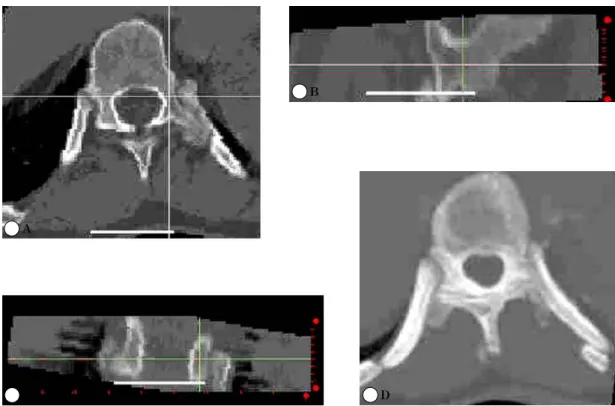
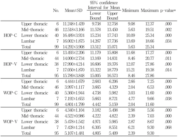
추경의 크기의 측정에서는 측정의 신뢰도를 높이기 위하여 본 연구진에 의해 개발된 Inner Space 3-D 프로그램을 이용하여 관상면․시상 면 및 회전 변형을 교정한 3차원 영상 재구성 화면에서 추경의 축방향에 수직인 관상면에서 가장 협소한 부위의 각각의 외피질골을 측정하 였고(Fig. 1, 2), 이 화면에 나타난 척추경의 모 양을 분석하였다. 2차원적 단순 측정을 위해서 는 각 척추체마다 컴퓨터 단층 촬영상에서 얻 어진 축면의 영상에서 척추경이 가장 좁게 측 정되는 부위를 척추경의 협부로 가정하여 그 부위의 외피질골을 측정하였다(Fig. 3). 통계적 해석을 위하여서는 SPSS 10.0 for Windows를 이용하였다. 군간의 차이를 확인하기 위하여서
는 일원변량분석법을 이용하였다. 상관관계 분 석을 위하여서는 T-test와 ANOVA (analysis Fig. 1. The narrowest pedicle size was measured by Inner Space 3D program. The size of pedicle was measured using coronal image of Inner Space 3-D program after determining the level of isthmic portion by compounding axial and sagittal image of Inner Space 3-D program.(A: Axial image; B: Sagittal image; C: Coronal image; D: reconstructed vertebra by Inner Space 3-D program).
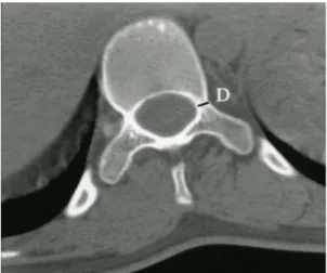
Fig. 2. Measurement technique for height (H) and width (W) of pedicle. Many of the pedicles show teardrop shape.
A
B
C D
of variance)를 이용하였다.
결 과
Cobb 방법
8)을 이용한 만곡의 크기를 측정한 결과 최저 18도에서 최고 68도이며 평균 36도 였다. 40∼49도가 7예(31.8%)로 가장 많았으며, 10∼19도가 3예(13.6%), 20∼29도가 5예(22.7%), 30∼39도가 5예(22.7%), 50도 이상이 2예(9%) 였다.
총 99개의 추체를 분석하였으며 흉추 3번이 1개, 흉추 4번이 5개, 흉추 5번이 9개, 흉추 6 번이 15개, 흉추 7번이 10개, 흉추 8번이 12개, 흉추 9번이 12개, 흉추 10번이 11개, 흉추 11번 이 10개, 흉추 12번이 7개였으며 요추 1번이 2 개, 요추 2번이 3개, 요추 3번이 2개였다.
Fig. 3. Transverse pedicle diameter(D) was measured at the mid-portion of the pedicle.
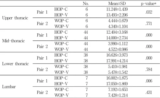
Table 1. Measurement of height and width of the pedicle on the concave and convex side related to its level of the apical vertebra
No. Mean±SD
95% confidence Interval for Mean
Minimum Maximum Lower
Bound
Upper Bound
HOP-C
Mid-thoracic 10 10.958±1.847 9.637 12.280 8.93 14.13 Lower thoracic 11 14.859±3.025 12.826 16.890 11.00 20.08
Lumbar 1 17.686 - - 17.69 17.69
Total 22 13.214±3.258 11.770 14.659 8.93 20.08
HOP-V
Mid-thoracic 10 13.548±2.216 11.962 15.133 9.69 16.08 Lower thoracic 11 16.121±2.709 14.301 17.941 13.56 20.75
Lumbar 1 19.150 - - 19.15 19.15
Total 22 15.089±2.842 13.829 13.349 9.69 20.75
WOP-C
Mid-thoracic 10 3.576±1.101 2.789 4.364 2.04 5.91 Lower thoracic 11 4.193±0.946 3.557 4.828 3.32 6.40 Lumbar 1 7.577 - - 7.58 7.58 Total 22 4.066±1.287 3.496 4.637 2.04 7.58
WOP-V
Mid-thoracic 10 4.796±1.065 4.034 5.558 3.60 7.03
Lower thoracic 11 4.408±1.034 3.713 5.103 2.87 6.62
Lumbar 1 7.342 - - 7.34 7.34
Total 22 4.718±1.173 4.198 5.238 2.87 7.34
HOP-C, Height of pedicle-concave side; HOP-V, Height of pedicle-convex side; WOP-C, Width of
pedicle-concave side; WOP-V, Width of pedicle-convex side; SD, Standard deviation.
첨부는 각 해당 만곡의 첨부 추체를 원칙으 로 하여 결정하였으며 첨부가 추간판일 경우에 는 상부 추체를 첨부 추체로 결정하였으나 본 연구에서는 추간판이 첨부가 되는 경우는 한 예도 없었다. 첨부 추체의 위치는 흉추 9번이 8개(36.4%), 흉추 8번이 7개(31.8%), 흉추 7번 이 4개(18.2%), 흉추 10번, 11번 요추 1번이 각 1개(4.5%)였다.
2 mm 간격으로 촬영한 컴퓨터 단층 촬영으 로 인해 3차원적 재구성이 불가능했던 척추경
을 제외한 분석 가능하였던 요측의 99개, 철측 의 95개의 척추경을 대상으로 분석을 실시하였 다. 상하 척추체의 추경에 대한 크기 분석을 위해 흉추 1번에서 4번까지를 상위 흉추, 5번 에서 8번까지를 중위 흉추, 9번에서 12번까지 를 하위 흉추로 구분하였다. 재구성이 가능하 였던 척추경을 가진 추체 중 첨부 추체는 중위 흉추에 11개, 하위 흉추에 10개, 요추에 1개가 있었으며, 첨부 추체를 제외한 추체는 상위 흉 추에 6개, 중위 흉추에 34개, 하위 흉추에 37
Table 2. Measurement of height and width of the pedicle on the concave and convex side related to its level of major curve
No. Mean±SD
95% confidence Interval for Mean
Minimum Maximum p-value*
Lower Bound
Upper Bound
HOP-C
Upper thoracic 6 11.248±1.439 9.738 12.758 9.08 12.37 .000 Mid-thoracic 46 12.518±3.166 11.578 13.450 5.63 19.51 .002 Lower thoracic 40 16.489±3.924 15.234 17.743 10.09 25.34 .000 Lumbar 7 16.002±1.875 14.267 17.736 13.69 18.66 .034 Total 99 14.292±3.908 13.512 15.071 5.63 25.34
HOP-V
Upper thoracic 6 13.493±2.206 11.179 15.808 11.68 17.27 .000 Mid-thoracic 44 14.000±2.734 13.169 14.831 8.46 20.77 .011 Lower thoracic 38 17.990±4.214 16.606 19.376 12.07 27.86 .000 Lumbar 7 17.050±1.870 15.322 18.779 15.21 19.38 .014 Total 95 15.789±3.848 15.005 16.573 8.46 27.86
WOP-C
Upper thoracic 6 4.444±1.679 2.683 6.206 2.66 7.25 .000 Mid-thoracic 46 3.997±1.117 3.665 4.329 2.04 6.53 .000 Lower thoracic 40 5.360±1.944 4.738 5.982 3.03 11.60 .000 Lumbar 7 7.192±1.653 5.663 8.721 4.77 9.66 .038 Total 99 4.801±1.799 4.442 5.159 2.04 11.60
WOP-V
Upper thoracic 6 4.340±1.104 3.182 5.498 2.98 5.56 .000 Mid-thoracic 44 4.522±0.986 4.222 4.822 2.39 7.03 .000 Lower thoracic 38 5.478±1.542 4.971 5.985 2.87 8.67 .000 Lumbar 7 7.428±1.214 6.305 8.551 6.21 9.30 .068 Total 95 5.107±1.481 4.805 5.409 2.39 9.30
HOP-C, Height of pedicle-concave side; HOP-V, Height of pedicle-convex side; WOP-C, Width of
pedicle-concave side; WOP-V, Width of pedicle-convex side; SD, Standard deviation; *Analysis of
Variance.
개, 요추에 6개가 있었다.
척추경의 평균 높이 및 폭은 첨부 추체에서 요측 13.2 mm, 4.1 mm와 철측 15.1 mm, 4.7 mm였다. 중위 흉추 첨부 추체 추경의 평균 높 이 및 폭은 요측 10.9 mm, 3.6 mm와 철측 13.5 mm, 4.8 mm였고 하위 흉추 첨부 추체 추경의 평균 높이 및 폭은 요측 14.9 mm, 4.2 mm와 철측 16.1 mm, 4.4 mm였다. 요추 첨부 추체의 높이 및 폭은 요측 17.7 mm, 7.6 mm 와 철측 19.1 mm, 7.3 mm였다(Table 1). 척추 경의 평균 높이와 폭은 상위 흉추에서는 요측 11.2 mm, 4.4 mm와 철측 13.5 mm, 4.3 mm였 고 중위 흉추에서는 요측 12.5 mm, 3.9 mm와 철측 13.9 mm, 4.5 mm였다. 하위 흉추에서는 요측 16.5 mm, 5.4 mm와 철측 18.0 mm, 5.5
mm였다. 요추에서는 요측 16.0 mm, 7.2 mm 와 철측 17.1 mm, 7.4 mm였다(Table 2). 상하 척추경의 크기 비교 시 상부 척추에서 하부 척 추로 내려올수록 척추경의 높이 및 폭이 통계 학적으로 유의하게 증가했다(Table 2).
측만된 척추체의 요철측 척추경을 비교 시 높이는 상위, 중위, 하위 흉추 및 요추 모두에 서 철측에 비해 요측이 통계적으로 유의하게 작았다(P=0.000). 폭은 상위 흉추, 하위 흉추 그리고 요추에서는 통계학적으로 유의한 차이 를 나타내지는 않았지만(P>0.05) 중위 흉추에 서는 통계적으로 유의하게 작았다(P=0.000) (Table 3). 첨부 추체의 경우에서도 높이(P=
0.000) 및 폭(P<0.01) 모두 철측에 비해 요측이 통계학적으로 유의하게 작았다(Table 4).
Table 3. Comparison of height and width of the each pedicles on the concave and convex side related to its level of major curve
No. Mean±SD p-value*
Upper thoracic
Pair 1 HOP-C 6 11.248±1.439 HOP-V 6 13.493±2.206 .032 Pair 2 WOP-C 6 4.444±1.679 WOP-V 6 4.340±1.104 .771
Mid-thoracic
Pair 1 HOP-C 44 12.404±3.168 HOP-V 44 14.000±2.734 .000 Pair 2 WOP-C 44 3.990±1.112 WOP-V 44 4.522±0.986 .000
Lower thoracic
Pair 1 HOP-C 38 16.626±3.973 HOP-V 38 17.991±4.214 .000 Pair 2 WOP-C 38 5.410±1.981 WOP-V 38 5.478±1.542 .784
Lumbar
Pair 1 HOP-C 7 16.002±1.875 HOP-V 7 17.050±1.869 .006 Pair 2 WOP-C 7 7.192±1.653 WOP-V 7 7.428±1.214 .431
HOP-C, Height of pedicle-concave side; HOP-V, Height of pedicle-convex side; WOP-C, Width of pedicle-concave side; WOP-V, Width of pedicle-convex side; SD, Standard deviation;
*T-test.
3차원적 재구성을 통해 얻어진 관상면에서 의 척추경의 폭과 2차원적인 컴퓨터 단층 촬영 을 통한 축면의 영상에서의 척추경의 폭을 비 교 시 척추체마다의 변이는 존재했지만 상위, 중위, 하위 흉추 그리고 요추 모두에서 3차원 적 재구성을 통해 얻어진 관상면에서의 척추경 의 폭이 통계학적으로 유의하게 작았다(P=0.01) (Table 5).
3차원적으로 재구성된 척추경의 모양을 관 상면에서 관찰한 바에 의하면 원주모양의 척추 경은 67개(34.5%)였고, 신장 모양 혹은 낙루 모양의 비원주형 구조를 가진 척추경이 127개 (65.5%)였다. 비록 개인간 및 상․하 부위별 변이가 심해 통계학적 유의성을 구할 수는 없 었지만 중위 흉추에서 이러한 형태가 많이 발 견되었고 하위 흉추 및 요추로 갈수록 원주형
Table 5. Comparison of width of pedicle by coronal image of 3-D reconstruction and axial CT image
No 3-D reconstruction Axial CT
p-value*
Mean±SD Mean±SD
WOP-C
Upper vertebra Middle vertebra Lower vertebra Lumbar vertebra
Total
6 46 40 7 99
4.444±1.679 3.997±1.117 5.360±1.944 7.192±1.653
4.590±1.327 4.214±0.977 5.461±1.495 7.434±1.803
.004 .004 .004 .004
WOP-V
Upper vertebra Middle vertebra Lower vertebra Lumbar vertebra
Total
6 44 38 7 95
4.340±1.104 4.522±0.986 5.478±1.542 7.428±1.214
4.627±1.116 4.746±0.902 5.809±1.551 7.779±1.514
.000 .000 .000 .000 WOP-C, Width of pedicle-concave side; WOP-V, Width of pedicle-convex side; SD, Standard deviation; *T-test.
Table 4. Comparison of height and width of each pedicles of apical vertebra on the concave and convex side relation to its level of major curve
No. of Apical
vertebra Mean±SD p-value*
Pair 1 HOP-C 22 13.214±3.258
HOP-V 22 15.089±2.842 .000
Pair 2 WOP-C 22 4.066±1.287
WOP-V 22 4.718±1.173 .001
HOP-C, Height of pedicle-concave side; HOP-V, Height of pedicle-convex side; WOP-C, Width of pedicle-concave side; WOP-V, Width of pedicle-convex side; SD, Standard deviation;
*T-test.
에 가까운 형태를 나타내는 경우가 많았다.
고 찰
특발성 척추측만증 치료는 크게 단순한 관 찰, 보조기 치료, 수술적 치료 3가지로 나눌 수 가 있다. 측만증의 치료에 있어 특히 유의해야 할 점은 측만증에서의 변형은 단순한 이차원적 인 변형이 아닌 추체의 회전 변형과 함께 시상 면상에서도 정상적인 만곡 상태가 소실되는 3 차원적인 변형이라는 점이다.
10, 11)과거에는 척 추측만증의 수술적 치료에 있어 후방 도달법을 이용한 Harrington 신연기기를 이용한 교정술 과 유합술이 많이 시행되었는데
12)이는 관상면 에서의 이차원적인 교정만이 가능하였고 이에 따라 평배(flatback)와 같은 부작용을 초래하였 다. 이에 최근에는 삼차원적 교정이 가능한 여 러 가지 기기술이 개발되어 왔는데 Cotrel과 Dubousset에 의한 C-D 기기술,
13)TSRH(Texas Scottish Rite Hospital) 등이 개발되었고, 특히 석 등,
3, 4)Liljenqvist 등
2)은 분절간 척추경 나 사못 기기술을 시행하여 기존의 갈고리 기기술 을 시행한 결과보다 좋은 치료 결과를 보고하 였다. 척추경 나사못은 삽입 후 모든 방향의 힘에 대해 안정된 상태를 보이고 강봉의 삽입 이 쉽고, 감염률이 낮고, 선택적인 신연 및 압 축력을 적용할 때 견고한 고정을 나타내며 수 술 시간을 줄일 수 있는 등 갈고리보다 좋은 점이 있으나
3)흉추에 있어서는 척추경의 내경 이 작아 신경 손상 및 척추경의 골절 등 합병 증의 위험성이 있다.
5-7)따라서 척추경의 폭보 다 굵은 나사못을 삽입 시 치명적인 합병증이 발생할 수 있으므로 척추측만증 환자에서 척추 경 나사못 삽입을 위한 척추경 크기의 측정 및
모양을 이해하는 것은 수술의 합병증 및 치료 의 예후를 위해 중요하다.
척추경 나사못을 삽입 시 그 내경을 직접 측정하는 것이 가장 좋은 방법이겠지만 그 방 법을 대체하기 위해 간접적인 척추경 측정법이 다양하게 보고되었다.
14-20)그 중에서도 컴퓨터 단층 촬영을 이용하여 척추경의 폭을 측정하는 방법은 여러 문헌에서 그 신뢰도에 대해 실측 치와 비교하여도 비교적 실측치에 가까운 값을 나타낸다고 알려져 있다.
17, 21)Misenheimer 등
22)은 사체를 이용한 그들의 연구에서 컴퓨터 단 층 촬영과 척추체의 실측값 사이에는 평균 0.33 mm의 차이밖에 나지 않음을 보고하였다.
본 연구에서는 실측치와 컴퓨터 단층 촬영을 이용한 방법과의 비교는 아니었지만 종래 사용 되어져 오던 컴퓨터 단층 촬영을 이용한 축면 영상의 척추경의 폭을 측정하는 것보다는
16, 23)3차원적 재구성을 통하여 좀 더 척추경의 단면 에 가까운 모양을 재현하여 그 폭을 측정하는 것이 실측치에 가까운 값을 구할 수 있었다.
그 이유는 척추측만증 자체가 척추체의 회전 변형 및 시상면에서도 변형을 가진 3차원적인 변형이라는 것뿐만 아니라 척추경의 단면 모양 이 단순한 원주 모양이 아닌 상․하 비대칭의 형 태로 기존의 측정 방법에 의한 원주형에서 가 장 넓은 부위가 측정되는 것과는 달리 이러한 척추경의 모양 자체도 척추경의 폭에 영향을 미칠 것으로 생각된다. 또한 술 전에 척추경의 크기를 측정할 시에는 3차원적인 재구성이 불 가능하다면 회전 변형 및 시상면에서의 변형을 완벽하게 교정할 수는 없지만 컴퓨터 단층 촬 영에서 관상면을 재형성한 후 관상면에서 측정 하는 것이 좀 더 정확한 측정법일 것이다.
O’Brien 등
23)은 척추측만증 환자를 대상으로
실시한 그의 연구에서 T5-T8사이의 중위흉추 에서 척추경의 폭이 가장 작았으며 T9-T12 사이의 하위흉추에서 폭이 가장 컸음을 보고하 였다. 본 연구에서도 척추경의 높이와 폭은 중 위흉추와 첨부추체 부위에서 가장 작게 나타났 다. 또한 척추경의 높이 및 폭은 평균값에 있 어 상부 흉추를 제외하고는 모두 요측이 철측 보다는 작았으며, 첨부 추체에 있어서도 1예가 있어 평균값을 구할 수 없었던 요추 첨부 추체 를 제외하고는 모두 요측이 철측보다 작았다.
Misenheimer 등
22)과 석과 김 등
24)의 연구에 따르면 척추경 외경의 80%(어른)에서 115%(청 소년)까지의 나사못 삽입이 가능하다고 하였 다. 또한 석 등
24)은 상위 흉추에 대해 4.5 mm, 중위 흉추에 대해 5∼5.5 mm, 그리고 하위 흉 추에 대해 5.5∼6 mm의 나사못 삽입이 가능하 다고 하였다. 본 연구에서는 척추경의 폭은 요 측의 중위 흉추(평균 3.97 mm)와 첨부 추체 (평균 4.07 mm)에서 가장 좁게 측정되었으며, 이는 4 mm 정도의 나사못 삽입 시에도 신경 손상의 방지를 위해 상당한 주의가 필요할 것 으로 사료되었다. 그러나 철측은 중위 흉추(평 균 4.52 mm)와 첨부 추체(평균 4.72 mm)에 4∼4.5 mm 정도의 나사못 삽입이 가능할 것으 로 생각되며 하위 흉추는 첨부 추체(평균 4.19 mm)를 제외하고는 요측이 평균 5.36 mm로 5∼5.5 mm 정도의 나사못 삽입이 가능하며 철 측은 평균 5.48 mm로 역시 5 mm 이상의 나 사못 삽입이 가능하다고 사료된다. 그러나 O’Brien 등
23)의 연구를 고려할 때 척추경-늑골 고정을 고려한다면 좀 더 넓은 범위의 나사못 선택도 가능할 것으로 사료된다.
그런데 척추경의 폭은 요측 흉추에서 2.04∼
6.53 mm(평균 3.99 mm)로 O’Brien 등
23)의 연
구에서의 5.2∼8.5 mm보다 작게 측정되었다.
이는 김 등
16)과 Hou 등
15)이 지적한 바와 같이 인종간의 차이이거나, 변형을 교정한 3차원적 영상을 이용했으므로 측정 방법의 차이로 인해 더 정확한 협부가 측정되었기 때문일 것이다.
일반적으로 척추경의 모양은 그 중심에 약 간의 해면골을 가지고 있는 원주형의 모양으로 알려져 왔다.
6)해부학적으로 신경근이 이 척추 경의 내하측을 지나 주행함으로 인해 척추경 나 사못을 삽입 시 신경근의 손상을 줄이기 위해 척 추경의 외상측 부위를 이상적인 삽입구로 선택 하고 있다. 하지만 Misenhimer 등
22)과 Kothe 등
25)은 척추경의 모양이 단순한 원주모양의 형 태가 아니라 낙루 혹은 신장 형태와 같이 상․
하, 좌․우 비대칭적 구조로 되어 있으며 가장 넓은 부위는 척추경의 아랫부분임을 보고하였 고 이러한 형태는 흉추에서 더 많이 관찰되고 있음을 지적하였다. 또한 척추경의 외측 피질 골의 두께가 내측 피질골보다 상당히 얇기 때 문에 척추경 나사못 삽입시 골절 부위가 주로 외측 피질골에서 많이 발생한다고 하였다. 본 연구에서도 마찬가지로 단순한 원주형태의 모 양보다는 낙루나 신장의 모양에 가까운 형태를 지닌 척추경이 더 많았다.
요 약
특발성 척추측만증 환자에서 3차원적인 재
구성을 이용한 관상면에서 척추경의 폭을 측정
하는 것이 오차를 줄일 수 있는 방법이고 척추
경 나사못의 삽입 시 척추경의 크기를 술 전에
측정한 후 그 값의 최대치보다 작은 최대 크기
의 원추형 나사못을 삽입하는 것이 안전한 방
법이라 사료된다. 척추경 나사못을 삽입할 시
에는 척추경의 모양이 원주 형태가 아닌 낙루 형태나 신장 형태 등 추체마다의 다양한 모양 을 고려하여 나사못 크기와 삽입 방법에 세심 한 주의가 필요할 것으로 사료된다.
참 고 문 헌
1. Hamill CL, Lenke LG, Bridwell KH, Chapman MP, Blanke K, Baldus C. The use of pedicle screw fixation to improve correction in the lumbar spine of patients with idiopathic scoliosis. Spine 1996 May 15;21(10):1241-9.
2. Liljenqvist U, Lepsien U, Hackenberg L, Niemeyer T, Halm H. Comparative analysis of pedicle screw and hook instrumentation in posterior correction and fusion of idiopathic thoracic scoliosis. Eur Spine J 2002 Aug;11(4):
336-43.
3. Suk SI, Lee CK, Kim WJ, Chung YJ, Park YB. Segmental pedicle screw fixation in the treatment of thoracic idiopathic scoliosis. Spine 1995 Jun 15;20(12):1399-405.
4. Suk SI, Lee CK, Min HJ, Cho KH, Oh JK.
Comparison of Cotrel-Dubousset pedicle screws and hooks in the treatment of idiopathic scoliosis. Int Orthop 1994 18(6):341-6.
5. Matsuzaki H, Tokuhashi Y, Matumoto F, Hoshono M, Kiuchi T, Toriyama S. Problems and solutions of pedicle screw plate fixation of lumbar spine. Spine 1990 Nov;15(11):1159 -65.
6. Roy-Mamille R, Saillant G and Mazel C.
Plating of thoracic, thoracolumbar and lumbar injuries with pedicle screw plates. Orthop Clin North Am 1986 Jan;17(1):147-59.
7. West JL III, Ogilvie JW, Bradford DS.
Complications of the variable screw plate pedicle screw fixation. Spine 1991 May;16(5):
576-9.
8. Cobb JR. Outline for the study of scoliosis.
Instructional course lectures, The American Academy of Orthopaedic Surgeons, Vol 5.
Ann Arbor; J. W. Edwards, 261-75, 1948.
9. King HA, Moe JH, Bradford DS, Winter RB.
The selection of fusion levels in thoracic idiopathic scoliosis. J Bone Joint Surg 1983 Dec; 65(9):1302-13.
10. Nash CL, Moe JH. A study of vertebral rotation. J Bone Joint Surg Am 1969 Mar;
51(2):223-29.
11. Winter RB. Congenital Scoliosis. Orthop Clin North Am 1988 Apr;19(2):395-408.
12. Harrington PR. Treatment of scoliosis.
Correction and internal fixation by spine instrumentation. J Bone Joint Surg Am 2002 Feb;84-A(2):316.
13. Cotrel Y, Dubousset J, Argenson C. First clinical evaluation of C-D vertebral screw.
Presented at the 5th proceeding of the international congresson Cotel-Dubousset instrumentation Paris, France, June 21, l988 14. Berry JL, Moran JM, Berg WS, Steffee AD.
A morphometric study of human lumbar and selected thoracic vertebrae. Spine 1987 May;
12(4):362-7.
15. Hou S, Hu R, Shi Y. Pedicle morphology of the lower thoracic and lumbar spine in a Chinese population. Spine 1993 Oct;18(13):1850 -5.
16. Kim NH, Lee HM, Chung IH, Kim HJ, Kim SJ. Morphometric study of the pedicles of thoracic and lumbar vertebrae in Koreans.
Spine 1994 Jun 15;19(12):1390-4.
17. Krag MH, Beynnon BO, Pope MH, Frymoyer JW, Haugh LD. An internal fixator for posterior application to short segments of the thoracic, lumbar, and lumbosacral spine. Design and testing. Clin Orthop 1986 Feb;(203):75-98.
18. Panjabi MM, Takata K, Goel V, Federico D, Oxland T, Duranceau J, et al. Thoracic human vertebrae: Quantitative three-dimensional anatomy. Spine 1991 Aug;16(8):888-901.
19. Scoles PV, Linton AE, Latimer B, Levy ME, Digiovanni BF. Vertebral body and posterior element morphology: The normal spine in middle life. Spine 1988 Oct;13(10):1082-6.
20. Zindrick MR, Wiltse LL, Doornik A, Widell EH, Knight GW, Patwardhan AG, et al.
Analysis of the morphometric characteristics of the thoracic and lumbar pedicles. Spine 1987 Mar;12(2):160-6.
21. Krag MH, Weaver DL, Beynnon BD, Haugh LD. Morphometry of the thoracic and lumbar spine related to transpedicular screw placement for surgical spinal fixation. Spine 1988 Jan;
13(1):27-32.
22. Misenheimer GR, Peek RD, Wiltse LL, Rothman SL, Widell EH Jr. Anatomic analysis of pedicle cortical and cancellous diameter as related to screw size. Spine 1989 Apr;14(4):
367-72.
23. O’Brien MF, Lenke LG, Mardjetko S, Lowe TG, Kong Y, Eck K, et al. Pedicle morphology in thoracic adolescent idiopathic scoliosis: is pedicle fixation an anatomically viable technique?
Spine 2000 Sep 15;25(18):2285-93.
24. Suk SI, Lee JH. A study of the diameter and change of the vertebral pedicle after screw insertion. Presented at the 3rd Intermeeting SIROT-Boston, Massachusetts, 1994.
25. Kothe R, O'Holleran JD, Liu W, Panjabi MM.
Internal architecture of the thoracic spine.
Spine 1996 Feb;21(3):264-70.