408
Successful Treatment of a Large Pulmonary Arteriovenous Malformation by Repeated Coil Embolization
Jimyung Park, M.D., Hyung-Jun Kim, M.D., Jee min Kim, M.D. and Young Sik Park, M.D.
Division of Pulmonary and Critical Care Medicine, Department of Internal Medicine, Seoul National University Hospital, Seoul, Korea
Pulmonary arteriovenous malformations (AVMs) are caused by abnormal vascular communications between the pulmonary arteries and pulmonary veins, which lead to the blood bypassing the normal pulmonary capillary beds.
Pulmonary AVMs result in right-to-left shunts, resulting in hypoxemia, cyanosis, and dyspnea. Clinical signs and symptoms vary depending on the size, number, and flow of the AVMs. Transcatheter embolization is the treatment of choice for pulmonary AVMs. However, this method can fail if the AVM is large or has multiple complex feeding arteries.
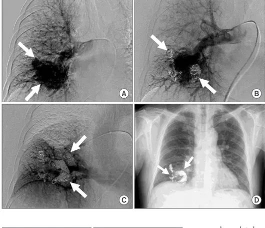
Surgical resection is necessary in those kind of cases. Here, we report the case of a patient with a 6-cm pulmonary AVM with multiple feeding arteries that was successfully treated by repeated coil embolization without surgery.
Keywords: Arteriovenous Malformations; Embolization, Therapeutic; Pulmonary Artery; Pulmonary Veins
considerably high rates of morbidity and mortality associated with the condition.
Before the 1980s, surgical resection was the only available treatment for AVMs. Since then, advances in treatments have led to the successful application of transcatheter emboliza- tion. Currently, transcatheter embolization is the treatment of choice for pulmonary AVMs
1. However, some pulmonary AVMs are not amenable to transcatheter embolization if they have a large size or are associated with multiple complex feeding arteries. Although surgical resection is occasionally recommended in patients with large pulmonary AVMs, no definite threshold for the surgical indication is present
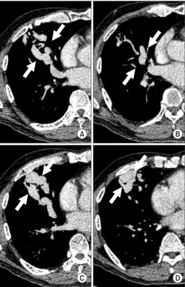
4,5. In the present report, we describe the case of a patient with a large (6 cm×3 cm) pulmonary AVM that was successfully treated by repeated transcatheter embolization without surgery.
Case Report
A 69-year-old man with a history of hypertension and pul- monary tuberculosis presented with chronic rhinorrhea and nasal obstruction. He was a former smoker (50 pack-year).
He was diagnosed with chronic sinusitis and scheduled to undergo surgical treatment for sinusitis. He had undergone chest radiography during the preoperative evaluation. The Copyright © 2015
The Korean Academy of Tuberculosis and Respiratory Diseases.
All rights reserved.
Introduction
Pulmonary arteriovenous malformations (AVMs) are caused by abnormal vascular communications between the pulmonary arteries and pulmonary veins, which lead to blood bypassing the normal pulmonary capillary beds. AVMs result in right-to-left shunts that subsequently cause hypox- emia
1. Pulmonary AVMs vary in size from 1 to 5 cm. Gener- ally, pulmonary AVMs <2 cm in size do not produce clinical symptoms
2,3. Despite the lack of clinical symptoms in most cases, pulmonary AVMs usually require treatment due to the
CASE REPORT
http://dx.doi.org/10.4046/trd.2015.78.4.408ISSN: 1738-3536(Print)/2005-6184(Online) • Tuberc Respir Dis 2015;78:408-411
Address for correspondence: Young Sik Park, M.D.
Department of Internal Medicine, Seoul National University Hospital, 101 Daehak-ro, Jongno-gu, Seoul 03080, Korea
Phone: 82-2-2072-7214, Fax: 82-2-762-9662 E-mail: [email protected]
Received: Jun. 25, 2015 Revised: Aug. 1, 2015 Accepted: Aug. 13, 2015
cc