R E S E A R C H Open Access
Clinical significance of focal ß-amyloid
deposition measured by 18 F-flutemetamol PET
Si Eun Kim 1,2 , Byungju Lee 3 , Seongbeom Park 1,4,5 , Soo Hyun Cho 1,6 , Seung Joo Kim 1,7 , Yeshin Kim 8 , Hyemin Jang 1,4,5 , Jee Hyang Jeong 9 , Soo Jin Yoon 10 , Kyung Won Park 11 , Eun-Joo Kim 12 , Na Yeon Jung 13 , Bora Yoon 14 , Jae-Won Jang 8 , Jin Yong Hong 15 , Jihye Hwang 16 , Duk L. Na 1,4,5,17,18
, Sang Won Seo 1,4,5,19,20
, Seong Hye Choi 21* and Hee Jin Kim 1,4,5*
Abstract
Background: Although amyloid PET of typical Alzheimer ’s disease (AD) shows diffuse ß-amyloid (Aß) deposition, some patients show focal deposition. The clinical significance of this focal Aß is not well understood. We examined the clinical significance of focal Aß deposition in terms of cognition as well as Aß and tau cerebrospinal fluid (CSF) levels. We further evaluated the order of Aß accumulation by visual assessment.
Methods: We included 310 subjects (125 cognitively unimpaired, 125 mild cognitive impairment, and 60 AD dementia) from 9 referral centers. All patients underwent neuropsychological tests and
18F-flutemetamol (FMM) PET.
Seventy-seven patients underwent CSF analysis. Each FMM scan was visually assessed in 10 regions (frontal, precuneus and posterior cingulate, lateral temporal, parietal, and striatum of each hemisphere) and was classified into three groups: No-FMM, Focal-FMM (FMM uptake in 1 –9 regions), and Diffuse-FMM (FMM uptake in all 10 regions).
Results: 53/310 (17.1%) subjects were classified as Focal-FMM. The cognitive level of the Focal-FMM group was better than that of Diffuse-FMM group and worse than that of No-FMM group. Among the Focal-FMM group, those who had FMM uptake to a larger extent or in the striatum had worse cognitive levels. Compared to the Diffuse- FMM group, the Focal-FMM group had a less AD-like CSF profile (increased Aß42 and decreased t-tau, t-tau/Aß42).
Among the Focal-FMM group, Aß deposition was most frequently observed in the frontal (62.3%) and least frequently observed in the striatum (43.4%) and temporal (39.6%) regions.
Conclusions: We suggest that focal Aß deposition is an intermediate stage between no Aß and diffuse Aß deposition. Furthermore, among patients with focal Aß deposition, those who have Aß to a larger extent and striatal involvement show clinical features close to diffuse Aß deposition. Further longitudinal studies are needed to evaluate the disease progression of patients with focal Aß deposition.
Keywords: ß-amyloid,
18F-flutemetamol PET, Alzheimer ’s disease, Cognition, Cerebrospinal fluid
© The Author(s). 2020 Open Access This article is distributed under the terms of the Creative Commons Attribution 4.0 International License (http://creativecommons.org/licenses/by/4.0/), which permits unrestricted use, distribution, and reproduction in any medium, provided you give appropriate credit to the original author(s) and the source, provide a link to the Creative Commons license, and indicate if changes were made. The Creative Commons Public Domain Dedication waiver (http://creativecommons.org/publicdomain/zero/1.0/) applies to the data made available in this article, unless otherwise stated.
* Correspondence: [email protected]; [email protected]
21
Department of Neurology, Inha University School of Medicine, Incheon, Korea
1
Department of Neurology, Samsung Medical Center, Sungkyunkwan University School of Medicine, 50 Ilwon-dong, Gangnam-ku, Seoul 135-710, Republic of Korea
Full list of author information is available at the end of the article
Background
Alzheimer’s disease (AD), the most common cause of dementia, is characterized by ß-amyloid (Aß) and tau ac- cumulation in the brain [1]. Aß accumulation starts ap- proximately 10 to 20 years before dementia symptoms begin. Thus, detecting the presence of Aß is essential for early diagnosis of AD. With recent advances in detecting Aß in vivo, the use of an Aß biomarker is clinically avail- able through PET imaging or CSF analysis [2, 3].
Although amyloid PET of typical AD shows diffuse Aß deposition, some patients show focal Aß deposition, the clinical significance of which is not well defined. Previ- ous studies lack pathological examination results of pa- tients with focal Aß deposition on PET imaging. Brain Aß burden on PET imaging may be quantitatively mea- sured in the research field using standardized uptake value ratios (SUVR) and studies showed that higher SUVR is correlated with poor clinical outcome [4–6].
Meanwhile, in clinical practice, interpretation of amyloid PET relies on dichotomous visual rating (positive or negative). According to visual interpretation guidelines of PET images such as
18F-florbetaben or
18F-flutemeta- mol (FMM), if any of the brain regions (frontal, parietal, precuneus/posterior cingulate (PPC), lateral temporal lobes, and striatum) is positive in either hemisphere, the scan is considered to be positive [7, 8]. However, since Aß deposition is a gradual process [9], a dichotomous visual rating may be misleading. Identifying the clinical significance of participants in the gray zone may help manage patients in clinical practice. Thus, this particular group needs to be well characterized.
In this study, we hypothesized that patients showing focal Aß deposition have unique clinical characteristics.
We examined patients showing focal Aß deposition on FMM PET scan. We compared cognition and CSF AD biomarkers between patients with No-, Focal-, and Diffuse-FMM uptake. We also aimed to assess whether the extent and region of focal FMM uptake are related to cognition. We further evaluated the order of Aß accu- mulation by visual assessment.
Methods Participants
We recruited 310 patients with cognitively unimpaired (CU; n = 125), mild cognitive impairment (MCI; n = 125), and AD dementia (ADD, n = 60) who underwent FMM PET between June 2015 and December 2017. The CU individuals had normal age-, sex-, and education- adjusted performance on standardized cognitive tests [10]. The participants with MCI met the criteria pro- posed by Petersen et al. [11]: (1) subjective memory complaints, (2) relatively normal performance in other cognitive domains, (3) normal activities of daily living (ADL), (4) objective memory impairment below − 1.5 SD
on either verbal or visual memory tests, and (5) not de- mented. The ADD patients met the criteria for dementia by the Diagnostic and Statistical Manual of Mental Dis- orders 4th Edition, Text Revision (DSM-IV-TR) [12] and were diagnosed with probable ADD according to the NIA-AA core clinical criteria [1]. The patients were re- cruited from 9 referral hospitals in South Korea (175 from Samsung Medical Center, 135 from Validation Co- hort of Korean Brain Aging Study for the Early Diagno- sis and Prediction of AD (KBASE-V) [13]). All participants underwent neurologic examination, neuro- psychological test, and Apolipoprotein E (APOE) geno- typing. We screened blood tests including a complete blood count, blood chemistry, thyroid function, vitamin B12, folate, and syphilis serology and excluded partici- pants with abnormal findings that could affect their cog- nition. Participants with previous or current neurological or psychiatric diseases such as brain tumors, encephal- itis, epilepsy, and depressive disorders that could affect cognitive function were also excluded. On MRI, patients with structural lesions such as hydrocephalus, brain tu- mors, or traumatic brain injuries were also excluded.
The Institutional Review Boards approved this study at all participating centers. We obtained written, informed consent from patients and caregivers.
18
F-flutemetamol PET acquisition and analysis
We performed FMM PET scans using a Discovery 600 PET/CT scanner (GE), Discovery 690 PET/CT scanner (GE), Discovery STE PET/CT scanner (GE), Biography MCT PET/CT scanner (Siemens) or Gemini TF PET/CT scanner (Philips) on a total number of 310 participants as described in a previous study [13]. The participants under- went a 20 min PET scan (4 × 5 min dynamic frames) starting at 90 min after intravenous injection of 185 MBq
± 10% of
18F-flutemetamol. Low-dose computed tomog- raphy was utilized for attenuation correction before scans.
We reconstructed the images with the Ordered Subsets Expectation Maximization algorithm using 4 iterations and 16 subsets.
Blinded visual interpretation
Visual interpretation of the FMM PET images was per- formed by two blinded neurologists (referred to as
“readers”) who successfully completed the manufacturing company’s electronic training program. Visual interpret- ation of FMM PET images was performed by systematic review of ten brain regions (frontal, parietal, PPC, stri- atum, and lateral temporal lobes in each hemisphere) [14].
For each region, the readers used dichotomous assessment
in classifying images as either normal or abnormal in a
rainbow color scale anchored to the pons. We defined
each region to be abnormal when there was increased cor-
tical gray matter signal (above 50–60% peak intensity)
and/or reduced (or absent) gray-white matter contrast [15]. Inter-reader agreement of interpretation of FMM PET was excellent (kappa score = 0.94).
We classified patients into three groups. No-FMM (no FMM uptake in any region), Focal-FMM (FMM uptake in 1–9 regions), and Diffuse-FMM (FMM uptake in all 10 regions). Examples of FMM PET images are shown in Additional file 1: Figure S1.
Cerebrospinal fluid collection, processing, and analysis CSF sampling was performed in 77 participants (49 CU, 16 MCI, and 12 ADD) by procedures as previously de- scribed [13]. We obtained CSF in 15 mL polypropylene tubes (Falcon, Corning Science, NY, USA) and centrifuged at 2000×g for 10 min at room temperature (RT). Approxi- mately 10 cc of the CSF supernatant was frozen and trans- ferred to the laboratory at Inha University. For measuring CSF biomarkers, the CSF was thawed and gently extracted into pipettes with polypropylene tips. A total of 0.4 mL ali- quots of CSF was frozen in polypropylene tubes (Sarstedt AG & Co., Nümbrecht, Germany) and stored at − 80 °C until analysis. We measured the level of CSF Aß 42, total tau (t-tau), and phosphorylated tau (p-tau) using the multiplex xMAP Luminex platform with INNO-BIA Alz- Bio3 kits. AlzBio3 kits (Fujirebio Europe, Ghent, Belgium) contained capture monoclonal antibodies for Aß 42, t-tau, and p-tau, which linked to two aqueous quality controls (a-QC) with pre-defined concentration ranges for the three biomarkers. The procedure is described elsewhere [16, 17]. To reduce the effects of sources of variability on the results [18], CSF analysis was followed by the manu- facturer’s instructions and standard of procedures that were previously described [19, 20].
Neuropsychological evaluation
All participants underwent a standardized neuropsycho- logical battery called the Korean version of the Consor- tium to Establish a Registry for Alzheimer’s Disease Assessment Packet (CERAD-K) [21] or Seoul Neuro- psychological Screening Battery (SNSB) [22], which con- sisted of tests of language, visuospatial, memory, and frontal/executive functions.
Tests in CERAD-K included the Korean version of the Boston Naming Test (K-BNT) to assess language func- tion; constructional praxis (copying figures) to assess visuospatial function; 10 word list recall (20-min delayed recall) and constructional recall (20-min delayed figure recall) to assess verbal and visual memory, respectively;
Controlled Oral Word Association Test (COWAT: ani- mal naming) and Stroop Test (color reading) to assess frontal/executive function; and the Mini-Mental State Examination (MMSE) to assess global cognitive function.
Tests in SNSB included the K-BNT to assess language function; Rey-Osterrieth Complex Figure Test (RCFT:
copying) to assess visuospatial function; Seoul Verbal Learning Test (20-min delayed recall) and RCFT (20- min delayed recall) to assess verbal and visual memory, respectively; COWAT (animal naming) and the Stroop Test (color reading) to assess frontal-executive function;
and the MMSE to assess global cognitive function. [23]
Tests were conducted by experienced staff and super- vised by board-certified neuropsychologists. The norms for each test were based on 1987 normal Korean partici- pants (for CERAD-K) or 1067 normal Korean partici- pants (for SNSB). In the analyses, we used the z-scores of each test, which were based on the mean and stand- ard deviation of each measure in the age- and education-matched norms.
Statistical analysis
For descriptive statistics, we used the chi-square test and analysis of variance (ANOVA) followed by Bonferroni’s post hoc analyses.
To compare the cognitive profile of the three groups (No-FMM, Focal-FMM, and Diffuse-FMM group), we used ANOVA followed by Bonferroni’s post hoc analyses. When we compared the cognitive profile of the Regional-FMM group with the No-FMM or the Diffuse-FMM group, we used ANOVA followed by Bonferroni’s correction for 30 multiple tests (5 regions and 6 cognitive tests). To evaluate the association between cognition and number of FMM up- take regions, we used linear regression analyses.
For comparison of the CSF profile of the three groups, we used analysis of covariance (ANCOVA) followed by Bonfer- roni’s post hoc analyses after controlling for age. To evaluate the association between CSF profile and number of FMM uptake regions, linear regression analyses were used after adjusting for age. All statistical tests were performed using MedCalc (MedCalc Software version 19, Ostend, Belgium).
To determine the spreading order of FMM uptake, we assumed that regions with earlier appearance of path- ology would show abnormal FMM uptake in a greater number of participants, as suggested by previous studies [24, 25]. The different frequency of regional involvement was assessed using a bootstrapping method with 1000 re-samples in R v3.1.3 (Institute for Statistics and Math- ematics, Vienna, Austria; www.R-project.org), which de- rived the estimates of 95% confidence intervals and standard error. We calculated asymptotic p values and corrected for multiple comparisons with Bonferroni’s method for all combinations of regional pairs.
Results
Characteristics of the participants
The demographic and clinical characteristics are pre-
sented in Table 1. Of all the participants, 17.1% (53/310)
patients were classified into the Focal-FMM group. The proportion in the Focal-FMM group and the extent of Focal-FMM uptake differed according to cognitive level.
13.6% (17/125) of CU, 16.0% (20/125) of MCI, and 26.7% (16/60) of ADD were classified into the Focal- FMM group. Among the Focal-FMM group, the median number of uptake regions was 1.0 (95% CI = 1.0–4.0) in CU, 3.5 (95% CI = 2.0–5.8) in MCI, and 6.0 (95% CI = 3.6–8.0) in ADD. The Focal-FMM group was older than the No-FMM group and had more APOE ε4 carriers compared to the No-FMM group. In addition, there were statistically significant differences in distribution of cognitive level across the groups.
Cognitive profiles of Focal-FMM group
Compared to the No-FMM group, the Focal-FMM group showed significantly lower performance in all cog- nitive domains except for visuospatial function. Com- pared to the Diffuse-FMM group, the Focal-FMM group showed better performance in verbal memory and visual memory functions. Global cognitive function (measured by MMSE) of the Focal-FMM group was better than the Diffuse-uptake group but worse than the No-FMM group (Table 2).
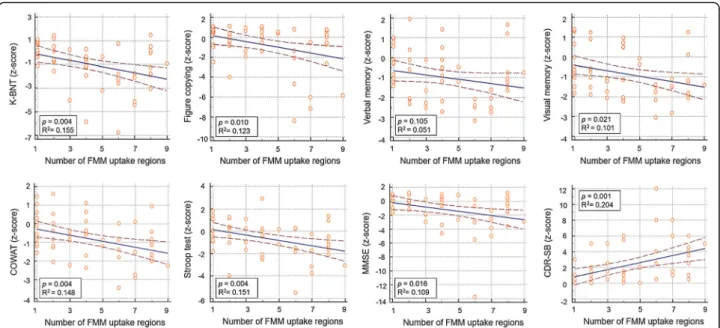
Among the Focal-FMM group, as the number of FMM uptake regions increased, z-scores decreased in all cognitive domains such as K-BNT (β = − 0.264, p = 0.004), visuospatial function (β = − 0.290, p = 0.010), ver- bal memory (β = − 0.105, p = − 0.105), visual memory (β = − 0.138, p = 0.021), COWAT (β = − 0.162, p = 0.004), Stroop Test (β = − 0.239, p = 0.004), and MMSE (β = − 0.306, p = 0.016) (Fig. 1.).
We further divided Focal-FMM group into patients with less FMM uptake (1–5 regions involved) group and patients with more FMM uptake (6–9 regions involved) group. Focal-FMM group with 1–5 regions involved did not show significant difference compared to the No- FMM group, while it showed better performance in all cognitive domains except for language when compared to Diffuse-FMM group. Focal-FMM group with 6–9 re- gions involved showed worse performance in all cogni- tive domains compared to No-FMM group, while it did not show significant difference when compared to Diffuse-FMM group (Additional file 1: Table S1).
Then, we compared each regional Focal-FMM group with the Focal- or Diffuse-FMM group to evaluate the regional effects of focal FMM uptake on cognitive func- tion. Compared to the Diffuse-FMM group, verbal mem- ory scores were higher in the Focal-FMM group with frontal, lateral temporal, parietal, or PPC regional in- volvement whereas no difference was found in the Focal-FMM group with striatal involvement. Compared to the Diffuse-FMM group, visual memory scores were higher in the Focal-FMM group with frontal, lateral tem- poral, or parietal regional involvement whereas no differ- ence was found in the Focal-FMM group with PPC or striatal involvement. Compared to the No-FMM group, verbal and visual memory scores were lower in the Focal-FMM group with PPC or striatal involvement (Table 2).
Comparisons of cognitive scores between the No-, Focal-, and Diffuse-FMM groups in each cognitive level (CU, MCI and ADD) are shown in Additional file 1:
Table S2). Among CU individuals, the cognitive score Table 1 Demographic characteristics of participants
No-FMM uptake
( n = 174) Focal-FMM uptake
( n = 53) Diffuse-FMM uptake
( n = 83) p
No vs focal uptake
p
No vs diffuse uptake
p
Focal vs diffuse uptake
Age (mean ± SD) 69.4 ± 8.6 73.5 ± 7.9 71.4 ± 8.5 0.008 0.253 0.494
Men (%) 73 (42.0) 15 (28.3) 37 (44.6) 0.075 0.692 0.058
Education, years (mean ± SD) 10.7 ± 5.0 10.3 ± 5.0 11.0 ± 4.5 1.000 1.000 1.000
APOE ε4 carrier (%) 21/166 (12.7) 23/53 (43.4) 42/81 (51.9) < 0.001 < 0.001 0.340
Disease duration (months) (Mean ± SD)
62.5 ± 52.9 50.3 ± 44.5 41.5 ± 41.6 0.455 0.009 1.000
Vascular risk factors
Hypertension (%) 80 (46.0) 23 (43.4) 29 (34.9) 0.742 0.095 0.324
Diabetes (%) 33 (19.0) 5 (9.4) 11 (13.3) 0.104 0.257 0.502
Hyperlipidemia (%) 57 (32.8) 18 (34.0) 11 (13.3) 0.871 0.001 0.004
Cognitive level < 0.001 < 0.001 < 0.001
CU (%) 102 (58.6) 17 (32.1) 6 (7.2)
MCI (%) 64 (36.8) 20 (37.7) 41 (49.4)
ADD (%) 8 (4.6) 16 (30.2) 36 (43.4)
Abbreviations: FMM18F-flutemetamol,APOE ε4 Apolipoprotein ε4, CU cognitively unimpaired, MCI mild cognitive impairment, ADD Alzheimer’s disease dementia
Table 2 Cognitive profile according to
18F-flutemetamol uptake regions Neurop sychol ogical tests of cogn itive dom ain No-FM M uptak e Focal-FMM uptake D iffuse-F MM uptak e No vs focal uptake No vs diffus e uptake
Focal vs diffuse uptake
(n = 174) Total (n = 53) Frontal (n = 33) Lat eral tempo ral (n = 21) Pari etal (n = 32 ) PPC (n = 32) Striatu m (n =2 3 ) (n = 83) Langu age K-BN T 0.02 ± 1.17 − 0.94 ± 1. 78 − 1. 33 ± 1.68* − 1.27 ± 1.93 − 1.40 ± 2.00* − 1.50 ± 1.97 * − 1.80 ± 2.00 * − 0.97 ± 1.50 < 0.001 < 0.001 1.000 Visuos patial Figure cop ying − 0.12 ± 1.31 − 0.71 ± 2. 19 − 1. 10 ± 2.54 − 0.55 ± 2.02 − 0.95 ± 2.55 − 1.37 ± 2. 53 − 1.53 ± 2.82 − 1.62 ± 3.76 0.324 < 0.001 0.089 Mem ory Verbal memor y − 0.46 ± 1.26 − 0.97 ± 1. 26 − 1. 12 ± 1.19
#− 0.97 ± 1.31
#− 1.05 ± 1.40
#− 1.32 ± 1. 27*
#− 1.63 ± 0.84 * − 2.12 ± 1.04 0.020 < 0.001 < 0.00 1 Visua l me mory − 0.18 ± 1.10 − 0.83 ± 1. 15 − 1. 08 ± 1.06*
#− 0.68 ± 1.05
#− 0.93 ± 1.19*
#− 1.09 ± 1. 21* − 1.34 ± 1. 08* − 1.77 ± 0.96 < 0.001 < 0.001 < 0.00 1 Frontal/ execut ive COW AT − 0.07 ± 1.10 − 0.78 ± 1. 11 − 0. 98 ± 1.08* − 1.09 ± 1.02* − 0.89 ± 1.30* − 1.17 ± 1. 04* − 1.32 ± 1.02 * − 1.09 ± 1.11 < 0.001 < 0.001 0.331 Stro op test − 0.06 ± 1.42 − 0.60 ± 1. 63 − 0. 93 ± 1.67 − 0.66 ± 1.57 − 0.91 ± 1.81 − 1.18 ± 1. 66* − 1.48 ± 1. 54* − 1.66 ± 1.77 0.080 < 0.001 < 0.00 1 Global MMS E − 0.31 ± 1.35 − 1.13 ± 2. 46 − 1. 65 ± 2.80 − 1.04 ± 1.86 − 1.35 ± 2.86 − 1.85 ± 2. 84 − 2.24 ± 3.21 − 2.57 ± 2.98 0.039 < 0.001 < 0.00 1 CDR -SB 0.80 ± 1.04 2.19 ± 2.63 2.94 ± 2.82* 2.55 ± 2. 43 2.50 ± 2. 87 3.17 ± 2.90* 3.52 ± 3.18* 3. 10 ± 2.68 < 0.001 < 0.001 0.024 Values are mean z-scores of neuropsychologi cal tests and raw score of CDR-SB (mean ± SD) *p < 0.05 between regional Focal-FMM-uptake group and No-FMM-uptak e group (after Bonferroni ’s correction for 5 regions and 6 cognitive tests) #p < 0.05 between regional Focal-FMM-up take group and Diffuse-FMM-uptake group (after Bonferroni ’s correction for 5 regions and 6 cognitive tests) Abbreviations :FMM
18F-flutemetamol ,COWAT The Controlled Oral Word Association Test, K-BNT Korean version-Boston Naming Test, MMSE Mini-Mental State Exam, CDR-SB Clinical Dementia Rating Scale Sum of Boxes
did not differ among the No-, Focal-, and Diffuse-FMM groups. However, among MCI patients, the Focal-FMM group showed better performance in verbal and visual memory function, as well as in MMSE score, compared to the Diffuse-FMM group. Among ADD patients, Focal-FMM patients performed worse in language and frontal function compared to those of the No-FMM group and performed better in verbal memory than those of the Diffuse-FMM group. We provided a break- down of cases by clinical designation and number of re- gions in Additional file 1: Table S3).
CSF amyloid and tau level of focal-FMM group
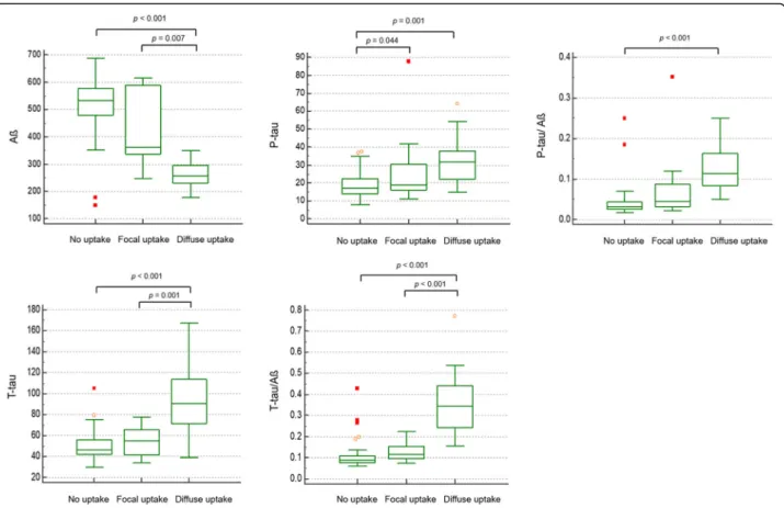
Levels of CSF AD biomarkers (Aß 42, p-tau, t-tau, p- tau/ Aß 42, and t-tau/ Aß 42) in No-, Focal-, and Diffuse-FMM groups are shown in Fig. 2. The Focal- FMM group showed increased levels of CSF Aß 42 and decreased levels of CSF t-tau and t-tau/Aß 42, compared to the Diffuse-FMM group. However, there were no dif- ferences between Focal-FMM-uptake and No-FMM- uptake groups except for p-tau level.
Spreading order of FMM-uptake among focal-FMM- uptake group
Among the Focal-FMM group, Aß deposition was most frequently observed in the frontal (62.3%, 95% CI = 48.8–75.8) followed by PPC (60.4%, 95% CI = 46.8–74.0), parietal (60.4%, 95% CI = 46.8–74.0), striatum (43.4%, 95% CI = 29.6–57.2), and lateral temporal (39.6%, 95%
CI = 26.0–53.2) regions (Fig. 3).
Discussion
In this study, we investigated the clinical significance of patients with Focal-FMM uptake, which consisted of 17.1% of all participants. Our major findings were as fol- low. First, cognitive function of patients with Focal- FMM uptake was in the intermediate stage between patients with No- and Diffuse-FMM uptake. Among Focal-FMM patients, the larger extent and striatal in- volvement of FMM uptake was associated with worse cognition. Second, CSF AD biomarkers of Focal-FMM group were less AD-like compared to the Diffuse-FMM group. Finally, among the Focal-FMM group, FMM up- take was most frequently observed in the frontal and least observed in the striatum and lateral temporal re- gions. Taken together, our findings suggest that patients with Focal-FMM uptake have unique clinical character- istics. Furthermore, among patients with focal FMM up- take, those who have larger extent and striatal involvement of FMM showed clinical features close to diffuse Aß uptake and, thus, might be considered as be- ing in more advanced stage of AD.
We found that 17.1% of all participants had Focal- FMM uptake. Focal-FMM consisted substantial portion of participants in each cognitive level: 13.6% (17/125) of UC, 16.0% (20/125) of MCI, and 26.7% (16/60) of ADD.
In clinical practice, interpretation of amyloid PET relies on visual assessment, which currently guides focal-FMM uptake to be read as positive for Aß. However, the clin- ical significance of focal Aß uptake is not well under- stood. Characterizing participants in this gray zone may help better manage patients.
Fig. 1 Cognitive profile according to number of FMM uptake regions among the Focal-FMM group. Solid blue line is the regression line. Brown
dotted lines indicate 95% confidence intervals. Abbreviations: FMM =
18F-flutemetamol; K-BNT = Korean version-Boston Naming Test; COWAT =
The Controlled Oral Word Association Test; MMSE = Mini-Mental State Exam
Our first major finding was that cognitive function of patients with Focal-FMM was in the intermediate stage between patients with No- and Diffuse-FMM uptake.
More importantly, the cognitive function differed ac- cording to the number and location of regions of focal FMM uptake. Among the Focal-FMM group, cognitive scores decreased with increasing number of FMM up- take regions. Our results are in line with previous studies using quantitative measurement of Aß burden [PIB [26–
28],
18F-florbetapir [29], or FMM [30] SUVR]. Subjects with higher PiB SUVR showed lower scores on episodic memory tests [27]. Higher FMM SUVR correlated with lower delayed memory index [30], and higher
18F-florbe- tapir SUVR correlated with lower MMSE scores [29].
Among the Focal-FMM group, those who had FMM up- take in the striatum had the worst cognitive scores. Al- though there have been numerous studies on the associations between quantitative Aß deposition and cognition, no study has reported the association between visually assessed Aß deposition and cognitive profiles.
As quantitative analysis is not widely used in clinical practice, studies on visual assessment is valuable for cli- nicians. Our results suggest that when managing patients showing focal FMM uptake, clinicians should consider
the number and location of regions with focal Aß deposition.
Our second major finding was that patients with focal Aß deposition on PET showed less AD-like CSF profiles compared to the Diffuse-FMM group. In addition, the increased number of FMM uptake regions significantly correlated with CSF biomarker levels toward a more AD-like pattern (increased Aß42 and decreased t-tau, t- tau/ Aß42). Our results are consistent with previous studies showing negative correlation between CSF Aß42 levels and PET-based quantitative uptake of
18F-florbeta- pir [31] or
18F-florbetaben [32].
Our third major finding was the order of Aß accu- mulation. Among the Focal-FMM-group, Aß depos- ition was most frequently observed in the frontal (62.3%) followed by the PPC (60.4%), parietal (60.4%), and least frequently observed in the stri- atum (43.4%) and lateral temporal (39.6%) regions.
Unlike the Thal stage of amyloid deposition [9], our data showed that striatal involvement preceded the involvement of lateral temporal region. However, our result generally reflects a downward spreading pat- tern of Aß, suggesting that Aß deposits first in the cortex followed by subcortical structures [9].
Fig. 2 Comparison of Alzheimer ’s disease biomarkers (Aß, P-tau, and T-tau) from cerebrospinal fluid among No-, Focal-, and Diffuse-FMM groups after adjusted for age. Box and whisker plots show medians, lower to upper quartile, and lines extending from minimum to maximum values.
Abbreviations: FMM =
18F-flutemetamol; P-tau = phosphorylated tau; T-tau = total tau
Furthermore, our data revealed that, among Focal- FMM patients, those with subcortical Aß involve- ment (striatum) showed lower cognition than those with cortical Aß involvement (frontal, lateral tem- poral, parietal, and PPC). Our findings are concord- ant with those of previous studies which found that subcortical Aß involvement, especially striatal Aß, was related to worse cognitive performance and fas- ter cognitive decline [33–35]. Patients with striatal involvement implies that they had higher Thal stage (Aß phase 3) and thus are more likely to have tau.
Therefore, we suggest that even when Aß involve- ment is focal, Aß deposition in the striatum might be a sign of possible worse clinical outcome.
However, the present study has some limitations.
First, our study used a cross-sectional design and, therefore, we do not know the cognitive trajectory of participants. Further longitudinal studies are war- ranted to evaluate the disease progression rate of the Focal-FMM group. Second, we lack pathological data on Focal-FMM patients. Amyloid PET-negative MCI or dementia patients in our data might have vascu- lar, hippocampal sclerosis, or other pathologies as the main etiology for cognitive impairment. Further studies that could exclude non-AD pathologies are necessary. Nevertheless, the strength of our current study is that we have identified the clinical signifi- cance of focal Aß deposition, which comprised a
substantial portion of participants in each cognitive level.
Conclusions
In the current study, we found that focal Aß deposition has unique clinical characteristics that differ from pa- tients with no or diffuse Aß deposition. We suggest that focal Aß deposition should be considered as an inter- mediate stage between no Aß and diffuse Aß deposition.
In addition, when managing patients showing focal Aß deposition, clinicians should consider the number and location of regions of focal Aß deposition. Those with more regions involved, especially in the striatum, show clinical features close to diffuse Aß deposition. Thus, cognitively unimpaired or MCI individuals with such signs might be more closely monitored for future cogni- tive decline. Further longitudinal studies are needed to evaluate the disease progression of patients with focal Aß deposition.
Supplementary information
The online version of this article (https://doi.org/10.1186/s13195-019-0577-x) contains supplementary material, which is available to authorized users.
Additional file 1: Table S1. Cognitive profiles according to the number of 18F-flutemetamol uptake regions. Table S2. Cognitive profile of No-, Focal-, and Diffuse-FMM groups in each cognitive level. Table S3.
Number of uptake regions in cognitively unimpaired, mild cognitive im- pairment, and Alzheimer ’s disease dementia. Figure S1. Examples of
18F- Fig. 3 Spreading order of FMM among Focal-FMM group ( N = 53). It shows the statistical significance in the comparison of the frequencies of FMM regional involvement between each pair of regions. The regional differences of uptake frequency display a stepwise pattern. Only the pairs of comparison passing Bonferroni ’s multiple comparisons are shown. Color bars represent logarithmic scale of p value (−log
10). Abbreviation:
PPC = precuneus/posterior cingulate
flutemetamol (FMM) PET scan in ‘No-FMM’, ‘Focal-FMM’ and ‘Diffuse- FMM ’ groups. Red arrows and dashed circles show FMM uptake, while white arrows and dashed circles indicate no FMM uptake. (DOCX 2813 kb)
Abbreviations
AD: Alzheimer ’s disease; ADD: AD dementia; Aß: ß-amyloid; ANOVA: Analysis of variance; ANCOVA: Analysis of covariance; a-QC: Aqueous quality controls;
CERAD-K: The Korean version of the Consortium to Establish a Registry for Alzheimer ’s Disease Assessment Packet; COWAT: Controlled Oral Word Association Test; CU: Cognitively unimpaired; CDR-SB: Clinical Dementia Rating Scale Sum of Boxes; DSM-IV-TR: Diagnostic and Statistical Manual of Mental Disorders 4th Edition, Text Revision; FMM:
18F-Flutemetamol; KBASE- V: Korean Brain Aging Study for the Early Diagnosis and Prediction of AD; K- BNT: Korean version-Boston Naming Test; MCI: Mild cognitive impairment;
MMSE: Mini-Mental State Examination; PPC: Precuneus/posterior cingulate; p- tau: Phosphorylated tau; RCFT: Rey-Osterrieth Complex Figure Test;
SUVR: Standardized uptake value ratios; SNSB: Seoul Neuropsychological Screening Battery; t-tau: Total tau
Acknowledgements Not applicable.
Authors ’ contribution
SEK, SHC, and HJK contributed to the conceptualization of the study, analysis and interpretation of data, and drafting. BL and SBP contributed to the analyses of imaging data and preparation of the figures and provided technical support. SHC, SJK, YK, HJ, JHJ, SJY, KWP, EJK, NYJ, and BY contributed to the data collection and interpretation of data. JJ, JYH, JH, DLN, and SWS contributed to the analysis and interpretation of data. All authors read and approved the final manuscript.
Funding
This research was supported by the Original Technology Research Program for Brain Science through the National Research Foundation of Korea (NRF) funded by the Korean government (MSIP) (No. 2014M3C7A1064752); by the National Research Foundation of Korea (NRF) grant funded by the Korea government (MSIP) (NRF- 2018R1A1A3A04079255); and a grant of the Korean Health Technology R&D Project, Ministry of Health & Welfare, Republic of Korea (HI18C1629 and HI18C0335).
Availability of data and materials
The datasets used and/or analyzed during the current study are available from the corresponding author on reasonable request.
Ethics approval and consent to participate
The Institutional Review Boards approved this study at all participating centers. We obtained written, informed consent from patients and caregivers.
Consent for publication Not applicable.
Competing interests
The authors declare that they have no competing interests.
Author details
1
Department of Neurology, Samsung Medical Center, Sungkyunkwan University School of Medicine, 50 Ilwon-dong, Gangnam-ku, Seoul 135-710, Republic of Korea.
2Department of Neurology, Inje University College of Medicine, Haeundae Paik Hospital, Busan, Korea.
3Department of Neurology, Yuseong Geriatric Rehabilitation Hospital, Pohang, Korea.
4Samsung Alzheimer Research Center, Samsung Medical Center, Seoul, Korea.
5
Neuroscience Center, Samsung Medical Center, Seoul, Korea.
6Department of Neurology, Chonnam National University Hospital, Chonnam National University Medical School, Gwangju, Korea.
7Department of Neurology, Gyeongsang National University School of Medicine and Gyeongsang National University Changwon Hospital, Changwon, Korea.
8Department of Neurology, Kangwon National University Hospital, Kangwon National University College of Medicine, Chuncheon, Korea.
9Department of Neurology, Ewha Womans University Mokdong Hospital, Ewha Womans
University School of Medicine, Seoul, Korea.
10Department of Neurology, Eulji University Hospital, Eulji University School of Medicine, Daejeon, Korea.
11
Department of Neurology, Dong-A Medical Center, Dong-A University College of Medicine, Busan, Korea.
12Department of Neurology, Pusan National University Hospital, Pusan National University School of Medicine and Medical Research Institute, Busan, Korea.
13Department of Neurology, Pusan National University Yangsan Hospital, Pusan National University School of Medicine and Research Institute for Convergence of Biomedical Science and Technology, Yangsan, Korea.
14Department of Neurology, Konyang University College of Medicine, Daejeon, Korea.
15Department of Neurology, Yonsei University Wonju College of Medicine, Wonju, Korea.
16Department of Neurology, Keimyung University Daegu Dongsan Hospital, Daegu, Korea.
17
Department of Health Sciences and Technology, SAIHST, Sungkyunkwan University, Seoul, Korea.
18Stem Cell & Regenerative Medicine Institute, Samsung Medical Center, Seoul, Korea.
19Department of Clinical Research Design and Evaluation, SAIHST, Sungkyunkwan University, Seoul, Korea.
20
Center for Clinical Epidemiology, Samsung Medical Center, Seoul, Korea.
21