Veterinary Science
http://dx.doi.org/10.4142/jvs.2014.15.1.73
Received: 13 Feb. 2013, Revised: 25 May 2013, Accepted: 28 Jun. 2013
Original Article
*Corresponding authors: Tel: +82-2-450-4139; Fax: +82-2-450-3037; E-mail: [email protected], [email protected]
ⓒ 2014 The Korean Society of Veterinary Science.
This is an Open Access article distributed under the terms of the Creative Commons Attribution Non-Commercial License (http://creativecommons.org/licenses/by-nc/3.0) which permits unrestricted non-commercial use, distribution, and reproduction in any medium, provided the original work is properly cited.
A simplified one-step nuclear transfer procedure alters the gene expression patterns and developmental potential of cloned porcine embryos
Sang Kyu Park
1, Sangho Roh
1,*, Jong-Im Park
2,*
1
Cellular Reprogramming and Embryo Biotechnology Laboratory, Dental Research Institute, Seoul National University School of Dentistry, Seoul 110-749, Korea
2
Department of Theriogenology, College of Veterinary Medicine, Konkuk University, Seoul 143-701, Korea
Various somatic cell nuclear transfer (SCNT) techniques for mammalian species have been developed to adjust species-specific procedures to oocyte-associated differences among species. Species-specific SCNT protocols may result in different expression levels of developmentally important genes that may affect embryonic development and pregnancy. In the present study, porcine oocytes were treated with demecolcine that facilitated enucleation with protruding genetic material.
Enucleation and donor cell injection were performed either simultaneously with a single pipette (simplified one-step SCNT;
SONT) or separately with different pipettes (conventional two-step SCNT; CTNT) as the control procedure. After blastocysts from both groups were cultured in vitro, the expression levels of developmentally important genes (OCT4,
NANOG, EOMES, CDX2, GLUT-1, PolyA, and HSP70) wereanalyzed by real-time quantitative polymerase chain reaction.
Both the developmental rate according to blastocyst stage as well as the expression levels CDX2, EOMES, and HSP70 were elevated with SONT compared to CTNT. The genes with elevated expression are known to influence trophectoderm formation and heat stress-induced arrest. These results showed that our SONT technique improved the development of SCNT porcine embryos, and increased the expression of genes that are important for placental formation and stress-induced arrest.
Keywords: CDX2, EOMES, HSP70, porcine, somatic cell nuclear transfer
Introduction
Although somatic cell nuclear transfer (SCNT) has been
used for various mammalian species, the efficiency of animal cloning is still very low [1-3]. In general, SCNT-generated embryos suffer from incomplete reprogramming with epigenetic modifications [4-7].
Additionally, embryos produced by SCNT or in vitro fertilization show different gene expression patterns when compared to their in vivo counterparts [8-11]. These effects are thought to result in abnormal development of the SCNT embryos during the peri- and post-implantation stages [10]. Several previous works have attempted to improve SCNT protocols by optimizing the culture medium composition, controlling activation conditions, and improving micromanipulation techniques [4,12-20].
Various SCNT protocols have yielded different levels of embryonic development and different gene expression patterns in cloned blastocysts [9,11,13,21-23].
The UV exposure of oocytes used to confirm the enucleation process is known to decrease the in vitro developmental potential of SCNT porcine embryos [17]. A novel UV exposure-free enucleation method using demecolcine, which induces oocytes to develop visible extrusions containing chromosomes with polar bodies into the surface, has been introduced for porcine SCNT programs [3,22]. Direct nuclear injection and cell fusion methods have been generally used for introducing a somatic cell nucleus into an enucleated oocyte [9,17,21,24]. Although both techniques result in similar developmental competence in vitro, the cell fusion method is more commonly performed for porcine species.
However, the cell fusion method using electric fusion
devices requires more complex and longer steps than the
direct injection protocol [9,16]. On the other hand, direct
injection has been developed into a one-step procedure for which the enucleation and injection steps are performed simultaneously with the same pipette. This ‘one-step SCNT’ method has been successfully adapted for rat cloning and has improved embryonic development for rhesus monkey cloning [5,23,25].
Several marker genes that are important for embryonic development are associated with pluripotency (OCT4 and NANOG), trophectoderm formation (EOMES and CDX2), stress (HSP70), metabolism (GLUT-1), and translation (PolyA). OCT4 and NANOG are expressed in the inner cell mass of blastocysts [26,27]. CDX2 and EOMES are expressed during formation of the blastocyst trophectoderm, which represents the first step in embryo differentiation [28]. PolyA polymerase plays a role in RNA processing [29]. The length of an mRNA poly(A) tail is crucial for stabilization of the 3’ end of the mRNA transcript. Thus, the expression of PolyA is important for mammalian embryonic development [29,30]. Expression of GLUT-1 that encodes the metabolic protein GLUT-1 is significantly decreased in morulae produced in vitro compared to those generated in vivo [31]. The heat shock protein (HSP) family of molecular chaperones is well known for its role in stress adaptation, and assisting in the folding of newly synthesized proteins and their transcripts [32]. HSP70 expression is an indicator of heat shock, and the absence of HSP expression indicates the maladjustment of embryos to environmental change such as heat stress.
In the present study, we assessed the development of porcine embryos produced via SCNT using two cloning method: simplified one-step SCNT (SONT) with demecolcine and conventional two-step SCNT (CTNT).
For this, the expression patterns of several marker genes important for embryonic development were analyzed.
Materials and Methods
All buffer solutions and culture media were obtained from Life technologies (USA). Other inorganic and organic compounds were obtained from Sigma-Aldrich (USA) unless otherwise specified.
Oocyte recovery and in vitro maturation (IVM) Ovaries were collected from 5- to 6-month-old prepubertal gilts (100 ± 10 kg body weight) from a local slaughterhouse, placed in saline at 30∼35
oC, and transported to the laboratory within 2 h. After washing with saline three times, cumulus-oocyte complexes (COCs) were recovered by aspiration of 2- to 5-mm follicles using an 18-gauge hypodermic needle attached to a 5 mL disposable syringe (Korea Vaccine, Korea). After washing three times in IVM medium, COCs that were enclosed by more than three layers of compact cumulus cells and an evenly granulated ooplasm were selected for in
vitro maturation. The 200∼250 selected COCs per were cultured in four-well culture dishes (Nunc, Denmark) containing 500 μL of IVM medium under warmed and gas-equilibrated mineral oil for 44 to 46 h at 38.5
oC and 5%
CO
2. The IVM medium for oocytes was composed of tissue culture medium 199 with Earle’s salts and L-glutamine (TCM199; Life technologies) supplemented with 26.2 mM NaHCO
3, 3.05 mM glucose, 0.91 mM sodium pyruvate, 0.57 mM L-cysteine, 10 ng/mL epidermal growth factor (EGF), 1 μg/mL insulin, 10 IU/mL equine chorionic gonadotropin (eCG) and human chorionic gonadotropin (hCG), and 0.1% (w/v) polyvinyl alcohol (PVA) [3].
Preparation of nuclear donor cells
Fibroblasts were recovered from one porcine conceptus, which was assumed to be between embryonic days 28 to 39 in age, collected from a local slaughterhouse. The head, extremities, and internal organs were removed, and the remaining tissues were cut into small pieces (less than 1mm cube). Cells were dispersed by incubation for 20 min at 38.5
oC with 0.25% (v/v) trypsin solution and cultured in Dulbecco’s minimum essential medium supplemented with 10% (v/v) fetal bovine serum for 4 day at 38.5
oC in incubator. For each passage, cells were cultured until confluent, disaggregated by incubation in a 0.05% (v/v) trypsin solution for 3 min at 38.5
oC, and dispersed into three new dishes. The cells were maintained in culture for at least eight passages. For long-term storage, cells were collected after trypsinization, frozen in freezing medium (DMEM with 20% FBS and 10% DMSO), and stored in liquid nitrogen.
Preparation of oocytes for enucleation
A demecolcine-derived enucleation technique [3,22] was used to minimize the loss of mRNA and ooplasmic factors.
Matured oocytes containing the first polar body were cultured for 2 h at 38.5
oC in TCM199 supplemented with 0.4 μg/mL demecolcine and 0.05 M sucrose.
CTNT Oocytes with protruding chromosomes (protruding rate,
approx. 70%; data not shown) were enucleated with a 12
μm beveled pipette (inner diameter, 10 to 12 μm, Humagen
Pipets; Origo, Denmark) in HEPES-buffered TCM199
(hTCM199) supplemented with 7.5 μg/mL cytochalasin B
(CB), 0.05 M sucrose, and 0.4 μg/mL demecolcine. The
enucleated oocytes (cytoplasts) were washed extensively in
hTCM199 and placed in fresh hTCM199 for nuclear
injection. The fetal fibroblasts used as donor cells were
transferred to a 10% (w/v) polyvinylpyrrolidone-
supplemented hTCM199 microdrop (PVP drop, 6 μL) and
gently mixed at room temperature. A microdrop of
hTCM199 (injection drop) with 15∼20 cytoplasts was
Fig. 1. Schematic diagram illustrating the SONT (B) and CTNT (A) protocols. The black arrowheads indicate the protruding chromosomes (oval) and first polar body (round). The black arrows indicate the nuclear donor fibroblasts (filled gray circle).
SCNT: somatic cell nuclear transfer.
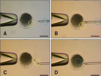
Fig. 2. Stereomicroscopic image of the SONT procedure. The yellow circle (A∼C) indicates the oocyte chromosomes and adjacent first polar body. The blue circle (A∼D) indicates the nuclear donor cell. Just after aspirating (B) and releasing (C) the oocyte chromosomes and polar body using a micropipette containing the nuclear donor fibroblast, the same pipette was used to immediately introduce the donor cell into the enucleated oocyte. Scale bars = 100 μm.
Fig. 3. Comparison of total cell numbers in blastocysts produced by SONT or CTNT. (A) Mean number of total cells in SONT and CTNT blastocysts. (B) Hoechst 33342 staining of the blastocysts (left: CTNT, right: SONT). Scale bar = 100 μm.
placed adjacent to the PVP drop, and the drops were covered with light mineral oil to prevent evaporation.
Individual fibroblasts in the PVP drops were aspirated into a beveled injection pipette (inner diameter, 10 to 12 μm, Humagen pipets; Origo). The cytoplasmic membrane of the fibroblasts was ruptured by repetitive pipetting. An injection pipette containing the ruptured fetal fibroblasts was moved to the injection drop, and a ruptured fibroblast was injected into an ooplast through the cytoplasmic membrane (Fig. 1A).
SONT Three to four PVP-treated donor cells were aspirated into a 12- μm beveled pipette after cytoplasmic membrane rupture. The demecolcine-treated oocytes with protruding chromosomes were enucleated with the pipette containing the donor cell, and the aspirated chromosomes from the metaphase II plate were expelled from the pipette. A donor karyoplast was injected into the cytoplasm through the hole that had been made during enucleation (Figs. 1B and 2).
Activation of reconstructed embryo
After incubation for 60 to 90 min in culture medium for stabilization, electrical activation of the reconstructed oocytes was performed at room temperature using a CF-150/B electro-cell fusion system (BLS, Hungary). The chamber contained two stainless steel electrodes that were 1.0 mm apart and was filled with with activation buffer;
0.26 M mannitol solution supplemented with 0.1 mM MgSO
4, 0.05 M CaCl
2, and 0.01% (w/v) PVA. The oocytes were activated with a 1.6 kV/cm DC pulse for 40 μseconds
in activation buffer. To prevent pseudo-polar body extrusion, the activated oocytes were treated for 5 h at 38.5
oC in North Carolina State University-23 medium (NCSU-23) supplemented with 5 μg/mL CB.
In vitro culture
The activated oocytes were cultured in in embryo culture medium NCSU-23 medium supplemented with both 1%
(v/v) essential and non-essential amino acids, 4 mg/mL
fatty acid-free Fraction V bovine serum albumin (BSA),
and 200 μM kinetin. The CB-treated oocytes were washed
nine times with embryo culture medium and cultured in 20
μL drops (10 to 15 oocytes per drop) of culture medium for
7 days at 38.5
oC and 5% CO
2.
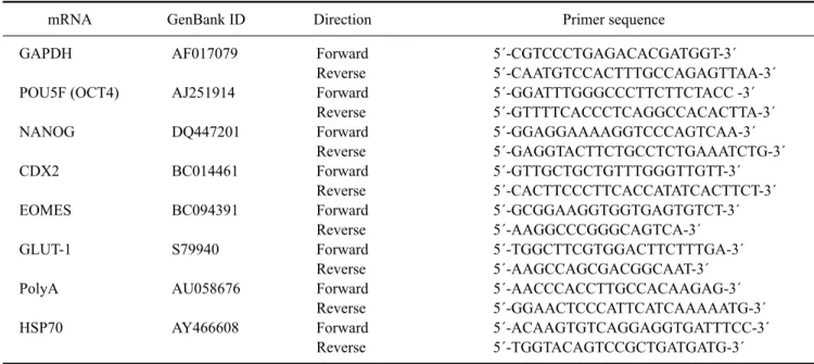
Table 1. Primers used for RT-PCR
mRNA GenBank ID Direction Primer sequence
GAPDH POU5F (OCT4) NANOG CDX2 EOMES GLUT-1 PolyA HSP70
AF017079 AJ251914 DQ447201 BC014461 BC094391 S79940 AU058676 AY466608
Forward Reverse Forward Reverse Forward Reverse Forward Reverse Forward Reverse Forward Reverse Forward Reverse Forward Reverse
5´-CGTCCCTGAGACACGATGGT-3´
5´-CAATGTCCACTTTGCCAGAGTTAA-3´
5´-GGATTTGGGCCCTTCTTCTACC -3´
5´-GTTTTCACCCTCAGGCCACACTTA-3´
5´-GGAGGAAAAGGTCCCAGTCAA-3´
5´-GAGGTACTTCTGCCTCTGAAATCTG-3´
5´-GTTGCTGCTGTTTGGGTTGTT-3´
5´-CACTTCCCTTCACCATATCACTTCT-3´
5´-GCGGAAGGTGGTGAGTGTCT-3´
5´-AAGGCCCGGGCAGTCA-3´
5´-TGGCTTCGTGGACTTCTTTGA-3´
5´-AAGCCAGCGACGGCAAT-3´
5´-AACCCACCTTGCCACAAGAG-3´
5´-GGAACTCCCATTCATCAAAAATG-3´
5´-ACAAGTGTCAGGAGGTGATTTCC-3´
5´-TGGTACAGTCCGCTGATGATG-3´
Total blastocyst cell count
Blastocysts were incubated in 500 μL of washing medium with Hoechst 33342 (1 μg/mL) for 15 min at RT. After staining, the whole blastocysts were mounted in glycerol on a glass slide (Paul Marienfeld, Germany) and flattened under a coverslip (Paul Marienfeld). Digital photographs were obtained using an inverted microscope (Carl Zeiss, Germany) capable of UV illumination and equipped with excitation filters (460 nm for fluorescence, Fig. 3B). The cells in the images were counted.
mRNA extraction and first-strand cDNA synthesis mRNA was recovered from five to 10 porcine blastocysts on day 7 using an oligo (dT)
25nucleotide attached to magnetic beads (Dynabeads mRNA Purification Kit;
Invitrogen) according to the manufacturer’s instructions.
Briefly, the embryos were resuspended in 100 μL lysis/binding buffer [100 mM Tris-HCl (pH 7.5), 500 mM LiCl, 10 mM EDTA (pH 8.0), 1% Li dodecylsulfate [LiDS], and 5 mM Dithiothreitol] and vortexed at room temperature for 5 min to facilitate embryo lysis and RNA release. An oligo (dT)
25magnetic bead suspension (50 μL) was added to the samples and incubated with shaking at room temperature for 5 min. The hybridized mRNA-oligo (dT) magnetic beads were washed twice with washing buffer A [10 mM Tris-HCl (pH 7.5), 0.15 M LiCl, 1 mM EDTA, and 1% LiDS] and then once with washing buffer B [10 mM Tris-HCl (pH 7.5), 0.15 M LiCl, and 1 mM EDTA]. Finally, the mRNA samples were eluted in 10 μ Diethyl Pyrocarbonate (DEPC) -treated double-distilled water. For first-strand cDNA synthesis, reverse transcription was performed for 1 h at 37
oC in a final reaction volume of 30
μL. The reaction contained purified mRNA, 6 μL of 5×
reaction buffer, 1 μL of dNTPs (1.25 mM each), 1 μL of a random hexamer (50 ng/ μL), and 1 μL of M-MuLV reverse transcriptase (200 U/ μL; Promega, USA).
Real-time quantitative PCR (RT-PCR)
cDNA produced from the blastocyst mRNA was analyzed using RT-PCR. The expression levels of genes associated with pluripotency (OCT4 and NANOG), trophectroderm formation (CDX2 and EOMES), metabolism (GLUT-1), translation (PolyA), and heat stress (HSP70) were evaluated (Table 1). For optimal quantification, primers were designed using Primer Express software (Applied Biosystems, USA). RT-PCR was performed using an ABI PRISM 7500 system and SYBR Green PCR Master Mix (Applied Biosystems). All points of the standard curve and all samples were run in triplicate as technical replicates.
Standard curves were generated using a verified DNA template for porcine GAPDH.
For each run, 1 μL of cDNA was used as template in a
reaction mixture containing 5 μL of double-distilled water,
2 μL of each forward and reverse primer (20 pmol/mL), and
10 μL of SYBR Green PCR Master Mix. The following
amplification protocol was performed: denaturation at
95
oC for 10 min, 40 cycles of amplification and
quantification at 94
oC for 15 seconds and 60
oC for 1 min
with a single fluorescence measurement, and a dissociation
curve stage (temperature increase of 0.1
oC every 30 sec
from 60
oC up to 95
oC with fluorescence measurements
taken). Data were analyzed with 7500 System Sequence
Detection software (Applied Biosystems). All samples had
the same starting quantities of all candidate reference genes
Table 2. In vitro development of cloned porcine embryos reconstructed by two different somatic cell nuclear transfer (SCNT) protocols
Group Number of reconstructed
oocytes
Number (% ± SEM) of embryos that developed into Two cells Blastocysts SONT
CTNT
110 140
53 (48.2 ± 3.5) 76 (54.3 ± 3.9)
16 (14.6 ± 1.2)*
15 (10.7 ± 1.1)*
Seven replicate experiments. *A significant difference was observed (p < 0.05). SONT: simplified one-step SCNT, CTNT:
conventional two-step SCNT.