AN UNUSUAL PRESENTATION OF OBSTRUCTED HEMIVAGINA AND IPSILATERAL RENAL ANOMALY SYNDROME: A CASE REPORT
Woo-Oh Kim, MD
1, Kyung Eui Park, MD
1, Seung-Yup Ku, MD, PhD
1,2, Seok Hyun Kim, MD, PhD
1,2, Young Min Choi, MD, PhD
1,2, Jung Gu Kim, MD, PhD
1, Shin Yong Moon, MD, PhD
1,21Department of Obstetrics and Gynecology, Seoul National University College of Medicine; 2Institute of Reproductive Medicine and Population, Medical Research Center, Seoul National University, Seoul, Korea
Most patients with syndrome of obstructed hemivagina and ipsilateral renal anomaly (OHVIRA) were presented with dysmenorrhea, pelvic mass and hematocolpos. These common symptoms of OHVIRA syndrome were aggravated soon after the menarche. Early diagnosis is important to relieve the symptom, to prevent injury of genital tract and to preserve fertility. We have experienced an unusual presentation of OHVIRA syndrome, which were presented with vaginal spotting and diagnosed at age 24. Therefore, we present it with a review of the literature.
Keywords:
Uterus didelphys; Obstructed hemivagina; Renal agenesis; Müllerian anomalies
Received: 2011. 5. 3. Accepted: 2011. 8. 8.
Corresponding author: Seung-Yup Ku, MD, PhD
Department of Obstetrics and Gynecology, Seoul National University Hospital, 28 Yeongeon-dong, Jongno-gu, Seoul 110-744, Korea
Tel: +82-2-2072-1971 Fax: +82-2-762-3599 E-mail: [email protected]
Th is is an Open Access article distributed under the terms of the Creative Commons Attribution Non-Commercial License (http://creativecommons.org/licenses/
by-nc/3.0/) which permits unrestricted non-commercial use, distribution, and reproduction in any medium, provided the original work is properly cited.
Copyright © 2011. Korean Society of Obstetrics and Gynecology 일측의 질폐쇄와 동측 신장의 무발생이 중복자궁과 동반된 증후군은
1922년 Purslow에 의해 최초로 보고되었고[1], 국내에는 1981년 Pyun 등[2]이 최초로 보고하였다. 이후 전 세계적으로 드물게 보고되고 있었 으나 최근 20년 동안 3차 의료기관을 중심으로 case series로 170예 이상 추가로 보고되었다. 대개의 경우 초경 이후 심해지는 생리통과 주 기적인 복통을 호소하는 사춘기 여성에서 골반 종괴 혹은 질벽의 팽윤 이 관찰되고 일측의 신장 무발생이 확인되는 경우 본 증후군을 의심할 수 있다[3,4]. 본 증후군은 자궁내 혈종이 진행하여 생식기관의 해부학 적인 변형을 초래하거나 자궁내막증, 골반내 유착 등의 동반 질환으로 인해 불임증을 유발할 수 있으므로 조기진단 및 치료가 중요하다[5].
초경 이후 특별한 증상 없이 지내오다가 질출혈로 검사하던 중 우연 히 발견된, 비전형적인 증상 발현을 보이는 일측 질폐쇄와 동측 신장 무발생을 동반한 중복자궁 1예를 경험하였기에 문헌 고찰과 함께 보고 하고자 한다.
증 례
환 자: 김 O 수, 24세.
산과력: 0-0-0-0
월경력: 초경은 13세에 있었고 최근 생리는 2009년 9월 7일에 있었 다. 생리 주기는 규칙적이었고 양은 보통이었다.
과거력 및 가족력: 특이사항은 없었다.
현병력: 초경 이후 특별한 증상 없이 지내오던 중 내원 3개월 전부터 질출혈 발생하였다. 6주 정도 지속되었고 한의원에서 약 복용 후 멈추 었으나 2009년 9월 7일 정상적으로 생리 한 이후에 9월 24일부터 10 월 말까지 질출혈이 다시 발생하였다. 인근 산부인과에서 자궁과 방광 사이에 혈종이 보인다고 들었고 수술적 치료 위해 큰 병원 권유받아 2009년 11월 6일 본원 외래로 내원하였다.
이학적 검사 소견: 전신 상태는 양호하였고 골반진찰 소견상 자궁경부 는 우측으로 치우쳐서 작은 크기로 촉지되었다. 질경검진상 좌측 질벽 http://dx.doi.org/10.5468/KJOG.2011.54.12.820
pISSN 2233-5188 · eISSN 2233-5196
이 팽윤되어 있었다.
검사 소견: 수술 전 검사로 시행한 혈액검사 및 요검사 소견은 정상 범 위 이내였다.
초음파촬영 소견: 경질 초음파 소견상 중복자궁의 소견을 보였고 양쪽 자궁 모두 자궁내막에 이상 소견은 보이지 않았다. 좌측 자궁 경부 인 근으로부터 4.78 × 3.74 cm 크기의 균일한 양상의 저에코성 병변이 보 였으며 질유혈증(hematocolpos)이 의심되는 소견이었다(Fig. 1).
경정맥 신우조영술 소견: 우측 신우 및 요관이 잘 조영되었고 좌측은 보이지 않아 좌측 신장무발생에 합당한 소견이다(Fig. 2).
자기공명영상 소견: 자궁과 자궁경부가 두 개인 중복자궁에 합당한 소 견을 보였다. 좌측 자궁경부와 연결된 질이 늘어나 있고 내부에는 혈종 으로 차있어 자궁내 혈종에 합당한데 질 하부는 좁아져 있어 이 부위의 폐색(obstruction)이 의심되고, 혹은 질폐쇄(vaginal atresia)의 가능성도 고려해야 하는 소견이었다. 좌측 신장은 보이지 않고 늘어난 좌측 질의 좌측으로 관상의 구조물(tubular structure)이 기시하는데 좌측 후위 요 관이 잔존한 것(remnant left distal ureter)이 질로 연결되어 있는 것으 로 생각되는 소견이었다(Fig. 3).
Fig. 1. Transvaginal sonographic fi ndings. (A) Uterine didelphys on coronal view. (B) Hematocolpos on sagittal view.
A B
Fig. 2. The left upper vagina was dilated and fi lled with hematoma which was seen as high signal intensity on T2 weighted magnetic resonance imaging. This fi nding caused by vaginal atresia was consistent with he- matocolpos. The annotated tubular structure arising from left upper side of affected vagina was presumed remnant left distal ureter opened to the vagina.
Fig. 3. The left upper vagina was dilated and fi lled with hematoma which was seen as high signal intensity on T2 weighted magnetic resonance imaging. This fi nding caused by vaginal atresia was consistent with he- matocolpos. The annotated tubular structure arising from left upper side of affected vagina was presumed remnant left distal ureter opened to the vagina.
식염수 주입 초음파자궁조영술(saline infusion sonohysterography) 소견: 자궁경부로 식염수를 주입한 후 우측 자궁내강이 조영되었고 좌 측 자궁과의 연결은 관찰되지 않았다(Fig. 4).
수술소견: 외래에서 상기의 검사 진행한 이후 입원하여 2010년 1월 15일에 전신 마취하 쇄석위로 진단 복강경을 시행하였고 중복자궁이
관찰되었다(Fig. 5). 좌측 난소는 좌측 자궁에, 우측 난소는 우측 자궁에 정상적으로 연결되어 있었다. 좌측 자궁 후벽에 하나의 작은 자궁내막 증 점병변이 관찰되어 소작술을 시행하였다. 이후 자궁내시경을 통해 질 내부를 관찰하였고 좌측 질벽이 팽윤되어 있어 좌측 질 폐색에 합당 한 소견이었다. 좌측 질 중격을 부분적으로 제거한 이후 좌측 질에 고 여 있었던 혈종을 모두 제거하였다. 좌측 질의 개존을 유지하기 위해 배액관을 삽입하였다.
수술 후 경과: 환자는 수술 이후 특별한 합병증 없이 수술 후 2일째 퇴 원하였다. 10개월 후 2010년 11월 17일 외래 경과 관찰 당시 질 중격 의 개존은 잘 유지되고 있었고, 초음파검사상 중복자궁 이외에 특이한 소견 보이지 않았다.
고 찰
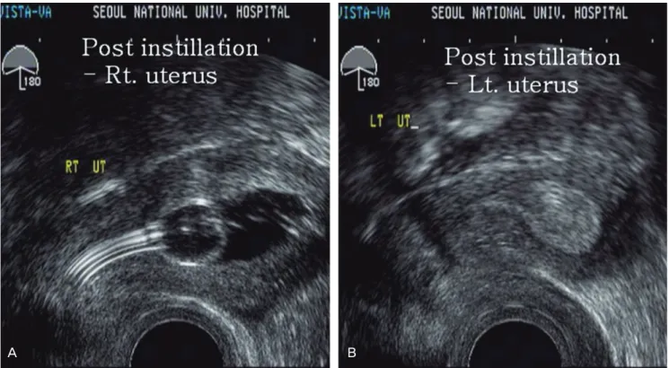
Müllerian 관의 기형은 대략 2-3%에서 나타난다[6]. 일측 질폐쇄와 동측 신장의 기형을 동반하는 증후군은 1922년에 Purslow에 의해 처 음 보고된 이래로 Herlyn-Werner-Wunderlich 증후군으로 불려왔다 [7]. 이후 60년 동안 세계적으로 115예가 보고되었는데 최근 20년 동 안 170예가 추가로 보고되었다[4]. 이는 본 질환에 대한 이해 및 진단 기법의 발달에 기인하는 것으로 생각되며 아직 이 증후군의 정확한 유 병률은 밝혀지지 않았다. 2007년에 Smith와 Laufer [4]는 현재까지 제 Fig. 4. Post-instillation images of saline infusion sonohysterography. (A) Endometrial cavity of right uterus was dilated normally. (B) But left side was not. There is no evidence of communication between both side of uterine cavity.
A B
Fig. 5. Laparoscopic fi nding. There were normal ovaries and tubes arising from each side of uterine cornus. Left lower side of uterine posterior wall was hyperemic and bulged due to hematocolpos. There was a small endo- metriotic spot on the affected peritoneal surface.
각기 다른 이름으로 보고된 사례들을 분석하여 진단 및 치료 방침의 유 사성을 확인하였고 본 증후군을 obstructed hemivagina and ipsilateral renal anomaly (OHVIRA) 증후군으로 부를 것을 제안하였다. OHVIRA 증후군은 보통 중복자궁에 동반하여 나타나는 경우가 많고 드물게는 중격자궁이 있는 경우에서 발생한 사례도 있었다.
OHVIRA 증후군은 태생 3주경 유전적 이상이나 기형발생인자 (teratogen)의 영향으로 태생기의 중신관(mesonephric duct) 형성이 불 완전하게 이루어지는 경우 동측의 신장이 발생하지 않게 되고, 요생식 동(urogenital sinus)에서 중신관이 불완전하게 형성됨으로써 중신방 관(paramesonephric duct)의 융합이 일어나지 않아 중복자궁이 발생 하게 된다[1]. OHVIRA 증후군에 동반하는 신기형의 대부분이 병변측 신장의 무발생이지만 중복신(renal duplication)이나 다낭성 신이형성 (multicystic dysplastic kidney)도 보고되고 있다[4].
2007년 Smith와 Laufer [4]의 연구에서 OHVIRA 증후군이 진단되는 연령의 중앙값은 14세이고 18세 이상에서 진단되는 경우는 총 27예 중 4예가 있었다. 주 증상으로는 생리통이 총 27예 중 23예로 가장 흔 하였고 부정출혈이 6예, 발열이 2예 있었다. 18세 이상에서 진단된 4 예는 타 병원에서 진단받은 이후 의뢰된 경우로 의무기록이 부실하여 발병 당시의 주소를 알 수 없었다[4]. 기존에 보고된 많은 연구들에서 도 전형적인 증상 발현으로 초경 이후 심해지는 생리통과 동반하는 하 복부 혹은 질의 팽윤을 들고 있으며 사춘기 무렵 일찍 진단되는 경우 가 가장 많다[1,2,4,7,8]. 비전형적인 증상 발현으로 보고된 사례를 보 면, 불완전 질폐색이나 완전 폐색에 동반하여 양쪽 자궁강의 교통이 있 는 경우 부정출혈로 발현하거나 정상쪽 질로 생리가 나와 병변측의 질 혈종이 심하지 않아 진단이 지연되는 경우가 있었고 화농성 질분비물 과 골반염에 합당한 소견을 보인 사례 또한 본 증후군으로 최종 진단된 경우가 있었다[5].
OHVIRA 증후군의 치료는 진단이 지연되어 질환이 진행할 경우 자 궁내막증이나 골반내 감염 등으로 불임증이 유발될 가능성이 있기 때 문에, 조기 진단을 통해 가능한 빨리 폐쇄된 질중격을 제거하여 혈종을 제거하는 것이 중요하다[4,5]. 이전의 연구들에서는 치료 성적과 관련 하여 조기에 폐쇄된 질중격만 제거하는 방법과 해부학적인 이상을 교 정하기 위해 편측 자궁절제술을 시행하는 방법 사이에 논란이 있었지 만, 개복수술을 통한 해부학적 이상의 교정은 패혈증이나 출혈 등의 위 험성이 증가하여 불필요하다는 보고가 있고[9] 골반염이나 혈종 등이 합병되지 않은 경우 폐쇄된 일측 자궁과 부속기를 남겨두더라도 임신 과 관련한 예후에 영향을 미치지 않는다고 한다[10]. 더불어 복강경 수 술을 통해 해부학적인 이상을 명확히 확인하고 영상검사로 진단하기 어려운 자궁내막증이나 골반내 유착 및 감염증을 진단 및 치료할 수 있 는 장점이 있어 수술적 치료 시 함께 시행할 것을 권고하고 있다[5,11].
본 증례는 부정출혈이 발생하여 시행한 진찰 및 검사를 통해 OHVIRA 증후군으로 진단되었다. 본 증례는 상기에 열거한 사례들과는 다르게 질의 완전 폐색과 함께 중복자궁 사이의 교통로가 없었던 경우 로 전형적인 증상을 보이지 않아 진단이 지연되었다. 본 증례의 초음파 검사 소견상 병변측 자궁의 자궁내막에 이상 소견은 보이지 않았고, 수
술 소견상 자궁 후벽에 자궁내막증 병변이 관찰되었으나 생리혈의 복 강내 역류가 있었다고 보기에는 의미가 거의 없을 정도의 작은 병변이 었다. 상기의 검사 소견 및 수술 소견을 종합해볼 때, 본 증례에서 초경 이후 증상이 심하지 않았고 진단이 지연된 원인은 검사로 증명되지 않 았지만 미세 천공을 통한 교통이 있다가 자연적으로 폐쇄된 후 뒤늦게 병변이 발생한 경우이거나 병변측 자궁내막의 기능이 정상측보다 저하 되어 있을 가능성도 고려해볼 수 있겠다.
본 증례와 같이 비전형적인 증상 발현을 보이는 경우 진단이 지연되 어 골반내 감염증이나 자궁내막증이 발생하거나 이후의 임신에 악영향 을 미칠 수 있어 OHVIRA 증후군이 의심되는 경우 세심한 문진, 진찰 및 검사를 시행하는 것이 중요한 임상적 의의를 지닌다고 판단하여 이 에 대한 문헌 고찰과 함께 보고하는 바이다.
References
1. Lee BH, Kim JW, Oh SI, Kim MH, Park NH, Lee JY, et al. 3 Cases of uterus didelphys with an obstructed hemivagina and ipsilat- eral renal agenesis. Korean J Obstet Gynecol 1997;40:1489-95.
2. Pyun JS, Kang H, Choi YW, Jin SJ. A case of uterus didelphys associated with unilateral imperforate vagina and ipsilateral renal agenesis. Korean J Obstet Gynecol 1981;24:519-84.
3. Gilliland B, Dyck F. Uterus didelphys associated with unilateral imperforate vagina. Obstet Gynecol 1976;48:5S-8S.
4. Smith NA, Laufer MR. Obstructed hemivagina and ipsilateral renal anomaly (OHVIRA) syndrome: management and follow- up. Fertil Steril 2007;87:918-22.
5. Zurawin RK, Dietrich JE, Heard MJ, Edwards CL. Didelphic uterus and obstructed hemivagina with renal agenesis: case report and review of the literature. J Pediatr Adolesc Gynecol 2004;17:137-41.
6. Shavell VI, Montgomery SE, Johnson SC, Diamond MP, Berman JM. Complete septate uterus, obstructed hemivagina, and ip- silateral renal anomaly: pregnancy course complicated by a rare urogenital anomaly. Arch Gynecol Obstet 2009;280:449-52.
7. Gholoum S, Puligandla PS, Hui T, Su W, Quiros E, Laberge JM.
Management and outcome of patients with combined vaginal septum, bifid uterus, and ipsilateral renal agenesis (Herlyn- Werner-Wunderlich syndrome). J Pediatr Surg 2006;41:987-92.
8. Yang KM, Chae NH, Bae DH, Lim JE, Lee YA, Kang OR, et al. 6 Cases of uterus didelphys associated with obstructed hemiva- gina and ipsilateral renal agenesis. Korean J Obstet Gynecol 1998;41:2022-6.
9. Hørding U, Legarth J. Uterus didelphys with a unilateral imper-
forate hemivagina and ipsilateral renal agenesis. Acta Obstet
Gynecol Scand 1987;66:277-8.
10. Stassart JP, Nagel TC, Prem KA, Phipps WR. Uterus didelphys, obstructed hemivagina, and ipsilateral renal agenesis: the Uni- versity of Minnesota experience. Fertil Steril 1992;57:756-61.
11. Asha B, Manila K. An unusual presentation of uterus didelphys with obstructed hemivagina with ipsilateral renal agenesis.
Fertil Steril 2008;90:849.e9-10.
비전형적인 증상 발현을 보이는 일측 질폐쇄 및 동측 신기형 증후군 1예
1서울대학교 의과대학 산부인과학교실, 2서울대학교 의학연구원 인구의학연구소 김우오1, 박경의1, 구승엽1,2, 김석현1,2, 최영민1,2, 김정구1, 문신용1,2
Obstructed hemivagina and ipsilateral renal anomaly 증후군은 중복자궁에 동반하여 편측 질폐쇄 및 동측 신장의 기형이 함께 나타나는 증후군으로 대개의 경우 초경 이후 심해지는 월경통 및 하복부 종괴, 질벽의 팽윤이 전형적인 증상으로 나타난다. 조기 진단 및 치료를 통 해 증상을 완화하고 생식기의 손상을 최소화하며 이후의 가임력을 유지할 수 있으므로 이는 예후에 중요한 영향을 미친다. 저자들은 초경 이후 특별한 증상 없이 지내오다가 질출혈로 검사하던 중 우연히 발견된, 비전형적 증상 발현을 보이는 일측 질폐쇄와 동측 신장무발생을 동반한 중복자궁 1예를 경험하였기에 문헌 고찰과 함께 보고하는 바이다.
중심단어: 중복자궁, 편측 질폐쇄, 신장무발생, 뮐러관 기형