Yonsei Med J http://www.eymj.org Volume 50 Number 4 August 2009 560
Seminal vesicle cyst is an extremely rare disease, occurring in 0.005% of the population.1It is often associated with other anomalies. Especially, Zinner2reported a seminal vesicle cyst in combination with ipsilateral renal agenesis in 1914, and the associated anomaly has been reported in literatures.3-5Treatment of the seminal vesicle cyst can be decided according to symptom existence. When surgical treatment for symptomatic seminal vesicle cyst is deemed necessary, conventional surgery is frankly invasive because of the deep location and dissection difficulty of the seminal vesicles in the retrovesical space. Recently, the laparoscopic approach has been advocated as an optimal minimally invasive technique for the surgical treatment of seminal vesicle pathology. It provides a good image and easy approach for the seminal vesicle.6,7
We present 4 cases of seminal vesicle cyst with ipsilateral renal agenesis, which were successfully treated by laparoscopy.
From May 2003 to January 2008, 4 patients with seminal vesicle cysts with ipsilateral renal agenesis were presented at our hospital. The mean age was 45.8 years (22 - 68). Chief complaints were perineal pain in two patients and hemato- spermia in two patients. Diagnostic procedures included digital rectal examina- tion, transrectal ultrasonography (TRUS), abdominopelvic computed tomography (CT), pelvic magnetic resonance imaging (MRI), and cystoscopy. TRUS showed
Original Article
DOI 10.3349/ymj.2009.50.4.560pISSN: 0513-5796, eISSN: 1976-2437 Yonsei Med J 50(4): 560-563, 2009
Congenital Seminal Vesicle Cyst Associated with Ipsilateral Renal Agenesis
Ill Young Seo,
1Hun Soo Kim,
2and Joung Sik Rim
1Departments of 1 Urology, 2 Pathology, Institute of Wonkwang Medical Science, Wonkwang University School of Medicine, Iksan, Korea.
Purpose:A seminal vesicle cyst in combination with ipsilateral renal agenesis is rarely encountered. We present cases of this disease entity with symptoms, which were treated with a laparoscopic approach as a minimally invasive surgical treatment. Materials and Methods:We experienced 4 patients with seminal vesicle cysts and ipsilateral renal agenesis. The mean age was 45.8 years. Chief complaints were perineal pain and hematospermia.
Seminal vesicle cysts and remnant ureters were excised by laparoscopic surgery with transperitoneal approaches.
Results:The mean operative time was 133.8 minutes. The mean hospital stay was 6.8 days. There were no operative complications or transfusions. Conclusion: In our report, patients of congenital seminal vesicle cyst associated with renal agenesis are presented. Laparoscopy is considered a minimal invasive management of these combined anomalies, providing a good image and an easy approach.
Key Words : Seminal vesicle, cyst, renal agenesis, laparoscopy
Received: August 20, 2008 Revised: November 7, 2008 Accepted: November 27, 2008 Corresponding author: Dr. Ill Young Seo, Department of Urology, Institute of Wonkwang Medical Science, Wonkwang University School of Medicine, 344-2 Sinyong-dong, Iksan 570-180, Korea.
Tel: 82-63-859-1333, Fax: 82-63-842-1455 E-mail: [email protected]
∙The authors have no financial conflicts of interest.
© Copyright:
Yonsei University College of Medicine 2009
INTRODUCTION
MATERIALS AND METHODS
cystic masses with heterogenous contents adjacent to the left seminal vesicle in all cases. Cyst sizes were 6.0×5.5 cm, 3.0×2.5 cm, 2.6×2.7 cm, and 5.5×5.0 cm (Table 1).
Pelvic MRI also showed round cystic masses in the left seminal vesicles with contents of high signal intensity in T1 weighted image and low signal intensity in T2 weighted image (Fig. 1). Abdominal CT scan revealed no left kidney and remnant ureter in the retroperitoneal space. The remnant ureter inserted into the seminal vesicle cyst (Fig. 2). Cysto- scopy revealed hemitrigones with no left ureteral orifices in all cases.
Seminal vesicle cysts and remnant ureters were excised by laparoscopic surgery under general anesthesia. Firstly, in a lithotomy position, the verumontanum and ejaculatory duct were observed with a 7 / 13.5F ureteroscope. Under the ureteroscopic guidance, a 0.035-inch guide wire was inserted into the seminal vesicle cyst and remnant ureter. A Foley catheter was placed into the bladder.
The patient was positioned in a supine position for a laparoscopic surgery. A transperitoneal access is achieved with Veress needle. Four ports were used, a 5/12-mm trocar at umbilicus, a 5/11-mm trocar at the right lateral border of the rectus muscle at the 5 cm lower level of the umbilicus, a 5/11-mm trocar at the left lateral border of the rectus muscle at the 5 cm lower level of the umbilicus, and a 5-mm trocar at the left lateral border of the rectus muscle at the 5 cm above level of the umbilicus.
Once the descending colon was mobilized medially, the left ureter was detected in the retroperitoneal space. It was dissected cephalad above the iliac vessel. There was no renal tissue in the end of the ureter. The lower ureter was dissect- ed to the bladder. The bladder was retracted anterior. A junction of the ureter and seminal vesicle was seen (Fig. 3).
The guide wire was detected in the communicated area.
The seminal vesicle was dissected and sectioned with Seminal Vesicle Cyst with Renal Agenesis
Yonsei Med J http://www.eymj.org Volume 50 Number 4 August 2009 561
Table 1. Patient Characteristics and Operative Results
Case 1 Case 2 Case 3 Case 4
Age 22 67 68 26
Chief complaint Perineal pain Perineal pain Hematospermia Hematospermia
(duration) (2 yrs) (7 days) (7 yrs) (4 months)
Cyst size (cm) 6.0 ×5.5 3.0 ×2.5 2.6 ×2.7 5.5 ×5.0
Laparoscopic Seminal Seminal Seminal vesicle Seminal vesicle
surgery vesicle cyst vesicle cyst cyst excision + cyst excision +
excision excision ureterectomy ureterectomy
Operating time
100 105 180 150
(mins) Estimated blood
30 70 50 190
loss (cc) Hospital stay
7 7 5 8
(days)
Complications None None None None
Fig. 1. Sagittal view of pelvic magnetic resonance imaging of case 1. A 6.0 ×5.5 cm sized, round cystic mass is seen with contents of low signal intensity in T2 weighted image (A) and high signal intensity in T1 weighted image (B).
Fig. 2. Abdominopelvic computed tomography of case 3. It shows no left kidney, a remnant left ureter, and a junction of the ureter inserted into the seminal vesicle (arrow).
A B
ultrasonic scissors and clips. The whole specimen was removed through the umbilical port.
The mean operative time was 133.8 minutes (range, 100 - 180). The mean hospital stay was 6.8 days (range, 5 - 8).
There were no operative complications and transfusions.
The mean estimated blood loss was 85 mL (range, 30 - 190) (Table 1). In all patients, oral intake and ambulation were resumed on postoperative day 1. The urethral Foley catheter was removed on postoperative day 1.
The pathological assessments of the specimens revealed benign seminal vesicle cyst in all cases. The cystic walls were composed of fibrous connective tissue. They were lined by columnar and basal cells with villous projections in hematoxyline and eosin stains. The ectopic ureter was inserted into the seminal vesicle cyst. The junction showed a transition of transitional epithelium to seminal vesicular
epithelium (Fig. 4).
Seminal vesicle cysts occur by congenital or acquired origin.
Most of them are congenital cause, which is believed to be secondary to obstruction of the ejaculatory duct. It is associated with anomalies of maldevelopment of the distal portion of the mesonephric duct, such as renal dysplasia, renal agenesis, and ectopic ureter into the seminal vesicle.8 Among the associated anomalies, ipsilateral renal agenesis was first described by Zinner. Shieh1noted 6 cases among 119 boys who were found to have ipsilateral renal agenesis.2 In cases of seminal vesicle cysts with ipsilateral renal agenesis, the ureter may insert into the prostatic urethra or seminal vesicle.
Seminal vesicle cysts are usually asymptomatic. However, the cyst can grow and induce inflammation and stimulate surrounding viscera, resulting in bladder irritating symp- toms, including urinary frequency and dysuria, suprapubic pain, hematospermia, and painful ejaculation. Initial diagnostic methods include an abdominal or transrectal ultrasonography. Abdominal computed tomography, magnetic resonance imaging, and seminal vesiculography are useful for the detection of accompanying deformity and differential diagnosis. A cystoscopy helps to confirm a hemitrigone, absence of ureteral orifice, and other anoma- lies in bladder.9-11
The seminal vesicle cyst is treated in patients with symp- toms, and simply observation is adequate in patients without pain or functional deformity. Conventional treatment methods, including an ultrasound guided cyst aspiration and a transurethral unroofing of ejaculatory duct and seminal vesicle cyst, are limited in treatment effect compared with radical resection, and there is a great danger of reappearance or infection. Recently, a laparoscopic surgery has appeared to be most suitable for surgical treatment of seminal vesicle cyst. Laparoscopic surgery, minimal invasiveness, has advantages of direct access to the seminal vesicle with an excellent image in deep, retrovesical field compared to open resection. Also, without damage of bladder and rectum, the seminal vesicle can be dissected from the peritoneum that is covering bladder and prostate.
In 1993, Kavoussi described a principle of laparoscopic access for seminal vesicle in patient with prostatic cancer, using a five-port transperitoneal approach.13Only 10 cases of laparoscopic excision for seminal vesicle cysts have been reported to date. Cherullo et al.12 reported 2 cases, which were treated with laparoscopic surgery in 2002. He used a four-port trasnperitoneal approach. Basillote et al.6reported Ill Young Seo, et al.
Yonsei Med J http://www.eymj.org Volume 50 Number 4 August 2009 562
RESULTS
DISCUSSION
Fig. 3. Laparoscopic finding of case 3. After dissection of the remnant ureter, the junction of the seminal vesicle and remnant ureter is seen (arrow).
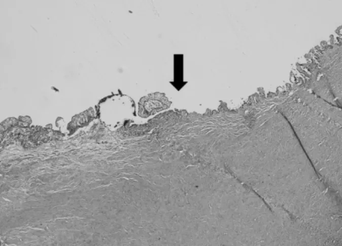
Fig. 4. Microscopic finding of the junction of the seminal vesicle and remnant ureter in case 3. Transition (arrow) of transitional epithelium to seminal vesicular epithelium is seen. The seminal vesicle is lined by columnar and basal cell with villous projections (H & E, ×40).
7-year old boy with seminal vesicle cyst, who was treated with laparoscopic excision. In this case, the cyst was detec- ted easily by filling the cyst with indigo carmine solution in laparoscopic view.
In our 4 cases, seminal vesicle cyst associated with renal agenesis was presented. They were treated with laparoscopic surgery, which was performed with a four-port transperi- toneal approach. It was a feasible and effective treatment for this disease, with advantages of magnification, good visualization, direct approach, and less invasiveness. Espe- cially, the junction of seminal vesicle cysts and remnant ureter was easily detected by an endoscopically inserted guide wire.
This paper was supported by Wonkwang University in 2008.
1. Sheih CP, Hung CS, Wei CF, Lin CY. Cystic dilatations within the pelvis in patients with ipsilateral renal agenesis or dysplasia. J Urol 1990;144:324-7.
2. Zinner A. Ein Fall von intravesikaler Samenblasenzyste. Wein Med Wochenschr 1914;64:605-9.
3. Donohue RE, Greenslade NF. Seminal vesicle cyst and ipsilateral renal agenesis. Urology 1973;2:66-9.
4. Roehrborn CG, Schneider HJ, Rugendorff EW, Hamann W.
Embryological and diagnostic aspects of seminal vesicle cysts associated with upper urinary tract malformation. J Urol 1986;
135:1029-32.
5. van den Ouden D, Blom JH, Bangma C, de Spiegeleer AH.
Diagnosis and management of seminal vesicle cysts associated with ipsilateral renal agenesis: a pooled analysis of 52 cases. Eur Urol 1998;33:433-40.
6. Basillote JB, Shanberg AM, Woo D, Perer E, Rajpoot D, Clayman RV. Laparoscopic excision of a seminal vesicle cyst in a child. J Urol 2004;171:369-71.
7. Ikari O, Castilho LN, Lucena R, D’Ancona CA, Netto NR Jr.
Laparoscopic excision of seminal vesicle cysts. J Urol 1999;162:
498-9.
8. Patel B, Gujral S, Jefferson K, Evans S, Persad R. Seminal vesicle cysts and associated anomalies. BJU Int 2002;90:265-71.
9. Beeby DI. Seminal vesicle cyst associated with ipsilateral renal agenesis: case report and review of literature. J Urol 1974;112:
120-2.
10. Fuselier HA Jr, Peters DH. Cyst of seminal vesicle with ipsilateral renal agenesis and ectopic ureter: case report. J Urol 1976;116:833-5.
11. Das S, Amar AD. Ureteral ectopia into cystic seminal vesicle with ipsilateral renal dysgenesis and monorchia. J Urol 1980;124:574-5.
12. Cherullo EE, Meraney AM, Bernstein LH, Einstein DM, Thomas AJ, Gill IS. Laparoscopic management of congenital seminal vesicle cysts associated with ipsilateral renal agenesis. J Urol 2002;167:1263-7.
13. Kavoussi LR, Schuessler WW, Vancaillie TG, Clayman RV.
Laparoscopic approach to the seminal vesicles. J Urol 1993;150:
417-9.
Seminal Vesicle Cyst with Renal Agenesis
Yonsei Med J http://www.eymj.org Volume 50 Number 4 August 2009 563