ISSN 0378-6471 (Print)⋅ISSN 2092-9374 (Online)
http://dx.doi.org/10.3341/jkos.2016.57.9.1484
Case Report
동측 일과성 흑암시를 보인 내경동맥 무발생증
Amaurosis Fugax Associated with Ipsilateral Internal Carotid Artery Agenesis
성재연1⋅김경남1⋅정혜선2⋅이연희1
Jae Yun Sung, MD1, Kyoung Nam Kim, MD, PhD1, Hye Seon Jeong, MD, PhD2, Yeon Hee Lee, MD, PhD1
충남대학교 의과대학 충남대학교병원 안과학교실1, 충남대학교 의과대학 충남대학교병원 신경과학교실2
Department of Ophthalmology, Chungnam National University Hospital, Chungnam National University School of Medicine1, Daejeon, Korea Department of Neurology, Chungnam National University Hospital, Chungnam National University School of Medicine2, Daejeon, Korea
Purpose: We report a case of amaurosis fugax associated with ipsilateral internal carotid artery agenesis.
Case summary: A 50-year-old woman presented with amaurosis fugax in her left eye; the frequency of episodes of the condition had recently increased to once a month. She had a history of hypertension and dyslipidemia, and was under medical therapy.
The visual acuity of both eyes was 20/20. Slit-lamp examination was normal except for pseudophakia. Ophthalmoscopy re- vealed a myopic tigroid fundus and a myopic tilted disc. No abnormalities were evident in fluorescein fundus angiography. Brain computed tomography showed that the left bony carotid canal was absent, and magnetic resonance angiography showed that the left internal carotid artery was also absent. She was diagnosed with left internal carotid artery agenesis. Other neurological and hematological parameters were within normal ranges. The amaurosis fugax spontaneously disappeared and has not re- curred over the past 12 months. Our case, although rare, suggests that amaurosis fugax may be associated with internal carotid artery agenesis.
J Korean Ophthalmol Soc 2016;57(9):1484-1488
Keywords: Agenesis, Amaurosis fugax, Internal carotid artery
■Received: 2016. 6. 2. ■ Revised: 2016. 7. 20.
■Accepted: 2016. 8. 29.
■Address reprint requests to Kyoung Nam Kim, MD, PhD Department of Ophthalmology, Chungnam National University Hospital, #282 Munhwa-ro, Jung-gu, Daejeon 35015, Korea Tel: 82-42-280-7600, Fax: 82-42-255-3745
E-mail: [email protected]
ⓒ2016 The Korean Ophthalmological Society
This is an Open Access article distributed under the terms of the Creative Commons Attribution Non-Commercial License (http://creativecommons.org/licenses/by-nc/3.0/) which permits unrestricted non-commercial use, distribution, and reproduction in any medium, provided the original work is properly cited.
일과성 흑암시는 망막의 허혈로 인하여 발생하는 단안의 일시적인 시력소실로 정의된다. 시력소실은 부분 또는 전 체 시야에서 나타나며, 대부분 수초에서 수분 이내에 정상 으로 회복된다.1 여러 원인에 의하여 일과성 흑암시가 발생 할 수 있는데 경동맥협착, 동맥경화성 질환 및 안구의 혈관 폐쇄성 질환이 가장 흔한 원인으로 알려져 있으며, 다른 질 환들과 감별하기 위하여 병력청취, 혈액검사, 안과적 검사,
뇌자기공명영상 및 혈관조영술 등이 필요하다.2-5
이 밖에도 드물지만 염증성 동맥염, 앞허혈시신경병증, 혈관연축, 뇌압상승, 겸상적혈구병, 전신성 홍반성 루푸스 등에 의하여 발생하기도 하며,1,6,7 운동이나 발살바 조작 후 에 일과성 흑암시가 유발되었다는 보고도 있다.8,9 저자들은 반복되는 단안의 일과성 흑암시를 주소로 내원한 환자에서 동측의 내경동맥 무발생을 진단하였으며, 국내는 물론 국 외에서도 아직까지 보고된 바 없어 문헌고찰과 함께 이를 보고하고자 한다.
증례보고
50세 여자가 3년 전부터 시작된 좌안의 일과성 흑암시를 주소로 내원하였다. 3-4개월에 1회 정도 발생하던 것이 최
Figure 1. Fundus photographs of the both eyes. Both eyes show myopic tigroid fundus and myopic tilted disc with peripapillary
atrophy.Figure 2. Fluorescein fundus angiography of the left eye. (A) Retinal artery filling was started at 10 seconds after injection of the
sodium fluorescein dye into the antecubital vein. (B) Retinal vein filling was completed at 18 seconds after injection.근에는 빈도가 잦아져서 1개월에 1회 정도라고 하였다. 시 력소실은 급작스럽게 가운데 초점을 제외한 주변시야 전체 가 까맣게 보이는 양상이었고, 5분 정도 지속되었다. 자세 변화나 기침 등의 특정 활동과 연관되어 나타나지는 않는 다고 하였고, 흑암시가 있는 동안 두통과 같은 다른 신경학 적 증상이 동반되지도 않았다. 혈압은 112/76 mmHg였고, 맥박수는 116회/분이었는데, 과거력상 3년 전부터 고혈압
과 고지혈증을 진단 받고 약물치료 중이었다. 양안 나안시 력은 20/20이었고, 안압은 골드만압평안압계로 20 mmHg 였다. 양안 모두 2년 전 백내장수술을 시행하여 인공수정체 안이었고 전안부에 다른 이상소견은 없었다. 안저검사에서 양안 모두 근시성 범무늬안저와 시신경유두주위위축을 동 반한 근시성 경사유두를 보였다. 우안은 상이측에 시신경 유두출혈이 있었다(Fig. 1). 시야검사결과는 양안 모두 정상
A B
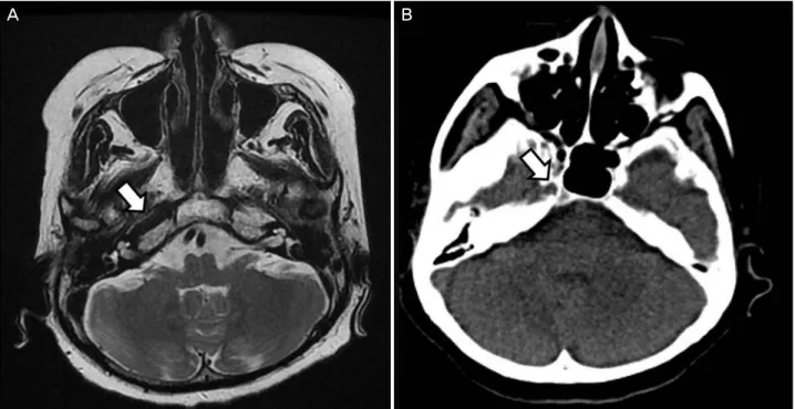
Figure 3. Magnetic resonance imaging (MRI) and computed tomography (CT). (A) MRI and (B) CT of the skull base showing a car-
otid canal on the right (arrows), but absent on the left.Figure 4. Magnetic resonance angiography (MRA). (A) MRA reveals the right internal carotid artery (arrow) and the absence of the
left internal carotid artery. (B) At the circle of W illis, the left middle cerebral artery and anterior cerebral artery (arrowheads) are filled via posterior communicating artery (arrow).이었으며, 형광안저혈관조영술에서 좌안 팔-망막순환시간 은 10초, 동정맥 통과시간은 약 8초로 정상이었다(Fig. 2).
일반혈액검사 및 항인지질 항체, 단백질 C, 단백질 S 등 의 혈액응고항진과 연관된 인자들에 대한 검사는 모두 정 상이었다. 혈관폐쇄성 질환을 감별하기 위해 시행한 뇌자 기공명영상에서 좌측 경동맥관이 관찰되지 않았고(Fig. 3A), 뇌자기공명혈관조영술에서도 좌측 내경동맥이 보이지 않
았으며(Fig. 4A) 좌측 앞대뇌동맥과 중대뇌동맥은 비대해 진 후교통동맥에서 기원하는 것을 확인하였다(Fig. 4B). 추 가적으로 시행한 전산화단층촬영검사에서 좌측 경동맥관 이 없는 것을 확인하였다(Fig. 3B). 좌측 내경동맥과 경동 맥관이 모두 존재하지 않는 좌측 내경동맥 무발생으로 진 단하였으며, 동측 내경동맥 무발생과 연관되어 나타난 일 과성 흑암시로 추정 진단하였다.
A B
A B
좌안에 간헐적인 일과성 흑암시 외에는 다른 신경학적 증상이나 이상소견을 동반하지 않아 추가적인 치료 없이 경과관찰하기로 하였다. 이후 일과성 흑암시는 저절로 소 실되었고 12개월 뒤까지도 재발하지 않았다. 환자는 타 병 원에서 항고혈압약과 항고지혈증약을 복용 중이었는데 an- giotensin II receptor antagonist 계열의 항고혈압약은 용량 을 줄였고, statin 계열의 항고지혈증약은 그대로 유지하고 있다고 하였다.
고 찰
내경동맥의 발생이상은 극히 드문 선천적 이상으로 발생 률이 0.01% 미만인 것으로 알려져 있으며, 방사선학적 검 사에서 우연히 발견되는 경우가 대부분이다.10 이러한 내경 동맥의 발생이상은 전산화단층촬영검사에서 경동맥관의 존 재유무 및 혈관조영술에서 내경동맥의 존재유무에 따라 무 발생(agenesis), 무형성(aplasia), 그리고 형성부전(hypoplasia) 으로 나눌 수 있다. 내경동맥과 경동맥관이 모두 존재하지 않는다면 내경동맥 무발생을 의미하며, 경동맥관의 흔적이 존재하는 경우는 무형성으로 진단할 수 있다. 그리고 형성 부전은 내경동맥이 존재하지만 발달이 불충분하여 혈관조 영술에서 측부순환이 관찰되는 경우이다.11 본 증례의 환자 는 좌측 내경동맥과 경동맥관이 모두 존재하지 않는 내경 동맥 무발생이었으며, 이 환자에서와 같이 좌측에 발생하 는 경우가 우측에 비해 약 3배 정도 많은 것으로 알려져 있 다.11 또한 윌리스 고리(circle of Willis)나 외경동맥 등을 통한 측부순환이 형성될 수 있는데, 본 증례와 같이 윌리스 고리의 후교통동맥을 통한 측부순환이 가장 흔한 것으로 알려져 있다.12
대부분의 내경동맥 발생이상 환자들은 잘 발달된 측부순 환 덕분에 나이가 들 때까지 무증상으로 지낼 수 있다. 하 지만 소수에서는 비정상적인 혈관에 의한 허혈성 손상이 발생하거나, 뇌동맥류의 동반이나 측부순환 혈관의 확장으 로 인한 압박성 손상이 발생했다고 보고된 바 있다.13,14 또 한 이와 같은 이차적인 해부학적 변화나 관류이상, 혈관운 동반응도의 이상 등이 발견되지 않으면서도 뇌출혈, 어지 럼증, 두통 등이 발현한 경우도 보고된 바 있다.15-17
지금까지 혈관폐쇄성질환을 포함하여 일과성 흑암시가 동반된 여러 질환들이 보고된 바 있는데 이들은 모두 일시 적인 안 혈류의 장애를 유발함으로써 단안의 일시적인 시 력소실을 일으키게 된다.2-9 대부분 흑암시 증상이 수 초에 서 수 분만 지속되고 발현시기를 예측하기 어렵기 때문에 형광안저혈관조영술이나 경동맥뇌혈관조영술 등을 통한 안혈류의 장애를 직접 확인하기는 어려우며, 본 증례에서
도 내원 당시에는 증상이 없었고 형광안저혈관조영술 결과 는 정상이었다. 본 증례에서 흑암시의 정확한 원인을 알기 는 어려우나 환자에서 경동맥협착과 같은 다른 뚜렷한 뇌 혈관의 이상이 없었고 망막혈관폐쇄의 증거도 보이지 않았 으며, 혈액학적 이상도 없었던 점으로 미루어 보아 안동맥 혈류를 공급해주던 측부순환의 일시적인 허혈로 인해 발생 했을 가능성을 생각해 볼 수 있다. 안동맥은 내경동맥의 첫 번째 분지로 안와와 안구의 주요 혈관공급을 담당한다. 안 동맥은 다양한 이상기시가 보고된 바 있는데, 후교통동맥 에서 기시하는 경우는 매우 드물고, 중간뇌막동맥과 앞대 뇌동맥에서 기시하는 경우가 가장 많은 것으로 알려져 있 으며 그밖에 뇌바닥동맥, 중뇌동맥, 외경동맥 등에서 기시 하는 경우도 보고된 바 있다.18 하지만 내경동맥의 발생이 상이 있는 경우에는 안동맥의 기시 부위가 일반적인 경우 와는 다를 것으로 예상되며 Naeini et al19은 우측 편두통을 동반한 동측 내경동맥의 무발생을 보고하였는데, 일반적인 경우에는 매우 드문 후교통동맥에서 안동맥이 이상기시하 였다. 아쉽게도 본 증례에서는 안동맥의 기시부위를 확인 할 수는 없었다.
내경동맥의 발생이상이 있는 환자에서 허혈성 손상이 발 생하는 기전은 아직까지 명확히 밝혀지지는 않았으나 영아 기를 거쳐 성장기 동안에는 측부순환도 점차 증가하면서 필요한 혈액을 공급할 수 있지만 나이가 더 들면서 측부순 환의 증가가 수요를 따라가지 못하는 보상부전에 의해 발 생한다는 설명이 가장 가능성 있는 가설로 받아들여지고 있다.20 또한 이런 불안정한 평형상태에서 간헐적인 혈압강 하나 심하지 않은 동맥경화도 촉발요인이 될 수 있는 것으 로 보고된 바 있다.20,21 본 증례의 환자는 고혈압에 대해서 3년 전부터 angiotensin II receptor antagonist를 복용 중이라 고 하였는데 내원시의 혈압은 정상범위였으나 심박수가 높 은 점으로 미루어보아 상대적인 저혈압의 가능성을 배제할 수 없으며, 초진 후 개인병원에서 복용 중이던 항 고혈압약 의 용량을 낮추었다고 하였고 이후 흑암시가 재발하지 않 은 점을 고려해 볼 때 측부순환의 간헐적인 보상부전이 흑 암시의 원인이었을 가능성을 생각해 볼 수 있다.
Pubmed 검색을 통한 문헌고찰에서 총경동맥의 무발생과 동반된 일과성 흑암시 1예를 찾을 수 있었으나,22 편측 내경 동맥의 발생이상과 동반된 흑암시는 보고된 바 없으며 국 내에도 없었다. 이 증례는 매우 드문 원인이기는 하지만 내 경동맥의 발생이상이 일과성 흑암시를 유발할 수 있음을 보여준다.
= 국문초록 =
동측 일과성 흑암시를 보인 내경동맥 무발생증
목적: 반복되는 일과성 흑암시를 보인 환자에서 동측 내경동맥 무발생을 진단하여 이를 보고하고자 한다.
증례요약: 50세 여자 환자가 좌안에 발생한 일과성 흑암시를 주소로 내원하였으며, 최근 빈도가 잦아져 1개월에 1회 정도로 반복되는 양상이었다. 과거력상 고혈압과 고지혈증을 진단 받고 약물치료 중이었다. 양안 나안시력은 20/20이었다. 세극등현미경검사에서 양 안 모두 인공수정체안이었고 이외 특이소견은 없었다. 안저검사에서 근시성 안저와 근시성 경사유두가 관찰되었다. 형광안저혈관조영 술 결과는 정상이었다. 전산화단층촬영검사에서 좌측 경동맥관이 관찰되지 않았고, 뇌자기공명혈관조영술에서 좌측 내경동맥이 보이 지 않아 내경동맥 무발생으로 진단되었다. 이외의 신경학적검사와 혈액검사는 모두 정상이었다. 이후 일과성 흑암시는 저절로 소실되 었으며 12개월 후까지도 증상의 재발은 없었다. 매우 드물지만 일과성 흑암시가 내경동맥의 발생이상과 연관될 수 있음을 보여주었다.
<대한안과학회지 2016;57(9):1484-1488>
REFERENCES
1) Current management of amaurosis fugax. The Amaurosis Fugax Study Group. Stroke 1990;21:201-8.
2) Burde RM. Amaurosis fugax. An overview. J Clin Neuroophthalmol 1989;9:185-9.
3) Hayreh SS, Zimmerman MB. Amaurosis fugax in ocular vascular occlusive disorders: prevalence and pathogeneses. Retina 2014;34:
115-22.
4) Kim NR, Chin HS. Progression of impending central retinal vein occlusion to the ischemic variant following intravitreal bevacizumab.
Korean J Ophthalmol 2010;24:179-81.
5) Lee DH, Lee SJ, Yoon IN. Clinical progress in impending central retinal vein occlusion. Korean J Ophthalmol 2010;24:83-8.
6) Shaw HE Jr, Osher RH, Smith JL. Amaurosis fugax associated with SC hemoglobinopathy and lupus erythematosus. Am J Ophthalmol 1979;87:281-5.
7) Heckmann JG, Gaul C, Neundörfer B, et al. Vasospastic amaurosis fugax. J Neurol Neurosurg Psychiatry 2003;74:149.
8) Jehn A, Frank Dettwiler B, Fleischhauer J, et al. Exercise-induced vasospastic amaurosis fugax. Arch Ophthalmol 2002;120:220-2.
9) Jo YJ, Yun YJ, Kwag JY, Kim JY. Valsalva maneuver-induced amaurosis fugax. J Korean Ophthalmol Soc 2010;51:779-83.
10) Afifi AK, Godersky JC, Menezes A, et al. Cerebral hemiatrophy, hypoplasia of internal carotid artery, and intracranial aneurysm. A rare association occurring in an infant. Arch Neurol 1987;44:232-5.
11) Graham CB 3rd, Wippold FJ 2nd, Capps GW. Magnetic resonance imaging in internal carotid artery agenesis with computed tomog- raphy and angiographic correlation-case reports. Angiology 1999;50:847-54.
12) Cali RL, Berg R, Rama K. Bilateral internal carotid artery agenesis:
a case study and review of the literature. Surgery 1993;113:227-33.
13) Mellado JM, Merino X, Ramos A, et al. Agenesis of the internal carotid artery with a trans-sellar anastomosis: CT and MRI find- ings in late-onset congenital hypopituitarism. Neuroradiology 2001;43:237-41.
14) Czarnecki EJ, Silbergleit R, Mehta BA, Sanders WP. Absence of the supraclinoid internal carotid artery in association with intra- cranial aneurysms. Neuroradiology 1998;40:11-4.
15) Jung BO, Lee JI, Choi JH, et al. Congenital hypoplasia of the bi- lateral internal carotid artery: a case report. J Korean Neurosurg Soc 2002;31:282-4.
16) Ito S, Miyazaki H, Iino N, et al. Unilateral agenesis and hypoplasia of the internal carotid artery: a report of three cases. Neuroradiology 2005;47:311-5.
17) Na SK, Park TH, Ahn JY, Kim MK. A case of unilateral agenesis of internal carotid artery with throbbing headache. J Neurocrit Care 2008;1:174-6.
18) Lasjaunias P, Berenstein A, Brugge KT. Clinical vascular anatomy and variations, 2nd ed. Berlin: Springer, 2001; 414-24.
19) Naeini RM, De J, Satow T, Benndorf G. Unilateral agenesis of in- ternal carotid artery with ophthalmic artery arising from posterior communicating artery. AJR Am J Roentgenol 2005;184:571-3.
20) Savastano S, Feltrin GP, Chiesura-Corona M, Miotta D. Cerebral ischemia due to congenital malformations of brachiocephalic ar- teries-case reports. Angiology 1992:43:76-83.
21) Sliwka U, Schmidt P, Reul J, Noth J. Agenesis of the internal car- otid artery: color Doppler, CT, and MR angiography findings. J Clin Ultrasound 1988;26:213-6.
22) Halstuk KS, Littooy FN, Baker WH. Absent common carotid ar- tery associated with amaurosis fugax: a case report. Surgery 1985;
97:502-6.