INTRODUCTION
Mammography, breast sonography, and magnetic resonance imaging are currently the preferred examination
Incidentally Detected Enhancing Breast Lesions on Chest Computed Tomography
Wen-Chiung Lin, MD
1, Hsian-He Hsu, MD
1, Chao-Shiang Li, MD
4, Jyh-Cherng Yu, MD
2, Giu-Cheng Hsu, MD
1, Cheng-Ping Yu, MD, PhD
3, Tsun-Hou Chang, MD
1, Guo-Shu Huang, MD
1Departments of 1Radiology, 2Surgery, 3Pathology, Tri-Service General Hospital and National Defense Medical Center, Taipei, Taiwan, ROC;
Department of 4Radiology, Renai Branch, Taipei City Hospital, Taipei, Taiwan
Objective: To evaluate the nature and imaging appearance of incidental enhancing breast lesions detected on a routine contrast-enhanced chest CT.
Materials and Methods: Twenty-three patients with incidental enhancing breast lesions on contrast-enhanced chest CT were retrospectively reviewed. The breast lesions were reviewed by unenhanced and enhanced CT, and evaluated by observing the shapes, margins, enhancement patterns and backgrounds of breast lesions. A histopathologic diagnosis or long-term follow-up served as reference standard.
Results: Sixteen (70%) patients had malignant breast lesions and seven (30%) had benign lesions. In 10 patients, the breast lesions were exclusively detected on contrast-enhanced CT. Using unenhanced CT, breast lesions with fi broglandular backgrounds were prone to be obscured (p < 0.001). Incidental primary breast cancer showed an non-signifi cant trend of a higher percentage irregular margin (p = 0.056). All of the four incidental breast lesions with non-mass-like enhancement were proven to be malignant.
Conclusion: Routine contrast-enhanced chest CT can reveal suffi cient details to allow for the detection of unsuspected breast lesions, in which some cases may be proven as malignant. An irregular margin of incidental enhancing breast lesion can be considered a suggestive sign of malignancy.
Index terms: Breast; Chest; Computed tomography (CT); Incidental breast lesions
Received July 9, 2010; accepted after revision September 24, 2010.
Corresponding author: Hsian-He Hsu, MD, Department of Radiology, Tri-Service General Hospital and National Defense Medical Center, No.325, Sec. 2, Cheng-Kung Rd, 114, Taipei City, Taiwan, R.O.C.
• Tel: (8862) 8792-7244 ext. 16480 • Fax: (8862) 8792-7245
• Email: [email protected]
This is an Open Access article distributed under the terms of the Creative Commons Attribution Non-Commercial License (http://creativecommons.org/licenses/by-nc/3.0) which permits unrestricted non-commercial use, distribution, and reproduction in any medium, provided the original work is properly cited.
pISSN 1229-6929 · eISSN 2005-8330 Korean J Radiol 2011;12(1):44-51
methods for the detection and characterization of breast diseases. Although dedicated breast computed tomography (CT) had been investigated for potential clinical
applications in the evaluation of breast (1), it was not considered the primary method to evaluate specifi c breast lesions.
Chest CT is routinely used for diseases of the lung,
mediastinum, pleura, chest wall, and diaphragm, but not
breast tissue. However, the entire breast tissue usually
appears on a chest CT. With the increased use of chest
CT, either for diagnostic or screening purposes, incidental
breast lesions are increasingly being encountered. In some
cases, CT may be the fi rst imaging modality to demonstrate
a breast lesion. Incidental breast lesions detected on
unenhanced or contrast- enhanced CT of chest have been
discussed in a few recent English literatures (2-8), which
300; Schering, Berlin, Germany) using a mechanical power injector at a rate of 2.0-3.0-mL/sec. The scanning was performed 50 seconds after the injection of the contrast medium. The imaging parameters were identical to those used for unenhanced CT.
In our study, only transverse images were reviewed, by reconstruction using a 5-mm thickness and 5-mm increments. The images were obtained by using a standard soft-tissue algorithm (window width, 350 Hounsfi eld unit [HU]; level, 40 HU) and a retrospective lung algorithm (window width, 1000 HU; level, -700 HU).
Image Interpretation
Of the 23 patients included in the study, all CT images were retrospectively reviewed by two experienced radiologists (with 11 and 21 years of experience in the interpretation of chest CT, respectively) in consensus. The reviewers had knowledge about the presence of breast lesions on chest CT; however, they were blinded to the locations and diagnosis of the breast lesions.
In each patient, unenhanced CT was reviewed to determine whether the breast lesion could be seen.
A contrast-enhanced CT was conducted immediately after interpretation of unenhanced CT. Reviewers were requested to describe the size, location, shape, margin characteristics, and enhancement patterns of the enhancing breast lesions detected on contrast-enhanced CT. An enhancing breast lesion referred to a breast lesion with attenuation higher than that of normal breast glandular tissue on contrast- enhanced CT.
The shape of the incidental enhancing breast lesion was described as a round, ovoid, lobulated, and irregular mass or non-mass. A breast lesion with a non-mass shape, which was also known as a non-mass-like enhancement, meant that the lesion could only be detected on contrast- enhanced CT, which appeared as regional enhancement of the breast. The area of enhancement did not appear as a mass-like confi guration, in which the internal features could be described as having a discretely different appearance from that of normal gland parenchyma. The margin of the incidental enhancing breast lesion was described as well- circumscribed, irregular, and spiculated.
Reviewers were also requested to determine the background of each breast lesion; namely, adipose or fi broglandular tissues. An adipose tissue breast lesion meant that more than half of the border was surrounded by adipose tissue. Similarly, a fi broglandular tissue breast evaluated the incidence and pathologic outcomes of
incidental breast lesions. We undertook a study emphasizing the discovery of incidental breast lesions with enhancement on routine contrast-enhanced chest CT, and evaluated the nature and imaging appearance of the incidental enhancing breast lesions.
MATERIALS AND METHODS
Patients
Our study has been approved by the institutional review board. All patients provided written informed consent for the diagnostic procedures performed.
From October 2006 to June 2009, a total of 2250 patients underwent routine contrast-enhanced chest CT at our institution, of which, 34 were identifi ed as having enhancing breast lesions. Of the 34 patients, seven cases were excluded from our study due to a known history of breast disease prior to CT (breast cancer, n = 6; breast abscess, n = 1); another four patients were excluded due to the absence of additional diagnostic confi rmation.
Ultimately, a total of 23 patients, all of which were female with a mean age of 59.2 ± 19.5 years (range, 22-85 years), were included as part of this retrospective study.
Clinical indications for chest CT in the 23 patients included a follow-up study in patients with lung cancer (n
= 7), abnormal chest radiograph (n = 6), follow-up study or a staging work-up for non-pulmonary malignancies (n
= 5; thyroid cancer, tongue cancer, lymphoma, renal cell carcinoma, cervical cancer - one for each type), abnormal nuclear medicine bone scan (n = 1), chest trauma (n = 2), a suspicious case of aortic aneurysm (n = 1), and hemoptysis (n = 1).
CT Technique
The chest CT was performed in the supine position, using
a 64-detector row scanner (Brilliance; Philips Medical
Systems, Cleveland, OH). Both unenhanced and contrast-
enhanced scans were performed and the standard protocol
at our institution included an unenhanced helical CT
from the lung apices through the adrenal glands, using
the following imaging parameters: a section thickness of
0.625 mm, a rotation speed of 0.75 second, a pitch of
1.05-1.25, and 120 kVp; effective tube current × time
product ranged between 150 and 200 mAs. Subsequently,
a contrast-enhanced helical CT was performed with the
intravenous administration of 100 mL iopromide (Ultravist
lesion referred to a lesion with more than half its border surrounded by fi broglandular tissue.
Final Diagnosis and Follow-Up Studies
All patients subsequently underwent sonography of the breast or mammography for the incidental breast lesions.
Sonographically-guided core-needle biopsy or surgery was performed for defi nitive diagnosis in 20 patients. The diagnoses in the three remaining patients were determined on the basis of long-term follow-up CT or sonography for at
least 12 months.
Statistical Analysis
The statistical analysis was performed with commercially available software (SPSS version 10.0, SPSS, Chicago, IL).
Continuous variables were expressed as mean ± standard deviation (SD); and the Student’s t-test was used for comparison of the two variables. The two-tailed Fisher’s exact test was used for analysis of the nominal variables. A p value of 0.05 was chosen as the threshold for statistical
B C
A
Fig. 1. 72-year-old female patient underwent chest CT for staging of lymphoma.
A. Contrast-enhanced CT showed focused image of regional non-mass-like enhancement in left breast (arrow); lesion was not detected on unenhanced CT. Lesion had fi broglandular tissue background. B. Craniocaudal view of left mammography showed cluster of microcalcifi cation in upper inner quadrant of left breast (arrow). C. Sonography of left breast at corresponding location showed hypoechoic mass with ill-defi ned margin (long arrows) and microcalcifi cations (short arrow). At surgery, confi rmed diagnosis was ductal carcinoma in situ.
Fig. 2. 76-year-old female patient underwent chest CT for staging of tongue cancer.
A. Contrast-enhanced CT showed right breast lesion with lobulated shape, irregular margin, and heterogeneous enhancement (arrow). Lesion had adipose tissue background. B. Sonography of right breast at corresponding location showed lobulated hypoechoic mass (arrows). Sonographically- guided core-needle biopsy confi rmed diagnosis of invasive ductal carcinoma.
B A
signifi cance.
RESULTS
Pathologic Outcomes
Sixteen (70%) of the 23 patients had incidental enhancing breast lesions which were subsequently proven to be malignant. Of the 16 patients, nine had invasive ductal carcinoma (IDC), four patients had ductal carcinoma in situ (DCIS), and one patient had two lesions located in bilateral breasts. The remaining three patients had breast metastases from lung cancer (n = 2) or renal cell carcinoma (n = 1) (Table 1). Sonographically-guided core-needle biopsy and/or mastectomy confi rmed the diagnoses in 15 patients. The remaining patient with renal cell carcinoma underwent a sonography of the breast, which disclosed
multiple breast tumors. However, she did not have further diagnostic work-up for the breast lesions due to her advanced stage of disease including multiple metastases of the lung and mediastinum. With chemotherapy, the follow- up CT performed six and 12 months later, showed complete remission of the breast lesions. The incidental enhancing breast lesions in this patient were assumed as metastases from renal cell carcinoma.
Our records showed a total of 2250 patients that
underwent routine contrast-enhanced CT of the chest during the study period. Malignant breast tumors were incidentally detected in 16 (0.7%) patients, whereas primary breast cancer was incidentally detected in 13 (0.6%) patients.
Seven (30%) patients had incidental enhancing benign breast lesions, including four fi broadenomas, one surgical scar, one breast abscess, and one lymphoid hyperplasia
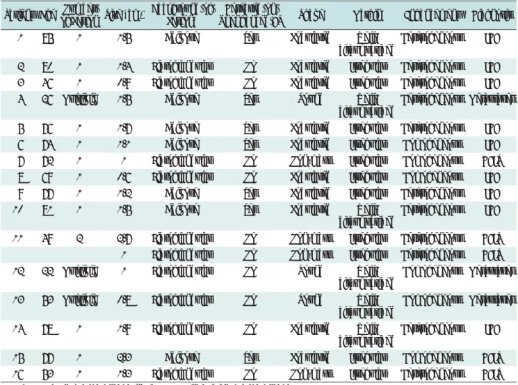
Table 1. Pathologic Outcomes and Imaging Appearance of 16 Patients with Malignant Incidental Enhancing Breast Lesions on Chest CT
Patient Age Number
of Lesion Size
§(cm) Background of Lesion
§Detected on
Unenhanced CT Shape Margin Enhancement Diagnosis
1 85 1 1.5 Adipose Yes Lobulated Well-
circumscribed
Heterogeneous IDC
2 80 1 1.4 Fibroglandular No Lobulated Irregular Heterogeneous IDC
3 46 1 0.9 Fibroglandular No Lobulated Irregular Heterogeneous IDC
4 26 Multiple 1.5 Adipose Yes Round Well-
circumscribed
Heterogeneous Metastases
5 76 1 1.7 Adipose Yes Lobulated Irregular Heterogeneous IDC
6 74 1 1.1 Adipose Yes Lobulated Irregular Homogeneous IDC
7 72 1 1 Fibroglandular No Non-mass Irregular Homogeneous DCIS
8 69 1 0.6 Fibroglandular No Lobulated Irregular Homogeneous IDC
9 77 1 1.2 Adipose Yes Lobulated Irregular Heterogeneous IDC
10 81 1 1.5 Adipose Yes Lobulated Well-
circumscribed
Heterogeneous IDC
11 49 2 2.7 Fibroglandular No Non-mass Irregular Heterogeneous DCIS
1 Fibroglandular No Non-mass Irregular Heterogeneous DCIS
12 22 Multiple 1 Fibroglandular No Round Well-
circumscribed
Homogeneous Metastases
13 53 Multiple 0.8 Fibroglandular No Round Well-
circumscribed
Homogeneous Metastases
14 78 1 1.9 Fibroglandular No Lobulated Well-
circumscribed
Heterogeneous IDC
15 77 1 2.3 Adipose Yes Lobulated Irregular Homogeneous DCIS
16 53 1 1.3 Fibroglandular No Non-mass Irregular Heterogeneous DCIS
Note.— DCIS = ductal carcinoma in situ, IDC = invasive ductal carcinoma.
§ In case of multiple lesions, only size or background of largest lesion was recorded.
(Table 2). Of the four patients with fi broadenomas, two had multiple lesions in bilateral breasts. Four of the seven patients underwent a sonographically-guided core-needle biopsy to confi rm the diagnosis. In the remaining three patients, a surgical incision and drainage was performed for one breast abscess. A comparison with prior chest CT at an interval of more than 12 months showed stability of the breast lesions in two patients with fi broadenoma and surgical scar, respectively.
The mean size of the malignant enhancing breast tumors was 1.4 ± 0.54 cm (range, 0.6-2.7 cm), while the mean size of the benign enhancing breast lesions was 1.5 ± 1.0 cm (range 0.5-3.4 cm). There was no signifi cant difference between the mean size of malignant and benign breast lesions (p = 0.68). All of the nine incidental IDCs were less than 2.0 cm (mean size, 1.3 ± 0.13 cm; range, 0.6-1.9 cm), and at stage T1, which is based on the TNM staging system for breast cancers.
The mean age of patients with incidental malignant enhancing breast tumors was 63.6 ± 19.7 years, whereas the mean age of patients with incidental benign enhancing breast lesions was 49.1 ± 15.9 years. There was no
signifi cant difference between the mean age of patients with malignant breast tumors and those with benign breast lesions (p = 0.06). However, the mean age of patients with incidental primary breast cancer (70.5 ± 12.8 years) was signifi cantly higher than that of patients with incidental benign breast lesions (p = 0.002).
Imaging Appearance of Incidental Enhancing Breast Lesions
With the unenhanced CT of chest, breast lesions could be detected in 13 (57%) of the 23 patients, which included seven (44%) of 16 malignant cases and six (86%) of seven benign cases. All of the lesions detected on unenhanced CT were enhanced on contrast-enhanced CT.
A total of 10 patients had incidental breast lesions exclusively detected on contrast-enhanced CT, and nine (90%) of the 10 patients had malignant breast lesions.
The backgrounds of incidental enhancing breast lesions were adipose tissues in 11 patients, and fi broglandular tissues in the remaining 12 patients (Figs. 1, 2). For the 13 patients with incidental breast lesions detected on unenhanced CT, only two (15%) had breast lesions with fi broglandular backgrounds; however, all (100%) of the 10 patients with incidental breast lesions that were exclusively detected on contrast-enhanced CT had breast lesions of fi broglandular backgrounds. In those patients with incidental breast lesions exclusively detected on contrast- enhanced CT, a signifi cantly high percentage of patients had incidental breast lesions of fi broglandular backgrounds (p < 0.001).
Of the 16 patients with malignant breast tumors,
heterogeneous enhancement of tumors were observed in 10 (63%) patients, and homogeneous enhancement was noted in the remaining six (38%). Of the seven patients with benign breast lesions, three (43%) showed heterogeneous enhancement and four (57%) showed homogeneous Table 2. Pathologic Outcomes and Imaging Appearance of Seven Patients with Benign Incidental Enhancing Breast Lesions on Chest CT
Patient Age Number of Lesion
Size
§(cm)
Background of Lesion
§Detected on
Unenhanced CT Shape Margin Enhancement Diagnosis
1 66 1 0.5 Fibroglandular No Lobulated Irregular Homogeneous Fibroadenoma
2 23 Multiple 2.4 Adipose Yes Lobulated Well-
circumscribed
Heterogeneous Fibroadenoma
3 62 Multiple 1.2 Fibroglandular Yes Lobulated Well-
circumscribed
Homogeneous Fibroadenoma
4 60 1 1 Adipose Yes Irregular Spiculated Homogeneous Surgical scar
5 33 1 3.4 Fibroglandular Yes Lobulated Well-
circumscribed
Heterogeneous Abscess of breast
6 50 1 0.8 Adipose Yes Lobulated Well-
circumscribed
Heterogeneous Lymphoid hyperplasia
7 50 1 1.5 Adipose Yes Ovoid Well-
circumscribed
Homogeneous Fibroadenoma
Note.— § In case of multiple lesions, only size or background of largest lesion was recorded.enhancement. There was no signifi cant difference in the percentage of heterogeneous or homogeneous enhancement between the malignant and benign incidental enhancing breast lesions (p = 0.65).
Incidental enhancing breast lesions with a lobulated shape were noted in 15 patients, in whom 10 (67%) had malignant breast lesions, and fi ve (33%) had benign breast lesions (Tables 1, 2). A total of four incidental enhancing breast lesions were categorized as non-mass breast lesions, and all of them were subsequently confi rmed to be malignant breast tumors (Fig. 1) (Table 1).
Irregular or spiculated margins were observed in 11 (79%) of the 14 incidental enhancing primary malignant breast tumors, and two (29%) of seven benign breast lesions (Tables 1, 2). Incidental primary breast cancer showed an insignifi cant trend of a higher percentage of irregular or spiculated margin (p = 0.056) (Fig. 2). Three patients of breast metastases showed multiple breast lesions that were round in shape and had a well-circumscribed margin.
Well circumscribed enhancing breast lesions were noted in fi ve (71%) of seven patients with benign breast lesions. The incidental enhancing benign breast lesion with a spiculated margin was proven to be a surgical scar from a past history of resection for fi broadenoma of the breast, which was obtained and the long-term stability of this lesion was confi rmed at follow-up CT. The benign breast lesion with an irregular margin was proven to be a 0.5-cm fi broadenoma by a sonographically-guided core-needle biopsy.
DISCUSSION
Although a routine chest CT was usually performed for indications other than breast disease, and the technology was not optimized for breast imaging, the incidental breast lesions could be occasionally detected. In some cases, incidental breast cancer could be detected on chest CT.
Swensen et al. (9) reported three (0.4%) cases of breast cancer from 735 women who underwent unenhanced low- dose chest CT for lung cancer screening. Shojaku et al.
(3) reported four (0.4%) cases of incidental breast cancer or metastatic breast carcinoma from 1008 patients who underwent unenhanced chest CT. In a recent study, eight (2%) incidental primary breast cancers were detected in 432 chest CT scans (6). We included only incidental enhancing breast lesions in our study, which amounted to 16 (0.7%) patients of 2250 who underwent routine contrast-enhanced chest CT.
A few researches have evaluated the pathologic outcomes of incidental breast lesions and determined the CT features of incidental breast lesions suggestive of malignancy (6-8).
In these studies, 24-32% of incidental breast lesions were subsequently proven to be malignant (6-8). Our study showed a much higher rate (70%) of malignancy. This higher result may be partly due to a selection bias from the retrospective study design, and the small number of cases in our study. However, it should be noted that we included only enhancing breast lesions in our study, and therefore some incidental breast lesions which were not enhanced on chest CT may not be included. Porter et al. (8) reported that 17 (50%) normal breast glandular “pseudo-masses”
and simple cysts, which may not be enhanced on contrast- enhanced CT, in a total of 34 incidental breast lesions.
By excluding the non-enhancing benign incidental breast lesions, the rate of malignant incidental breast lesions likely causes an increase in the rate of malignancy.
The imaging protocol of routine chest CT may be a reason for the relatively small number of benign incidental breast lesions in our series. The routine contrast-
enhanced chest CT was performed at 50 seconds after intravenous administration of contrast medium, and the entire scanning would be completed within two minutes after administration of the contrast medium. This period corresponded to the early arterial phase of a dynamic breast CT. Most malignant breast lesions had strong enhancement in the early arterial phase, and benign breast lesions were well-enhanced in late phase of the dynamic CT study (10, 11). As a result, breast malignancy may be incidentally detected at routine chest CT by the early enhancement of tumor, and some benign breast lesions may not be detected due to lack of suffi cient enhancement in the early phase.
By using dynamic CT of breast, the reported signs of malignancy included early enhancement of breast tumors and washout or plateau patterns of the time-density curve for three to eight minutes after injection of the contrast medium (10, 11). In our study, the incidental breast lesions were detected by routine contrast-enhanced chest CT. The contrast-enhanced study was performed at 50 seconds after the injection of contrast medium, and not a dynamic study.
Therefore, the analysis of the enhancement patterns at dynamic breast CT study could not apply to a routine chest CT.
The reported CT features suggestive of malignant
incidental breast lesions on routine chest CT included rim
enhancement of breast tumors, an irregular or spiculated
margin, and an axillary lymphadenopathy (5, 7, 8). In our study, the incidental primary breast cancer showed a higher probability of an irregular margin, with an insignifi cant trend. However, we have observed that incidental IDC may also demonstrate well-circumscribed margins. Therefore, it would seem reasonable to suggest that well-circumscribed incidental breast lesions should be referred for dedicated assessment, as recommended by previous authors (2-5, 8). We also noted that the age of patients with incidental primary breast cancer was higher than that of patients with incidental benign breast lesions. This may be consistent with the fact that risk of breast cancer rises with increased age.
Incidental malignant and benign breast lesions may share overlapping enhancement characteristics in our study. Heterogeneous or homogeneous enhancement was not a reliable sign to distinguish malignant from benign incidental breast lesions on chest CT. The size of incidental breast lesions was also not a reliable feature to distinguish malignancy from benignity. There was no case demonstrating axillary lymphadenopathy in our study;
however, this may be due to small number of cases or early stage of disease. All of the nine incidentally detected IDC in our study were at the T1 stage of breast cancer.
Some incidental breast lesions were exclusively detected on contrast-enhanced CT in our study. The adipose or fi broglandular backgrounds of breast lesions may be one reason for neglected breast lesions on unenhanced CT. Also, all breast lesions not detected on unenhanced CT were of fi broglandular background. In these cases, there was not enough of a difference in the attenuations between a breast lesion and adjacent fi broglandular tissue; therefore, the breast lesions could not be detected on unenhanced CT.
Three patients with four incidental breast lesions showed an interesting imaging fi nding of non mass-like enhancement. All of the four lesions were subsequently proven to be malignant. The description “non mass-like enhancement” had been used in the interpretation of breast magnetic resonance (MR) imaging, which meant that enhancement occurred in an area of fi broglandular tissue that otherwise appeared as normal on unenhanced images (12). Non mass-like enhancement on breast MR imaging may represent DCIS, lobular cancer, mastopathic changes (focal adenosis), hormonal stimulation, or infl ammatory changes (12).
Three patients with a known history of non-mammary malignancy showed multiple, well-circumscribed breast
tumors. These tumors were confi rmed as breast metastases.
Although not common, the possibility of breast metastases should be considered in patients with multiple well- circumscribed breast nodules and non-mammary malignancy.
There were limitations in our study; fi rst, the small number of patients making up the study population and retrospective nature of the study design may reduce the power of this study. A routine chest CT is less sensitive than dedicated breast CT or MR imaging in detecting breast lesions (10, 12). Second, it would have been preferable if all the patients had histopathologic diagnoses.
However, the 12-month follow-up studies in two patients with benign breast lesions were probably suffi cient for diagnosis of benignity. Widespread metastases occurred in one patient with breast metastases; therefore, only the breast sonography was performed, while the further histopathologic examination was reasonably not performed.
In conclusion, it is important for general radiologists to pay attention to the breasts on CT performed for other thoracic indications. Routine contrast-enhanced chest CT can reveal suffi cient details to allow the detection of unsuspected breast lesions, in which some cases may be subsequently proven to be malignant. A low threshold for referral of patients with routine chest CT-detected incidental enhancing breast lesions to a breast clinic for assessment is advocated. Irregular margins of incidental enhancing breast lesion can be considered a suggestive sign of a malignant lesion.
REFERENCES
1. Lindfors KK, Boone JM, Nelson TR, Yang K, Kwan AL, Miller DF. Dedicated breast CT: initial clinical experience. Radiology 2008;246:725-733
2. Kim SM, Park JM. Computed tomography of the breast.
Abnormal fi ndings with mammographic and sonographic correlation. J Comput Assist Tomogr 2003;27:761-770 3. Shojaku H, Seto H, Iwai H, Kitazawa S, Fukushima W, Saito K.
Detection of incidental breast tumors by noncontrast spiral computed tomography of the chest. Radiat Med 2008;26:362- 367
4. Yi JG, Kim SJ, Marom EM, Park JH, Jung SI, Lee MW. Chest CT of incidental breast lesions. J Thorac Imaging 2008;23:148- 155
5. Harish MG, Konda SD, MacMahon H, Newstead GM. Breast lesions incidentally detected with CT: what the general radiologist needs to know. Radiographics 2007;27:S37-S51 6. Hussain A, Gordon-Dixon A, Almusawy H, Sinha P, Desai
A. The incidence and outcome of incidental breast lesions detected by computed tomography. Ann R Coll Surg Engl
2010;92:124-126
7. Moyle P, Sonoda L, Britton P, Sinnatamby R. Incidental breast lesions detected on CT: what is their signifi cance? Br J Radiol 2010;83:233-240
8. Porter G, Steel J, Paisley K, Watkins R, Holgate C. Incidental breast masses detected by computed tomography: are any imaging features predictive of malignancy? Clin Radiol 2009;64:529-533
9. Swensen SJ, Jett JR, Hartman TE, Midthun DE, Sloan JA, Sykes AM, et al. Lung cancer screening with CT: Mayo Clinic experience. Radiology 2003;226:756-761
10. Miyake K, Hayakawa K, Nishino M, Nakamura Y, Morimoto T,
Urata Y, et al. Benign or malignant?: differentiating breast lesions with computed tomography attenuation values on dynamic computed tomography mammography. J Comput Assist Tomogr 2005;29:772-779
11. Inoue M, Sano T, Watai R, Ashikaga R, Ueda K, Watatani M, et al. Dynamic multidetector CT of breast tumors: diagnostic features and comparison with conventional techniques. AJR Am J Roentgenol 2003;181:679-686
12. Kuhl C. The current status of breast MR imaging. Part I. Choice of technique, image interpretation, diagnostic accuracy, and transfer to clinical practice. Radiology 2007;244:356-378