© 2014 The Korean Ophthalmological Society
This is an Open Access article distributed under the terms of the Creative Commons Attribution Non-Commercial License (http://creativecommons.org/licenses /by-nc/3.0/) which permits unrestricted non-commercial use, distribution, and reproduction in any medium, provided the original work is properly cited.
Original Article
Comparison of Injection of Intravitreal Drugs with Standard Care in Macular Edema Secondary to Branch Retinal Vein Occlusion
Kyungmin Lee, Heeyoung Jung, Joonhong Sohn
HanGil Eye Hospital, Incheon, Korea
Purpose: To compare the long-term efficacy and safety of intravitreal triamcinolon with or without rescue laser therapy (intravitreal triamcinolone injection [IVTA] group), bevacizumab with or without rescue laser treatment (intravitreal bevacizumab injection [IVB] group), or a combination of both with or without rescue laser therapy (IVTA + IVB group), with standard care for patients with macular edema secondary to branch retinal vein oc- clusion (BRVO).
Methods: We reviewed the medical records of 151 patients treated with intravitreal injection with or without res- cue laser for treatment of macular edema caused by BRVO, and who were followed up at 1, 3, 6, 12, and 24 months. During the observation period, rescue grid laser or repeated intravitreal injection with initial drug was performed if recurrence of macular edema was confirmed. Visual acuity, change in visual acuity, and intraocu- lar pressure were compared in each phase.
Results: Totals of 16%, 5.6%, and 0% of participants in the three groups showed significant visual loss of more than three lines of the Snellen chart at last follow-up. The IVTA group was the least effective treatment modali- ty, with statistical significance. The development rates of elevated intraocular pressure were similar among the groups.
Conclusions: Although IVTA yielded effects similar to those of standard grid photocoagulation based on the Standard Care vs Corticosteroid for Retinal Vein Occlusion study, IVB or IVTA + IVB with or without rescue laser treatment resulted in improvement in visual acuity at 24 months after the start of treatment and was as- sociated with few serious adverse side effects. Thus, these approaches could be useful for treating macular edema arising secondary to BRVO.
Key Words: Intravitreal injection, Macular edema, Retinal vein occlusion
Branch retinal vein occlusion (BRVO) is the second most common major retinal vascular disease after diabetic reti-
nopathy [1]. Epidemiologic studies found that the prevalence of BRVO ranged from 0.3% [2] to 1.6% [3], and major risk factors for the condition were increasing age, hypertension, and co-existing cardiovascular disease [2-6].
Macular edema is a major cause of visual acuity loss at- tributable to BRVO [7-9]. The Branch Vein Occlusion Study [7] found that grid photocoagulation was effective for treat- ment of macular edema. The Standard Care vs Corticosteroid for Retinal Vein Occlusion (SCORE) project showed that grid photocoagulation was the best option for standard care
Received: May 11, 2012 Accepted: April 16, 2013
Corresponding Author: Joonhong Sohn, MD, PhD. HanGil Eye Hosiptal,
#35 Bupyeong-daero, Bupyeong-gu, Incheon 403-722, Korea. Tel: 82-32- 503-3322, Fax: 82-32-504-3322, E-mail: [email protected] This article was presented as free paper on 11th EURETINA Congress at London in May 2011.
when used to treat macular edema secondary to BRVO [10].
In recent decades, several novel treatments for BRVO have been introduced in major studies such as the BRAVO [11] and GENOVA studies [12]. However, the effects of such treatments remain unclear [1,9,13]. Furthermore, the cited works did not mimic the true clinical situation in that in- travitreal bevacizumab injections (IVBs) were consecu- tively administered over a 3-month period to form a load- ing dose, the treatment strategy to be adopted after initial treatment failure was not considered, and lastly, no study has compared intravitreal triamcinolone acetate, bevaci- zumab, and drug combination injections for the treatment of macular edema secondary to BRVO.
In the present study, we reviewed the medical records of patients treated for visual loss caused by macular edema secondary to BRVO 1) to evaluate whether intravitreal tri- amcinolone acetate, bevacizumab, and a combination of tri- amcinolone and bevacizumab injections, with or without rescue laser therapy, improved visual acuity compared with standard care of the type administered in the SCORE study, and 2) to determine whether such treatment modalities re- duced unfavorable outcomes in patients with macular edema caused by BRVO, compared with those of the SCORE study.
Materials and Methods
All patients were evaluated and treated by three experi- enced retinal specialists. Diagnosis of BRVO was based on clinical examination, and macular edema was identified by optical coherence tomography (OCT). The possible treat- ment options for BRVO associated with macular edema were explained to potential study candidates in accordance
with the ethical standards laid down in the 1964 Declara- tion of Helsinki. No institutional review board approval was required because of the retrospective study design.
The treatment options were suggested included intravitreal triamcinolone acetonide injection, bevacizumab injection, and injection of a combination of triamcinolone acetonide and bevacizumab by doctor’s preference. The “off-label”
use of bevacizumab and the potential side-effects, particu- larly the possibility of development of thromboembolic events and uveitis, were extensively discussed with all pa- tients prior to treatment. Any patient with a recent history of myocardial infarction or cerebral vascular accident, and/
or uncontrolled hypertension, which were detected by questionnaires, electrocardiogram, and sphygmomanome- ter, was not offered bevacizumab. Patients were specifical- ly informed about the process of intravitreal injection and the potential risks of endophthalmitis, uveitis, cataract de- velopment, ocular hypertension, and retinal detachment.
This consecutive, retrospective, nonrandomized clinical interventional study included patients from January 2000 through December 2009 at HanGil Eye Hospital, Incheon, Korea. A total of 1,028 patients received one of the various interventions mentioned above, but only 151 patients were enrolled in our study after application of the inclusion and exclusion criteria (Table 1). The injection was performed at least 1 month after symptoms appeared. However, when patients were unsure about the initiation of symptoms, in- jection was performed as soon as the macular edema was detected. The regimens were determined by the treatment trend of macular edema associated with BRVO at that time, surgeon’s preference, and the patient’s status, such as economics, systemic health status, and ocular status.
Thirty-one patients (31 eyes) were selected without ran-
Table 1. Inclusion and exclusion criteria used in the present study Inclusion criteria Exclusion criteria
Best corrected visual acuity in Snellen chart ≤20 / 40, or ≥20 / 400
Center-involved macular edema secondary to BRVO*present on clinical examination Media clarity, papillary dilation, and subject cooperation sufficient for adequate fundus
photographs
Macular edema due to a cause other than BRVO*
An ocular condition such that visual acuity would not improve from resolution of the edema Substantial cataract estimated to have reduced visual acuity by ≥3 lines
Prior treatment with intravitreal corticosteroid at any time History of focal/grid macular photocoagulation
Prior pars plana vitrectomy
Major ocular surgery including cataract extraction within the prior 6 months
Yttrium aluminum garnet capsulotomy performed within 2 months prior to the BRVO* event IOP† ≥25 mmHg, open-angle glaucoma, or steroid-induced IOP† elevation that required IOP†- lowering treatment or pseudoexfoliation
Aphakia
*BRVO = branch retinal vein occlusion; †IOP = intraocular pressure.
domization and placed in the intravitreal triamcinolone in- jection (IVTA) group; each received a single intravitreal in- jection of 4 mg (0.1 mL) crystalline triamcinolone acetonide (Kenacort-A 40 mg/mL; Bristol-Myers Squibb, Singapore).
Another 95 patients (95 eyes), who received a single intravit- real injection of 2.5 mg (0.1 mL) bevacizumab (Avastin 25 mg/mL; Genentech, San Francisco, CA, USA) were placed in the IVB group. The remaining 25 patients (25 eyes), who received a combination of 1.25 mg (0.05 mL) bevacizumab and 2 mg (0.05 mL) triamcinolone acetonide, were placed in the IVTA + IVB group. In the combination group, an addi- tional anterior chamber paracentesis was performed with 20 gauge microincisional blade after the intravitreal injection in order to prevent intraocular pressure increase. During fol- low-up, rescue grid laser or repeated intravitreal injection with initial drug was performed if recurrence of macular edema was confirmed by fundus examination or OCT.
Each patient underwent a complete ophthalmological ex- amination including standardized visual acuity measure- ments using Snellen charts, slit-lamp biomicroscopy, Gold- mann applanation tonometry, and ophthalmoscopy, at baseline and at 1, 3, 6, 12, and 24 months after treatment.
At baseline we performed fluorescein angiography to iden- tify macular ischemia by detecting the enlargement of fo- veal avascular zone. There were no patients with baseline macular ischemia. Visual acuity, change in visual acuity, and intraocular pressure were compared in each treatment phase. The change in visual acuity was divided into 5 groups associated with the amount of change in visual acu- ity: visual acuity increased more than 3 lines in Snellen chart, increased 1 to 3 lines, did not change, decreased 1 to 3 lines, or decreased more than 3 lines.
Potential corticosteroid-induced and injection-related complications, if any, were also recorded. No patient had baseline ocular hypertension. Topical anti-glaucomatous medication was prescribed if intraocular pressure was more than 21 mmHg at any follow-up visit. Information on medical history (e.g., hypertension or diabetes) was ob- tained by chart review. All visual acuity measurements were converted to the logarithm of the minimum angle of resolution scale prior to statistical analysis.
All data are expressed as mean ± standard deviations.
The Kruskal-Willis test was used to perform statistical comparisons among the three groups. Analyses were achieved using SPSS ver. 14.1 (SPSS Inc., Chicago, IL, USA). A p-value at 1 month, 3 months, 6 months, 12
months, and 24 months are expressed as p1mon, p3mon, p6mon, p12mon, and p24mon. A p-value less than 0.05 was considered statistically significant.
Results
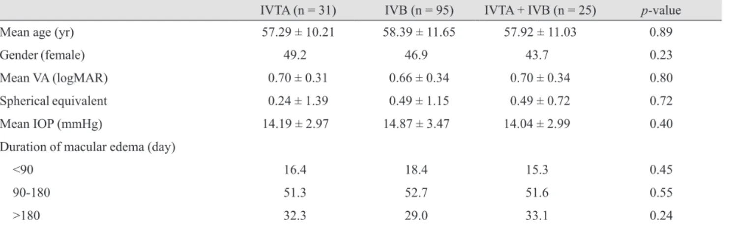
A total of 151 eyes of 151 patients with BRVO associat- ed with macular edema were included in the present study in which patients were divided into three groups based on intravitreal injection regimen. Mean patient age was 57 ± 10 years in the IVTA group, 58 ± 11 years in the IVB group, and 58 ± 11 years in the IVTA + IVB group, and the proportions of females were 49.2%, 46.9%, and 43.7%, respectively. There were no statistically signifi- cant differences among groups in terms of age, gender, preoperative best spectacle-corrected visual acuity, pre- operative intraocular pressure, or duration of macular ede- ma (p > 0.05 for all comparisons). No patient dropped out of the study and patient demographics are shown in Table 2.
Visual acuity and intraocular pressure
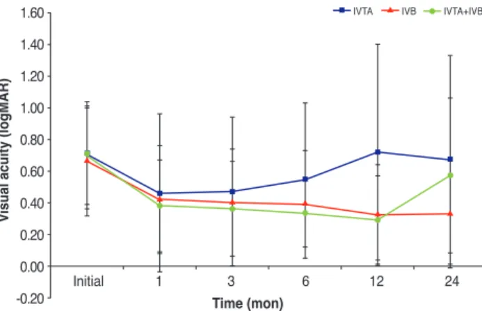
There were no statistically significant differences in postoperative visual acuity among the three groups at 1, 3, 6, or 24 months postoperatively (p > 0.05). However, at the 12-month follow-up, the IVTA group showed a sta- tistically significant decrease in visual acuity (p = 0.01) (Fig. 1). There was no significant among-group differ- ence in postoperative intraocular pressure at any fol- low-up time (p1mon = 0.07, p3mon = 0.07, p6mon = 0.63, p12mon
= 0.97,and p24mon = 0.23) (Fig. 2).
Change in visual acuity
There was no statistically significant difference in change in visual acuity among the three groups at 1, 3, 6, or 24 months postoperatively. However, at the 12-month follow-up, the IVTA group showed less visual improve- ment than did the other groups, with statistical signifi- cance (p = 0.01) (Table 3). The distribution of patients as- sociated with the amount of visual change in each group is schematized in Figs. 3, 4, and 5. Totals of 16%, 5.6%, and 0% of participants in the three groups showed sig- nificant visual loss of more than three lines of the Snellen chart at the final follow-up examination.
Length of follow-up and additional treatment
The average length of follow-up for each group is shown in Table 3. IVTA patients were followed-up for longer than the other two groups, with statistical significance. Totals of 25.81% of patients in the IVTA group, 32.63% in the IVB group, and 36% in the IVTA + IVB group required addi- tional treatment during follow-up. The groups did not dif- fer in the frequency of additional treatment or in the number of patients who were referred for laser treatment (p
> 0.05), but did vary when the times at which such treatment was needed were compared (p = 0.00) (Table 3).
Discussion
Macular edema is a major cause of visual acuity loss in BRVO patients. Thus, several treatment modalities for macular edema have been developed. One representative strategy is grid laser photocoagulation [10] which is the oldest form of treatment for this condition. As the patho- physiology of macular edema became better understood, several useful drugs were developed. Steroids, including triamcinolone acetonide, and anti-vascular endothelial growth factor (VEGF) antibodies (e.g., bevacizumab) are Table 3. Post-injection parameters of the treatment groups
IVTA (n = 31) IVB (n = 95) IVTA + IVB (n = 25) p-value
Terms of FU (mon) 34.61 ± 18.46 10.60 ± 8.58 11.52 ± 8.90 0.00*
Visual change at 1st month FU (logMAR) -0.23 ± 0.52 -0.24 ± 0.33 -0.32 ± 0.22 0.57 Visual change at 3rd month FU (logMAR) -0.22 ± 0.54 -0.26 ± 0.35 -0.35 ± 0.27 0.47 Visual change at 6th month FU (logMAR) -0.14 ± 0.52 -0.25 ± 0.36 -0.33 ± 0.21 0.30 Visual change at 12th month FU (logMAR) -0.04 ± 0.71 -0.33 ± 0.36 -0.40 ± 0.27 0.01* Visual change at 24th month FU (logMAR) -0.35 ± 0.62 -0.30 ± 0.41 -0.31 ± 0.26 0.23 Final visual change (logMAR) -1.37 ± 0.54 -0.34 ± 0.35 -0.40 ± 0.30 0.06 Visual acuity at last FU (logMAR) 0.53 ± 0.59 0.33 ± 0.34 0.31 ± 0.31 0.04* No. of patients who needed additional treatment (%) 8 (25.81) 31 (32.63) 9 (36) 0.08 No. of patients who got laser treatment (%) 3 (9.68) 5 (5.26) 2 (8) 0.67 No. of additional treatments 1.29 ± 5.29 1.44 ± 7.68 1.56 ± 8.70 0.39
Terms between treatments (mon) 12.63 ± 8.52 3.48 ± 2.24 6.22 ± 3.50 0.01*
IVTA = intravitreal triamcinolone injection; IVB = intravitreal bevacizumab injection; FU = follow up; logMAR = logarithm of the min- imum angle of resolution.
*Statistically significant by Kruskal-Willis test.
Table 2. Baseline characteristics of the treatment groups
IVTA (n = 31) IVB (n = 95) IVTA + IVB (n = 25) p-value
Mean age (yr) 57.29 ± 10.21 58.39 ± 11.65 57.92 ± 11.03 0.89
Gender (female) 49.2 46.9 43.7 0.23
Mean VA (logMAR) 0.70 ± 0.31 0.66 ± 0.34 0.70 ± 0.34 0.80
Spherical equivalent 0.24 ± 1.39 0.49 ± 1.15 0.49 ± 0.72 0.72
Mean IOP (mmHg) 14.19 ± 2.97 14.87 ± 3.47 14.04 ± 2.99 0.40 Duration of macular edema (day)
<90 16.4 18.4 15.3 0.45
90-180 51.3 52.7 51.6 0.55
>180 32.3 29.0 33.1 0.24
Values are presented as mean ± SD or percent.
IVTA = intravitreal triamcinolone injection; IVB = intravitreal bevacizumab injection; VA = visual acuity; logMAR = logarithm of the minimum angle of resolution; IOP = intraocular pressure.
widely used today.
In many studies, IVTA, IVB, and the combination of both treatments have shown beneficial effects in treatment of macular edema secondary to BRVO. Guthoff et al. [14]
compared the intravitreal bevacizumab with triamcino- lone acetonide. They concluded that both treatments are effective in reducing central macular thickness, but IVB is more effective in improving visual acuity for 2 months.
After 3 months, there is no difference in either group.
Ehrlich et al. [15] reported the clinical results of com- bined treatment using intravitreal bevacizumab and in- travitreal triamcinolone in retinal vein occlusion. They
0.00 20.00 40.00 60.00 80.00 100.00
10.34 10.00 12.00 16.67 30.00 16.13
1 3 6 12 24 Last FU (mon)
VA loss more than
3 lines VA loss 1-3 lines No change
VA gain 1-3 lines VA gain more than
3 lines 3.45
24.14
51.72 10.00 30.00
40.00 8.00 24.00
44.00 12.50 16.67 37.50
0.00 25.00
40.00 9.68
10.34 10.00 12.00
16.67 5.00
3.23
32.26
38.71
Fig. 3. Patient distribution in the intravitreal triamcinolone in- jection group with respect to change in visual acuity (VA) differ- ences as measured using a Snellen chart at each follow-up (FU) interval. The levels reflect gain or loss of Snellen chart lines com- pared with baseline VA.
0.00 20.00 40.00 60.00 80.00
100.00 4.35 6.38 8.62 4.76 10.00 5.26
1 3 6 12 24 Last FU (mon)
VA loss more than
3 lines VA loss 1-3 lines No change
VA gain 1-3 lines VA gain more than
3 lines 15.22
31.52
43.48 11.70 32.98
46.81 10.34 24.14
53.45 7.14 16.67
66.67 20.00 20.00
50.00
5.43 2.13 3.45 4.76 0.00 9.471.05
26.32
57.89
Fig. 4. Patient distribution in the intravitreal bevacizumab injec- tion group with respect to change in visual acuity (VA) differ- ences as measured using a Snellen chart at each follow-up (FU) interval. The levels reflect gain or loss of Snellen chart lines com- pared with baseline VA.
0.00 20.00 40.00 60.00 80.00
100.00 0.00 0.00 0.00 0.00 0.00 0.00
1 3 6 12 24 Last FU (mon)
VA loss more than
3 lines VA loss 1-3 lines No change
VA gain 1-3 lines VA gain more than
3 lines 8.00
16.00
72.00 8.00 28.00
64.00 11.76 23.53
64.71 0.00 14.29
78.57 40.00
20.00 20.00
4.00 0.00 0.00 7.14 8.00
20.00 4.00
12.00
76.00
Fig. 5. Patient distribution in the intravitreal triamcinolone injec- tion + intravitreal bevacizumab injection group with respect to change in visual acuity ( VA) differences as measured using a Snellen chart at each follow-up (FU) interval. The levels reflect gain or loss of Snellen chart lines compared with baseline VA.
1.60
Visual acuity (logMAR)
Time (mon) 1.40
1.20 1.00 0.80 0.60 0.40 0.20 0.00
-0.20 Initial 1 3 6 12 24
IVTA IVB IVTA+IVB
Fig. 1. Visual acuity as measured by a Snellen chart at each fol- low-up interval. The three groups showed no statistically signif- icant difference in visual acuity in most of the follow-up interval except for the 12-month follow-up, at which time the intravitreal triamcinolone injection (IVTA) group showed a statistically sig- nificant decrease in visual acuity (graphed on the logarithm of the minimum angle of resolution [logMAR] scale). IVB = intrav- itreal bevacizumab injection.
IVTA IVB IVTA+IVB
20 15 10 5 0
Intraocular pressure (mmHg)
Time (mon)
1 3 6 12 24
Initial
Fig. 2. Intraocular pressure at each follow-up interval. The three groups showed no statistically significant differences in intraocu- lar pressure at any follow-up interval. IVTA = intravitreal triam- cinolone injection; IVB = intravitreal bevacizumab injection.
concluded that combination treatment showed beneficial effects in improving structural outcomes such as central macular thickness, but had no advantage over results with IVB alone. Cekic et al. [16] compared the three different intravitreal treatment modalities, including intravitreal bev- acizumab, intreavitreal triamcinolone, and the combination of both, as in our study. They concluded that all treatment groups had similar therapeutic effects at one month, but at six months, the IVB group had better visual acuity. Howev- er, they only analyzed the short term effects of each treat- ment modality and they did not discuss any visual acuity changes according to treatment used. Of these studies, none were focused on whether a selected treatment might de- crease unfavorable outcomes compared with the standard treatment which the SCORE study [10] defined as focal grid photocoagulation.
In the present study, we compared the effects on visual acuity of different treatment regimens in macular edema patients with BRVO. Each treatment tested was similar in effect until 1 year of follow-up. At that time, triamcinolone acetonide was less effective than was bevacizumab or combination therapy when the extent of macular edema was assessed. This conclusion was in line with the finding that additional treatment was needed at approximately 12 months when IVTA was employed. Final visual acuity val- ues were significantly lower in the IVTA group than in the other patients, though this difference may be associated with the fact that the IVTA group had a longer period of follow-up. Thus, a negative selection bias may have been present with respect to IVTA-treated patients.
Patients receiving each of the three treatments required similar numbers of additional injections after the first treatment. Thus, the percentages of patients who received additional treatments, such as another injection or rescue laser therapy, were similar among the three groups. This finding indicates that an initial intravitreal injection did not greatly affect the natural course of disease.
To determine if any chosen treatment decreased unfa- vorable outcomes, we compared the groups in terms of im- provement or deterioration in visual acuity, as was per- formed in the SCORE study [10]. In the cited work, scoring increments were set by each five letters of the Early Treat- ment Diabetic Retinopathy Study (ETDRS) chart, equiva- lent to a single line of difference in a Snellen chart. The data are shown in Figs. 3, 4, and 5. In the SCORE study, 14.9% of patients in the standard care group, 11.6% in the 1
mg IVTA group, and 12% in the 4 mg IVTA group showed decreases in visual acuity of more than three lines of the Snellen chart. In the present study, 16.67% of patients in the IVTA group, 4.76% in the IVB group, and 0% in the IVTA + IVB group showed deterioration in visual acuity of more than three lines at the 12-month follow-up. The re- sults of the IVTA group were similar to those of the SCORE study [10], indicating that additional drug or res- cue laser treatment had less effect on visual improvement in this group. In the IVB and IVTA + IVB groups, howev- er, visual loss was much less than that of the standard group in the SCORE study [10], with statistical signifi- cance. This difference may reflect a valuable effect of bev- acizumab or the fact that rescue laser treatment worked well in both groups. However, as the final visual acuity values at 24 months of follow-up did not differ significant- ly among the three groups, bevacizumab appeared to max- imally influence the other treatment such as triamcinolone acetonide or rescue laser therapy to work well, but not change the natural course of the disease.
The present study had some limitations. First, we did not include a standard care group, but rather obtained such data from the SCORE study [10]. The second limitation is that the number of patients receiving 24-month follow-ups differed among the groups, with more patients available in the IVB group than in the other two groups. This imbal- ance may have biased our interpretation of the results. Re- cently, bevacizumab has become a drug of choice for treat- ment of several retinal diseases and many clinicians choose bevacizumab rather than triamcinolone acetonide because the risks of development of cataracts or glaucoma are low- er when the former drug is employed. The third limitation is that OCT data were not compared among groups be- cause, during the follow-up period, OCT techniques evolved rapidly and we switched from use of time domain (TD)-OCT to spectral domain (SD)-OCT. As there is no reliable method to convert TD-OCT data to SD-OCT re- sults, data comparison would be meaningless.
Much effort has been devoted to seeking a cure for mac- ular edema associated with BRVO because the condition is commonly associated with deterioration in visual acuity.
One advance in treatment has been the development of an- ti-VEGF agents, which increase vascular permeability and promote absorption of edema. We compared the effects of triamcinolone acetonide, bevacizumab, and a combination of the two drugs, injected into the vitreous cavity, with or
without additional drug treatment or rescue laser therapy, in macular edema patients with BRVO. Although IVTA yielded outcomes similar to those of standard grid photo- coagulation in the SCORE study, intravitreal bevacizumab only, or a drug combination with or without rescue laser therapy, resulted in significantly lower visual loss in the present work. Therefore, these two treatment modalities could be used as a substitute for grid laser photocoagula- tion. However, further clinical studies are warranted to compare the benefits of and tolerance to drug mixing, and application of laser therapy, as well as to gauge the effec- tiveness of treatment with respect to the size of the foveal avascular zone. Future work with a larger study population and longer follow-up is needed to achieve definite results.
Conflict of Interest
No potential conflict of interest relevant to this article was reported.
References
1. Hayreh SS. Prevalent misconceptions about acute retinal vas- cular occlusive disorders. Prog Retin Eye Res 2005;24:493-519.
2. Wong TY, Larsen EK, Klein R, et al. Cardiovascular risk factors for retinal vein occlusion and arteriolar emboli: the Atherosclerosis Risk in Communities & Cardiovascular Health studies. Ophthalmology 2005;112:540-7.
3. Mitchell P, Smith W, Chang A. Prevalence and associations of retinal vein occlusion in Australia: the Blue Mountains Eye Study. Arch Ophthalmol 1996;114:1243-7.
4. Klein R, Klein BE, Moss SE, Meuer SM. The epidemiolo- gy of retinal vein occlusion: the Beaver Dam Eye Study.
Trans Am Ophthalmol Soc 2000;98:133-41.
5. Klein R, Moss SE, Meuer SM, Klein BE. The 15-year cu- mulative incidence of retinal vein occlusion: the Beaver Dam Eye Study. Arch Ophthalmol 2008;126:513-8.
6. Cugati S, Wang JJ, Rochtchina E, Mitchell P. Ten-year inci-
dence of retinal vein occlusion in an older population: the Blue Mountains Eye Study. Arch Ophthalmol 2006;124:726-32.
7. Argon laser photocoagulation for macular edema in branch vein occlusion. The Branch Vein Occlusion Study Group.
Am J Ophthalmol 1984;98:271-82.
8. Chang MA, Fine HF, Bass E, et al. Patients’ preferences in choos- ing therapy for retinal vein occlusions. Retina 2007;27:789-97.
9. McIntosh RL, Mohamed Q, Saw SM, Wong TY. Interven- tions for branch retinal vein occlusion: an evidence-based systematic review. Ophthalmology 2007;114:835-54.
10. Scott IU, Ip MS, VanVeldhuisen PC, et al. A randomized trial comparing the efficacy and safety of intravitreal tri- amcinolone with standard care to treat vision loss associat- ed with macular Edema secondary to branch retinal vein occlusion: the Standard Care vs Corticosteroid for Retinal Vein Occlusion (SCORE) study report 6. Arch Ophthalmol 2009;127:1115-28.
11. Campochiaro PA, Heier JS, Feiner L, et al. Ranibizumab for macular edema following branch retinal vein occlusion:
six-month primary end point results of a phase III study.
Ophthalmology 2010;117:1102-12.e1.
12. Haller JA, Bandello F, Belfort R Jr, et al. Dexamethasone intravitreal implant in patients with macular edema related to branch or central retinal vein occlusion twelve-month study results. Ophthalmology 2011;118:2453-60.
13. Rogers SL, McIntosh RL, Lim L, et al. Natural history of branch retinal vein occlusion: an evidence-based systematic review. Ophthalmology 2010;117:1094-101.e5.
14. Guthoff R, Meigen T, Hennemann K, Schrader W. Compari- son of bevacizumab and triamcinolone for treatment of mac- ular edema secondary to branch retinal vein occlusion in a pair-matched analysis. Ophthalmologica 2010;224:319-24.
15. Ehrlich R, Ciulla TA, Moss AM, Harris A. Combined treatment of intravitreal bevacizumab and intravitreal triamcinolone in patients with retinal vein occlusion: 6 months of follow-up.
Graefes Arch Clin Exp Ophthalmol 2010;248:375-80.
16. Cekic O, Cakir M, Yazici AT, et al. A comparison of three dif- ferent intravitreal treatment modalities of macular edema due to branch retinal vein occlusion. Curr Eye Res 2010;35:925-9.