Ⅰ.
서 론
악안면 영역의 다양한 조직 결손부를 회복하기 위한 방 법으로 자가조직 이식술이 선호되어 왔으나 공여부 채취 를 위한 2차적인 수술이 필요하고, 이로 인해 환자에게 신 체적, 경제적 고통을 가중시키는 단점이 있어 최근에는 조 직공학을 이용하여 결손된 조직을 회복하려는 연구들이 김 종 렬
626-770 경상남도 양산시 물금읍 범어리
부산대학교 치의학전문대학원 구강악안면외과학교실 Jong-Ryoul Kim
Department of Oral and Maxillofacial Surgery, School of Dentistry, Pusan National University
Beomeo-ri, Mulgeum-eup, Yangsan, Gyeongsangnam, 626-770, Korea TEL: +82-55-360-5103 FAX: +82-2-364-0992
E-mail: [email protected]
미성숙 매복지치의 치낭, 치수, 치근유두 조직에서 다능성 줄기세포의 분리와 특성화에 대한 연구
송정호1∙박봉욱2∙변준호2∙강은주3∙노규진3∙신상훈1∙김욱규1∙김종렬1
1부산대학교 치의학전문대학원 구강악안면외과학교실,
경상대학교2의학전문대학원 치과학교실 구강악안면외과, 건강과학연구원, 3수의과대학 수의산과학
Isolation and characterization of human dental tissue-derived stem cells in the impacted wisdom teeth: comparison of dental follicle, dental pulp, and root apical papilla-derived cells
Jung-Ho Song1, Bong-Wook Park2, June-Ho Byun2, Eun-Ju Kang3, Gyu-Jin Rho3, Sang-Hun Shin1, Uk-Kyu Kim1, Jong-Ryoul Kim1
1Department of Oral and Maxillofacial Surgery, School of Dentistry, Pusan National University, Yangsan, Korea
2Department Oral and Maxillofacial Surgery, School of Medicine and Institute of Health Science, 3College of Veterinary Medicine, Gyeongsang National University, Jinju, Korea
Introduction:The first aim of this study was to isolate the dental tissue-derived stem cells from the dental follicle (DF), dental pulp (DP), and root apical papilla (RAP) of the extracted wisdom teeth. Second was to evaluate their characterization with the expressions of transcription factors and cell surface markers. Finally, their ability of the in vitro multi-lineage differentiations into osteogenic and adipogenic cells were compared, respectively.
Materials and Methods:Dental tissues, including dental follicle, dental pulp, and root apical papilla, were separated in the extracted wisdom teeth.
These three dental tissues were cultured in Dulbecco’s modified Eagle’s medium (DMEM) with supplements, respectively. After passage 3, the homogeneous shaped dental tissue-derived cells were analyzed the expression of transcription factors (Oct-4, Nanog and Sox-2) and cell surface mark- ers (CD44, CD90 and CD105) with reverse transcription polymerase chain reaction (RT-PCR) and fluorescence-activated cell sorting (FACS) analy- sis. In order to evaluate in vitro multi-lineage differentiations, the culture media were changed to the osteogenic and adipogenic induction mediums when the dental tissue-derived cells reached to passage 3. The characteristics of these three dental tissue-derived cells were compared with immuno- histochemistry.
Results:During primary culture, heterogenous and colony formatted dental tissue-derived cells were observed in the culture plates. After passage 2 or 3, homogenous spindle-like cells were observed in all culture plates. Transcription factors and mesenchymal stem cell markers were positively observed in all three types of dental tissue-derived cells. However, the quantity of expressed transcription factors was most large in RAP-derived cells.
In all three types of dental tissue-derived cells, osteogenic and adipogenic differentiations were observed after treatment of specific induction media.
In vitro adipogenic differentiation was similar among these three types of cells. In vitro osteogenic differentiation was most strongly and frequently observed in the RAP-derived cells, whereas rarely osteogenic differentiation was observed in the DP-derived cells.
Conclusion:These findings suggest that three types of human dental tissue-derived cells from extracted wisdom teeth were multipotent mesenchy- mal stem cells, have the properties of multi-lineage differentiations. Especially, stem cells from root apical papilla (SCAP) have much advantage in osteogenic differentiation, whereas dental follicle cells (DFCs) have a characteristic of easy adipogenic differentiation.
Key words:Human dental tissue-derived stem cells, Dental pulp stem cells (DPSCs), Dental follicle cells (DFCs), Stem cells from root apical papilla (SCAP), Mesenchymal stem cells (MSCs)
[원고접수일 2010.4.5 / 1차수정일 2010.4.30 / 2차수정일 2010.5.7 / 게재확정일 2010.5.20]
Abstract (J Korean Assoc Oral Maxillofac Surg 2010;36:186-96)
비교적 활발히 진행되고 있다. 조직공학이란 생명과학과 공학의 기본 개념과 기술을 통합 응용하여 생체조직의 구 조와 기능 사이의 상관관계를 이해하고 응용하여 생체조 직의 대체물을 만들어 이식함으로써 신체의 기능을 유지, 향상 또는 복원시키는 것을 목적으로 하는 학문이다. 이러 한 조직공학을 이용한 재생의학을 위해서는 다양한 줄기 세포 또는 미분화 전구세포가 필수적으로 필요하다. 줄기 세포에 대한 연구는 1981년 Evans 등1이 쥐의 배아줄기세 포를 확립하는데 성공한 후, 줄기세포의 무한한 자가재생 능과 모든 세포로 분화가 가능한 전능성에 대한 연구가 본 격적으로 진행되었다. 현재 이러한 줄기세포는 기원에 따 라 크게 배아줄기세포, 태아줄기세포, 성체줄기세포로 분 류된다2. 배아줄기세포는 착상 전후의 수정란이 배아에서 분리하는 반면, 성체줄기세포는 골수, 췌장, 지방, 근육, 혈 액, 제대혈, 골막, 모낭 및 피부, 그리고 치아조직 등의 다양 한 조직에서 추출과 배양이 연구되었다1-3. 성체줄기세포는 기존의 배아줄기세포와는 다른 배양조건과 그 가능성 때 문에 많은 관심과 연구가 이루어지며, 특히 종교 및 윤리적 인 면에서 배아줄기세포보다 자유로울 수 있어 줄기세포 를 이용한 조직재생에 대한 연구는 성체줄기세포가 주를 이루고 있다1,4,5.
최근 치의학 분야에서도 성체줄기세포를 이용한 결손조 직 재생에 많은 관심이 집중되고 있으며, 주로 결손된 골 및 신경조직 재생이 중점적으로 연구되고 있다. Yamada 등6,7 과 Fuerst 등8은 골수유래 간엽줄기세포(mesenchymal stem cell, MSC)를 이식재와 혼합하여 실험동물의 상악동저에 주입한 결과 자가골을 이식한 경우와 같은 효과를 얻었다 고 보고하였으며, Schimming 등9은 인간의 하악골 골막조 직을 골세포로 분화 증식시킨 후 상업적 비계(scaffold)와 같이 혼합하여 상악동저 증강술에 사용한 증례들도 발표 하였다. 이와 같은 인공치아 매식술을 위한 치조골 증강술 뿐 아니라 구개열을 비롯한 선천기형, 외상 및 종양 절제에 의한 후천적인 악골결손 등 악안면 영역에서의 골재생술 의 필요성은 점진적으로 증가된다고 할 수 있다. 또한, 악 안면 부위는 악골결손 외에도 다양한 원인에 의해 신경과 구강점막 및 피부 등 복합적인 조직의 재생이 필요할 수 있 다. 현재까지는 이러한 조직결손의 회복을 위해 자가조직 이식술이 널리 이용되었으나 공여부 수술에 따른 합병증 의 위험과 이식량이 제한적인 단점이 있다. 따라서, 자가조 직을 소량 채취하여 이에서 줄기세포를 추출 배양한 후 이 를 이용하는 조직공학적 조직재생법은 기존의 여러 가지 조직 이식술의 단점을 극복할 수 있는 획기적인 방법이 될 것으로 여겨진다.
조직공학을 위해 필요한 줄기세포는 전술한 바와 같이 다양한 조직에서 추출 및 배양이 가능하며, 각각의 조직기 원에 따라 장, 단점 등의 특징이 구분된다. 이들 중 현재까 지는 골수나 지방유래 성체줄기세포가 비교적 활발히 연
구되고 있다. 하지만, 이러한 신체 내부의 조직을 획득하기 위한 과정이 또한 침습적일 수 있기에 최근에는 보다 쉽게 얻을 수 있으면서 다능성이 뛰어난 조직에서 줄기세포를 추출하기 위한 노력이 있어왔고, 치아조직유래 줄기세포 도 이의 일환으로 연구되었다. 치아줄기세포(dental stem cells)의 가장 큰 장점은 비교적 성인이 된 후에도 미분화 조직이 잔존하기에 원시적이고 다능성인 줄기세포의 추출 이 가능하고, 발치 후 폐기되는 매복지치나 유치의 조직을 줄기세포 공여부로 이용할 수 있다는 점이다.
인간의 치아줄기세포에 대한 연구는 2000년 Gronthos 등
10,11이 매복지치의 치수에서 성체줄기세포(dental pulp stem
cells, DPSCs)를 추출하여 골수유래 줄기세포와 유사한 특 성을 발견한 것이 처음이었다. 이후 몇몇의 연구자들이 영 구치 외에 탈락하는 유치(stem cells from human exfoliated deciduous teeth, SHED)12,13나 과잉치14등의 치수에서도 다능 성의 특징을 가진 줄기세포의 추출을 성공적으로 보고하 였다. 또한, 치수 외에 치낭(dental follicle cells, DFCs)15,16, 치 근유두(stem cells from root apical papilla, SCAP)17, 치주인대 (periodontal ligament stem cells, PDLSCs)18등의 조직에서도 치아줄기세포들의 추출이 성공하였고 그 특징들이 부분적 으로 연구되고 있다. 현재 이러한 치아줄기세포들간의 특 성의 차이에 대해 조심스럽게 연구되고 있는데, Sonoyama 등19이 치근유두유래 줄기세포가 치수유래 줄기세포에 비 해 성장률과 상아질 같은 광화 기질 생성률이 높은 것을 관 찰하였고, 최근에는 Morsczeck 등20이 유치치수유래 줄기 세포와 치낭유래 줄기세포의 in vitro에서의 신경분화능력 을 비교하여 두 치아줄기세포가 완전히 다른 분화능력과 특성을 가진다고 하였다. 이와 같이 같은 개체에서 발생한 치아조직이지만 그 줄기세포의 추출 및 분화 능력은 서로 다를 수 있다. 하지만, 현재까지는 상기의 몇몇 제한된 연 구 외에는 다양한 치아조직유래 줄기세포들간의 특성의 차이에 대한 연구는 부족한 실정이다.
이에 본 연구는 같은 개체의 미성숙 매복지치에서 치낭 (dental follicle, DF), 치수(dental pulp, DP), 치근유두(root apical papilla, RAP) 조직을 분리하고, 이들 조직에서 각각 치낭유래 세포, 치수유래 세포, 치근유두유래 세포를 추출 한 후 이들의 특성을 비교 분석하여 차후 조직공학에 이용 시 어떤 치아조직유래 세포를 가장 유용하게 사용할 수 있 을 것인지를 조사하고자 시행하였다. 이를 위해 연구 지원 자로부터 발치한 하악 매복지치에서 3가지 치아조직을 분 리하고 초기 전사인자인 Oct-4, Nanog, Sox-2의 발현과 간 엽성 줄기세포의 표시자인 CD44, CD90, CD105의 발현 정 도를 관찰하여 이들 세포들이 다능성(pluripotent or multi- potent)의 특징을 가진 줄기세포인지 검증하였다. 또한, 각 각의 치아조직유래 세포들을 골세포 및 지방세포로 각각 분화 유도하여 각 치아조직유래 세포들간의 분화 정도의 차이를 비교 분석하였다.
Ⅱ.
연구대상 및 방법
1. 치아조직유래 세포의 추출 및 증식치근이 미성숙된 하악 매복지치 발치(germinectomy of lower wisdom teeth) 환자 중 본 연구를 이해하고, 조직 공여 에 동의한 4명의 환자(남자 2명, 여자 2명, 17-19세)에서 발 치한 하악 매복지치로부터 즉시 치낭, 치근유두, 그리고 치 수조직을 멸균상태에서 각각 분리하였다. 채취한 치아조 직은 신선한 상태로 실험실로 옮겨 100 U/mL penicillin과 100 μg/mL streptomycin이 함유된 소독된 Dulbecco’s phos- phate buffered saline (DPBS) (Invitrogen, MD, USA)로 수 차 례 세척한 후 각각의 조직을 약 1-3 mm2의 크기로 잘게 분 리하였다.
잘게 분리된 각각의 치아조직을 collagenase로 digestion시 키기 위하여, 먼저 calcium과 magnesium이 함유되지 않고 type I collagenase (Sigma, MO, USA)가 함유된 DPBS에 담 근 후 37℃에서 30분간 배양(incubation) 하였다. 이후 PBS 로 2-4회 세척하고, PBS에 37℃에서 10분간 배양한 후 10%
fetal bovine serum (FBS) (Invitrogen)을 배양액에 첨가시키 고 500 ×g에서 5분간 원심분리 하였다. 이후 상층액을 제 거하고 DPBS를 1 mL 첨가한 후 100 μm 크기의 nylon cell strainer를 통과시켜서 세포덩어리들을 제거하였다. 다시 500 ×g에서 5분간 원심분리하고, 상층액을 제거한 후 잔 존 세포들을 10% FBS, 100 U/mL penicillin (Sigma), 그리고 100 μg/mL streptomycin (Sigma)을 첨가한 ADMEM (Sigma) 에 부유시켜 35 mm 또는 60 mm 크기의 조직배양판(tissue culture plates) (Nunc, Denmark)에 넣고 37℃, 5% CO2와 humidity 조건에서 조직배양하였다. 이 시기에는 배지를 하 루에 한번씩 교체하고, 3일 후부터는 3일에 한번씩 배지를 교체하며, confluent cells은 0.25% trypsin-ethylenediaminete- traacetic acid (EDTA) (Invitrogen)으로 처리 후 계대배양 (subculture) 하였다. 일차배양(primary culture)은 2주 동안 이루어지며, 이후 1주마다 계대배양이 이루어졌다.(Fig. 1)
2. 배양된 치아조직유래 세포의 세포 수 증식률(prolifer- ation rate) 비교
세포의 증식률은 선행연구21를 참조하여 이루어졌다. 이 차 계대배양을 마치고 3차 계대배양(passage 3)이 이루어진 시점에서 passage 3의 첫째 날(day 1), 셋째 날(day 3), 여섯 째 날(day 6), 그리고 아홉째 날(day 9)에 각각 배양되는 세 포들을 일정한 비율로 사진을 채득한 후 cm2당 세포 수를 계산하였다. 각각 측정일의 배양판을 40배의 현미경으로 보이는 세포 수를 계산하고 이를 다시 cm2당 세포 수로 환 산하여 각각의 시기마다 세포 수의 증감을 각각 비교하였 다. 측정은 각 시기마다 각기 다른 부위를 최소 3회 이상 실 시하여 평균 수를 계산하였다.
3. 배양된 치아조직유래 세포에서 RT-PCR을 통한 초기 전사인자의 발현검사
배양된 3종류의 치아조직유래 세포들에서 줄기세포 특 성을 확인하기 위해 각각을 3번의 계대배양(passage 3)후 치아조직유래 세포를 배지에서 추출하여 초기 전사인자인 Oct-4, Nanog, 그리고 Sox-2에 대한 발현을 reverse transcrip- tion polymerase chain reaction (RT-PCR)을 통해 검사하였 다. 배양된 세포에서 RNeasy Mini Kit (Qiagen, Valencia, CA, USA)을 이용하여 total RNA를 추출하고, PCR 증폭을 위한 template인 cDNA는 oligo-dT primer를 가진 Omniscript Reverse Transcription Kit (Qiagen)를 이용하여 55℃에서 30 분간 합성하였다. PCR은 Maxime PCR Premix (iNtRON Biotechnology, Korea)을 이용하여 시행하였다. PCR은 95�
C 에서 3분간 denaturation과 60℃에서 30초간 annealing, 72
℃에서 45초간 elongation, 그리고 72℃에서 10분간 exten- sion을 35-40 cycles로 실시하였다. 이후 1% agarose gel과 1 μg/mL ethidium bromide에서 전기영동(electrophoresis)으로 분리하고 자외선 조명에서 사진촬영하였다. 사용한 초기 전사인자(Oct-4, Nanog, 그리고 Sox-2)의 primer들은 Table 1에 나타나 있다.
Table 1.RT-PCR primers used for evaluating transcription factors in dental tissue-derived stem cells
Gene Sequence of primer (5’- 3’) Amplification size Tm (℃) Locous GAPDH F - GAGTCAACGGATTTGGTCGT
238 60 AB062273
R - TTGATTTTGGAGGGATCTCG 04-Oct F - GATCCTCGGACCTGGCTAAG
213 60 AM851115
R - GACTCCTGCTTCACCCTCAG Nanog F - CAAAGGCAAACAACCCACTT
218 60 AB093576
R - TCTGGAACCAGGTCTTCACC Sox-2 F - TCACGTACACTGCCCTGAAG
175 60 Z31560
R - TGCAACGGATTGTGTTGTTT
(RT-PCR: reverse transcription polymerase chain reaction, GAPDH: glyceraldehyde-3-phosphate dehydrogenase)
4. 배양된 치아조직유래 세포에서 fluorescence-activated cell sorting (FACS) 분석을 통한 간엽성 줄기세포 표 시자의 발현검사
세 가지 치아조직유래 세포들에서 각각의 세포표면 표시 자(cell surface markers)를 검사하기 위해서 3차 계대배양을 마친 세포들을 flow cytometer (BD FACS Calibur, Becton Dickinson, USA)을 이용하여 분석하였으며, 각각의 분석은 3회 반복하여 오차를 줄였다. 3차 계대배양한 세포가 약 90%의 confluence에 도달하면 0.25% EDTA를 이용하여 세 포를 분리하고 10% FBS가 함유된 DPBS로 2회 세척하고 간엽성 줄기세포의 표시자인 CD44, CD90, 그리고 CD105 의 발현 정도를 관찰하였다. 이들 항체는 모두 fluorescein isothiocyanate (FITC)가 복합된(FITC conjugated CD makers) 것으로 Rat anti-mouse CD44 (1:100, BD Pharmingen, CA, USA), Mouse anti-human CD90 (1:100, BD Pharmingen), 그 리고 Goat anti-Mouse CD105 (1:100, BD Pharmingen)를 각 각 사용하였다.
5. 치아조직유래 세포의 골 및 지방세포로의 분화
추출 및 배양한 3종류의 치아조직유래 세포들을 골세포 로 분화시키기 위해 3차 계대배양한 치아조직유래 세포가 약 70% confluence에 도달하면 10 nM dexamethasone, 10 mM sodium β-glycerophosphate, 그리고 50 μM ascorbic acid 를 첨가한 Dulbecco’s modified Eagle’s medium (DMEM) 배 지 (골세포 유도배지)로 교체하였다. 지방세포 유도를 위 해서도 추출한 치아조직유래 세포를 10 nM dexamethasone, 0.5 mM indomethacin, 5 μg/ml insulin, 100 mM 3-sodium isobutyl-1-methylxantine, 그리고 10% FBS를 첨가한 DMEM 배지(지방세포 유도배지)로 교체하였다. 각각의 배지는 1 주에 2번씩 교체하면서 3주 동안 배양하고, 분화된 세포는 조직화학염색과 RT-PCR을 통해 확인하였다.
6. 면역조직화학염색을 이용한 골 및 지방세포의 평가
각각의 치아유래 줄기세포에서 골세포로 유도된 배지의 골기질을 평가하기 위하여 von Kossa 염색을 실시하였으 며, 이를 위해 3주 동안 골세포로 유도 배양한 세포를 PBS 로 세척하고, 4% 중성 formaldehyde에 30분간 실온에서 고 정하였다. 증류수로 세척 후 5% silver nitrate solution (Sigma)을 처리하고 암실에서 1시간 유지하였다. 증류수로 세척하여 여분의 silver nitrate solution을 제거하고, 색의 발 현을 위해 sodium carbonate/formaldehyde solution을 수분간 처리하였다. 잔존의 silver nitrate는 5% sodium thiosulfate를 처리하여 중화시켰다. 세포핵은 hematoxylin액으로 5분간 대조염색하였다.
지방세포로 유도 배양된 세포는 oil-red O 염색으로 지방
세포 유무를 확인하였으며, 이를 위해 지방세포 분화배지 에서 3주간 배양한 세포를 PBS로 세척하고 4% formalde- hyde로 고정한 후 6 mL의 stock oil-red O (Sigma)에 4 mL의 이중 증류수를 첨가한 oil-red O working solution을 처리하 고 실온에서 1시간 유지하였다. 증류수로 수 차례 세척과 건조 후 광학현미경으로 관찰하였다.
Ⅲ.
연구 결과
1. 치아조직유래 세포의 추출 및 증식세 종류의 치아조직을 잘게 분리한 후, collagenase type I 효소를 이용해 단일세포(single cell)로 만든 직후에는 배양 판에 부착되지 않은 둥근 세포들을 드물게 관찰할 수 있었 다. 이후 치아조직에서 추출한 세포는 초기 배양기간(약 10-14일) 동안 다양한 크기와 형태의 세포들이 관찰되면서 세포군락(colony) 형성도 관찰되었다. 초기배양 시 다양한 모양을 보이던 세포들은 2차 및 3차 계대배양 이후 비교적 형태가 균일하고 일정한 증식률을 보이는 세포들로 관찰 되었다.(Figs. 1, 2)
2. 세포 수 증식률(proliferation rate)
2차 계대배양을 마치고, 3차 계대배양 시 3종류의 치아조 직유래 세포들의 증식률을 비교하였다. 3차 계대배양 첫째 날, 셋째 날, 여섯째 날, 그리고 아홉째 날의 cm2당 세포 수 를 계산하여 세포 수가 증가하는 비율을 구하였으며, 치근 유두유래 세포가 가장 빠르게 증식하는 것이 관찰되었다.
특히 치근유두유래 세포는 셋째 날에서 여섯째 날 사이에 가장 급격히 증가하였다. 다음으로 치낭유래 세포가 빨리 증식하였고, 치수유래 세포의 증식률이 가장 낮게 나타났 다.(Figs. 3, 4)
3. 초기 전사인자의 발현
삼차 계대배양한 3가지 치아조직유래 세포에서 초기 전 사인자인 Oct-4, Nanog, 그리고 Sox-2의 발현이 RT-PCR로 모두 관찰되었으나, Sox-2 유전자는 치수유래 세포에서는 거의 관찰되지 않았다. 세 종류의 치아조직유래 세포 중에 서는 특히 치근유두유래 세포에서 가장 많은 전사인자의 발현이 관찰되었다.(Fig. 5)
4. 간엽성 줄기세포 표시자의 발현
삼차 계대배양한 3종류의 치아조직유래 세포 모두에서 간엽성 줄기세포의 표시자인 CD44, CD90, 그리고 CD105 의 발현을 FACS 분석으로 관찰할 수 있었다. 각각의 세포 군에서 특별한 발현율의 차이는 없었지만, CD105는 치수
Fig. 2.Isolation and primary culture of dental tissue-derived cells. Microphotographs of dental follicle-derived cells (DF), root apical papilla-derived cells (RAP), and dental pulp-derived cells (DP) at 1st and 10th days later of primary culture (passage 0). At 1st day of primary culture, round shaped unattached floating primary cells were rarely observed in the culture media.
When the culturing cells reached nearly 70-80% confluence of primary culture (10th day of primary culture), remarkably increased cell number and irregular heterogeneous shaped dental tissue-derived cells were observed. In this primary culture period, there are no definitely morphological differences between these three dental tissue-derived cells.
DF, ×200 Day 1 DF, ×40 Day 10
RAP, ×200 Day 1 RAP, ×40 Day 10
DP, ×200 Day 1 DP, ×40 Day 10
Fig. 1. A. Schematic illustrations of dental pulp (DP), root apical papilla (RAP), and dental follicle (DF) in the extracted impacted wisdom tooth. B. Photograph of the obtained dental tissues after treatment of mechanical dissociation and enzyme (collagenase I) digestion.(Pa: root apical papilla tissue, F: dental follicle tissue, Pu: dental pulp tissue)
Fig. 3. Sequential cell proliferations of the third passaged dental tissue-derived cells.(original magnification x100) RAP- derived cells showed the most prominent proliferation rate, especially between 3rd and 6th days of subculture, there is remarkable increase of cell number. However, DF and DP-derived cells showed the remarkable cell number increasing between 6th and 9th days of subculture.
(DP: dental pulp, RAP: root apical papilla, DF: dental follicle)
Day 1 Day 3 Day6 Day 9
DFRAPDP
Day 1 Day 3 Day 6 Day 9
700.00 600.00 500.00 400.00 300.00 200.00 100.00 0.00
DF RAP DP
Fig. 4.Graphs showed the increasing numbers of in vitro cultured cells as time passed. The most prominent cell proliferation rate was observed in the RAP-derived cells.
(DP: dental pulp, RAP: root apical papilla, DF: dental follicle)
또는 치근유두유래 세포들에서보다 치낭유래 세포에서 가 장 많은 발현이 관찰되었다.(Fig. 6)
5. 골세포 및 지방세포로의 분화
세 종류의 각기 다른 치아조직유래 세포를 4주 동안 골세 포로 유도 배양한 후 von Kossa와 alizarin red 염색으로 골 기질을 염색하였다. 치낭 및 치근유두유래 세포에서는 모 두 활발한 골기질 형성을 관찰할 수 있었고, 특히 치근유두 유래 세포에서 골기질 형성이 가장 활발함을 관찰할 수 있 었다. 이에 반해, 치수유래 세포에서는 같은 기간 동안 골 세포로 유도한 배지에서 다른 치아조직 유래 세포에서 보 다 비교적 약한 골기질 형성을 관찰할 수 있었다.(Fig. 7)
모든 3종류의 치아조직유래 세포를 지방세포로 4주간 유 도 배양한 후 oil-red O 염색 결과 붉게 양성으로 나타나는 지방세포를 관찰할 수 있었다. 이 시기 가장 강하게 지방세 포으로의 분화를 보이는 세포는 치낭유래 세포로 다른 두 종류의 치아조직유래 세포에서보다 월등히 많은 지방세포 를 관찰할 수 있었다.(Fig. 8)
Ⅳ.
총괄 및 고찰
줄기세포란 단일세포로서 자가재생 능력과 성숙된 전구 세포 및 최종 목적세포로 분화할 수 있는 능력을 가진 세포 이다. 줄기세포는 기원에 따라 크게 배아줄기세포, 태아줄 기세포, 성체줄기세포로 분류되고, 분화능력에 따라 totipotent (모든 배아 및 배아 외적 세포로 분화할 수 있는 능력), pluripotent (배아상태의 모든 세포로 분화할 수 있는 능력), multipotent (2가지 및 3가지 배엽의 세포로 분화할 수 있는 능력), oligopotent (multipotent 줄기세포보다 더 제 한적인 배엽의 세포로 분화할 수 있는 능력), unipotent (오 직 1가지 성숙된 세포로 분화할 수 있는 능력) 등으로 분류 할 수 있다2.
Oct-422, Sox-223, 그리고 Nanog24는 다능성 배아줄기세포 (pluripotent embryonic stem cells)에 강하게 발현되는 초기 전사인자로, 지금까지 연구에는 인간 및 생쥐의 배아줄기 세포에서만 강하게 발현되고, 이를 제외한 postnatal stem cells인 골수, 제대혈, 양수, 지방, 피부 등의 성체줄기세포 에서는 강하게 발현되지 않는다고 알려져 있다. 이 3가지
Oct-4 Nanog Sox-2
25.00
20.00
15.00
10.00
5.00
0.00
Pulp papilla follice
Fig. 5. Expression of transcription factors, Oct-4, Nanog, and Sox-2 in the cultured dental tissue-derived cells of passage 3 by RT-PCR. A. Although Sox-2 gene was weak- ly observed, these transcription factors were all detected in the dental tissue-derived cells. B. Quantification of tran- scription factors expressions. Transcription factors were most strongly detected in the root apical papilla-derived cells.(RAP)
(GAPDH: glyceraldehyde-3-phosphate dehydrogenase, DP: dental pulp, RAP: DF: dental follicle, RT-PCR: reverse transcription polymerase chain reaction)
Relative abundance of mRNA
B
GAPDH Oct-4 Nanog Sox-2
DP
RAP
DF
A
Fig. 6.Expressions of cell surface markers with FACS analy- sis. Mesenchymal stem cell marker proteins were detected in the all three types of dental tissue-derived cells.
A. Dental follicle cells. B. Dental pulp cells. C. Root apical papilla cells.
(FACS: fluorescence-activated cell sorting)
CD44 98%
CD90 98%
CD105 85%
CD44 98%
CD90 96%
CD105 48%
CD44 100%
CD90 99%
CD105 49%
100 101 102 103 104 FL1-H
100 101 102 103 104 FL1-H
100 101 102 103 104 FL1-H
100 101 102 103 104 FL1-H
100 101 102 103 104 FL1-H
100 101 102 103 104 FL1-H
100 101 102 103 104
FL1-H 10
0 101 102 103 104
FL1-H 10
0 101 102 103 104
FL1-H 0 40 80 120 160 200 0 40 80 120 160 200 0 40 80 120 160 200
0 40 80 120 160 200 0 40 80 120 160 200 0 40 80 120 160 200
0 40 80 120 160 200 0 40 80 120 160 200 0 40 80 120 160 200
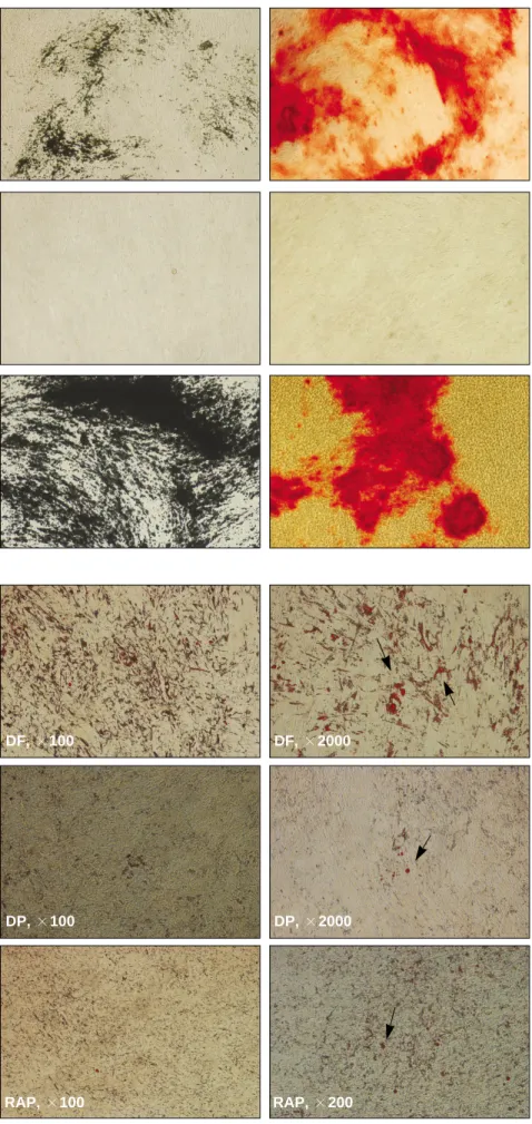
Fig. 7. Osteogenic differentiations of dental tissue-derived stem cells for 4 weeks. The most abundant bone matrix formation was observed in the RAP cells, whereas it was rarely detected in the DP cells by von Kossa and alizarin red stainings.
(DP: dental pulp, RAP: root apical papilla)
Fig. 8. Adipogenic differentiation of dental tissue-derived stem cells for 4 weeks.(origi- nal magnification x100) Small red color stained lipoid nodules were detected in all three types of dental stem cells by oil-red O staining.(arrows) Most actively adipogenic differentiation was showed in the dental fol- licle-derived cells.
Von Kossa Alizarin Red
Dental follicleDental pulpRoot apical papilla
DF, ×100 DF, ×2000
DP, ×100 DP, ×2000
RAP, ×100 RAP, ×200
전사인자는 최소한 2,260개의 인간 유전자의 프로모터 (promoter)에 관여하며, 3전사인자가 공동으로 관여하는 유 전자도 353가지나 된다25. 따라서 이들 3전사인자는 공동으 로 또는 각자 단독으로 인간 배아줄기세포의 다능성 (pluripotency)과 자가재생성(self renewal)을 조절하며, 줄기 세포의 전사에 중요한 역할을 하는 것으로 보인다26,27. 흥미 로운 것은 최근의 연구들에 의하면, 이들 전사인자가 배아 줄기세포에서뿐만 아니라, 골수유래 간엽성 줄기세포28, 지 방유래 간엽성 줄기세포29, 치수유래 줄기세포30, 태반기질 세포27, 그리고 피부유래 줄기세포21,31-33등과 같은 태생 후 성체줄기세포(postnatal adult stem cells)들에서도 발현되는 것이 관찰되었다는 점이다. 즉, 이는 초기 전사인자들은 성 체줄기세포에 거의 발현이 되지 않고 배아줄기세포에서만 강한 발현이 관찰된다는 기존의 가설을 바꾸는 연구들인 것이다. 본 연구에서도 3종류의 치아조직유래 줄기세포 모 두에서 초기 전사인자인 Oct-4, Nanog, 그리고 Sox-2의 발 현을 RT-PCR을 통해 관찰할 수 있었으나, Sox-2 유전자는 상대적으로 약하게 발현되었고, 특히 치낭유래 세포에서 는 Sox-2의 발현을 거의 관찰할 수 없었다. 또한, 치근유두 유래 세포에서 가장 강한 전사인자들의 발현을 관찰하였 고, 반대로 치낭유래 세포에서 가장 약한 전사인자의 발현 을 관찰하였다. 이러한 결과는 이들 치아조직유래 세포 중 치근유두유래 세포가 가장 원시적이고 다능성(primitive pluripotent or multipotent)의 특징을 가진다는 의미로 해석 될 수 있다. 이러한 결과는 Sonoyama 등19의 연구에서 치수 유래 줄기세포보다 치근유두유래 줄기세포에서 다능성이 더 관찰되고 분화능력이 더 우수하다는 연구결과와 부분 적으로 일치되는 결과이다. 또한, 선행된 연구에서는 인간 과 유사한 유인원의 치수유래 줄기세포에서 Oct-4 같은 전 사인자의 미약한 발현을 관찰하였는데30, 본 연구에서는 이 와는 다르게 치수유래 세포에서도 상당히 많은 양의 전사 인자들의 발현을 관찰할 수 있었다. 즉, 비록 정도의 차이 는 있지만, 본 연구에서 추출되고 배양된 3종류의 치아조 직유래 세포 모두 다능성을 가지는 원시세포의 특징을 가 진다고 볼 수 있을 것이다.
최근에 조직공학적 조직재생에 대한 요구가 증가하면서 성체줄기세포 중 간엽성 줄기세포(MSC)에 대한 관심이 증 가하고 있다. 현재까지 간엽성 줄기세포는 전술한 바와 같 이 인간이나 동물의 골수, 지방, 근육, 제대혈, 순환혈액, 피 부, 치아조직 등에서 추출 및 연구되었다. 이러한 간엽성 줄기세포는 몇 가지 특징을 가지는데, 첫 번째 특징은 일반 적인 배양조건에서 배양판에 부착되어(plastic-adherent cul- ture) 배양된다는 점이며, 두 번째 특징은 특정한 세포표면 표시자(cell surface marker, CD marker)의 발현이 관찰된다 는 점이다. 즉, CD13, CD29, CD44, CD73, CD90, CD105, 그 리고 CD166 같은 표시자가 양성으로 관찰되고, CD45, CD34, CD14, 또는 CD11b 같은 표시자는 거의 관찰되지 않 는 특징을 보인다. 마지막으로 세 번째 특징은 간엽성 줄기
세포를 특정 분화조건을 가진 배지상에서(in vitro culture) 분화시키면 골세포, 지방세포, 연골세포 등으로 성공적인 분화가 일어난다는 점이다34,35. 따라서, 이러한 특징을 바탕 으로 간엽성 줄기세포는 배양되는 세포의 물리적, 형태학 적 특징과 몇 가지 표시자의 발현여부, 그리고 다양한 세포 로의 분화가능여부 등으로 정의 될 수 있다. 본 연구에서도 매복지치 발치 후 분리한 치낭, 치수, 치근유두유래 세포 모두에서 CD44, CD90, CD105의 발현을 관찰할 수 있었으 며, 이러한 결과를 토대로 이들 3종류의 치아조직유래 세 포 모두 간엽성 줄기세포의 특징을 가진다고 말할 수 있다.
현재까지 치아조직유래 줄기세포는 치수, 치낭, 치근첨 부의 치유두(dental papilla), 그리고 치주인대 조직에서 추 출되었다. 치수유래 줄기세포는 Gronthos 등10에 의해서 2000년 처음 발표되었으며, 치수조직은 발생학적으로 신 경능(neural crest) 기원이지만, 골수유래 간엽성 줄기세포 와 상당히 유사한 특성을 가진다고 하였다. 이러한, 치수유 래 줄기세포는 매복지치, 하악 유전치, 그리고 정중과잉치 등의 치수에서 추출되었는데, 같은 치수유래 줄기세포도 그 기원에 따라 특성의 차이가 있음이 관찰되었다. 가장 큰 차이점은 유치의 치수유래 줄기세포는 hydroxyapatite/tri- calcium phosphate와 같이 면역억제 쥐(nude mouse)의 피부 하에 식립하면 골형성을 관찰할 수 있는데 반해12,13, 성인 매복지치의 치수유래 줄기세포는 같은 조건에서 상아질과 치수유사 구조물이 만들어 진다는 점이다10. 치낭유래 줄기 세포는 Handa 등36이 송아지의 치배에서 추출한 세포를 생 체 내에 이식하여 백악질유사 기질(cementum-like matrix) 형성을 보고한 것이 처음이며, 이후 Morsczeck 등15에 의해 서 인간의 지치 발치 후 치낭 조직에서 다능성 특징을 가진 줄기세포의 추출이 보고되었다. Seo 등18은 치주인대유래 줄기세포를 추출하여 생체 내 이식 시 백악질과 치주조직 이 형성됨을 관찰하여 2004년 처음 보고하였다. 치근유두 줄기세포는 Sonoyama 등19에 의해서 보고되면서, 치수유래 줄기세포보다 분화능력이나 상아질 형성 능력이 더 뛰어 나다고 하였다.
흥미로운 것은 몇 가지 선행된 연구에서 치수유래 줄기 세포는 골수유래 간엽성 줄기세포나 다른 성체줄기세포보 다 분화능력이 낮으며, 특히 지방세포로의 분화능력이 현 저히 떨어진다고 하였다10,11,31. 이에 비해 치근유두나 치주 인대 줄기세포는 좀 더 활발한 분화능력을 보이며, 특히 골 세포 분화와 백악질 등의 치주조직 분화에 장점을 가진다 고 하였다18,19. 본 연구에서도 선행된 연구들과 유사한 결과 들을 관찰할 수 있었는데, 특히 치수유래 줄기세포는 골세 포 분화능력이 현저히 떨어짐을 관찰할 수 있었다. 또한, 지방세포로의 분화에서는 선행된 연구에서와 같이 활발한 지방세포로의 분화는 관찰할 수 없었지만, 3종류의 치아조 직 줄기세포 중 치낭 줄기세포에서 가장 활발한 지방세포 분화를 관찰할 수 있었다. 즉, 3종류의 치아유래 줄기세포 모두에서 정도의 차이는 있지만, 골세포와 지방세포로의
성공적인 분화가 일어남을 관찰하였고, 이는 3종류의 치아 조직유래 줄기세포가 다능성의 특징을 가졌다는 의미로 해석할 수 있다.
본 연구로 그 동안 발치 후 버려지는 지치 주변 조직에서 줄기세포의 추출과 배양이 가능함을 관찰하였고, 특히 치 근유두유래 세포에서 강한 초기 전사인자와 간엽성 줄기 세포 표시자의 발현이 관찰되는 것으로 보아 가장 원시성 이 높지 않나 추측한다. 또한, 분화실험에서 치근유두유래 세포는 골세포로 분화되는데 가장 활발하고, 치낭유래 세 포는 지방세포로 분화되는데 장점이 있음이 관찰되었다.
향후 추가적인 연구가 진행되어야 하겠지만, 이러한 결과 를 바탕으로 다양한 재생의학에 치아줄기세포를 활용할 수 있을 것이며, 특히 발치한 치아조직을 장기 보존한 후 이에서 줄기세포를 추출할 수 있는 연구가 수행된다면, 현 재의 제대혈 보존과 같이 치아조직 또는 치아조직유래 줄 기세포를 장기 보존하여 유용하게 사용할 수도 있을 것으 로 기대된다.
Ⅴ
. 결 론
저자는 본 연구를 이해하고 조직제공에 동의한 4명의 환 자에서 미성숙 하악 매복지치를 발치한 후 치낭, 치수, 치 근유두 조직을 각각 분리하고, 이를 배양하여 각각의 치아 조직유래 세포를 추출하였다. 세 종류의 치아조직유래 세 포의 특성을 규명하기 위하여, 초기 전사인자의 발현과 세 포표면 표시자의 발현을 조사하였다. 세 종류의 치아조직 유래 세포에서 초기 전사인자인 Oct-4, Nanog, Sox-2의 발 현을 관찰할 수 있었고, 또한 간엽성 줄기세포 표시자인 CD44, CD90, CD105의 발현을 관찰할 수 있었다. 따라서, 본 연구에서 미성숙 매복지치 조직으로부터 추출한 세포 는 모두 미분화 원시적인 특성을 가지는 간엽성 줄기세포 라 할 수 있다. 이들 3종류의 치아조직유래 줄기세포를 각 각 골세포 및 지방세포로 유도 분화시킨 실험에서 모든 종 류의 치아조직유래 줄기세포에서 골 및 지방세포로의 분 화를 관찰할 수 있었다. 세 종류의 치아조직유래 줄기세포 중 치근유두유래 줄기세포에서 가장 활발한 골세포로의 분화를 관찰할 수 있었으며, 치수유래 줄기세포에서 가장 낮은 골세포로의 분화능력을 관찰하였다. 지방세포로의 분화 실험에서는 치낭유래 줄기세포에서 가장 활발한 지 방세포로의 분화를 관찰하였으며, 이들 3종류의 치아조직 유래 줄기세포에서 각각 분화능력에 차이가 있음을 관찰 하였다.
References
1. Evans MJ, Kaufman MH. Establishment in culture of pluripoten- tial cells from mouse embryos. Nature 1981;292:154-6.
2. Wagers AJ, Weissman IL. Plasticity of adult stem cells. Cell 2004;116:639-48.
3. Mao JJ, Giannobile WV, Helms JA, Hollister SJ, Krebsbach PH, Longaker MT, et al. Craniofacial tissue engineering by stem cells. J Dent Res 2006;85:966-79.
4. Almeida-Porada G, Porada C, Zanjani ED. Adult stem cell plas- ticity and methods of detection. Rev Clin Exp Hematol 2001;5:
26-41.
5. Temple S. Stem cell plasticity-building the brain of our dreams.
Nat Rev Neurosci 2001;2:513-20.
6. Yamada Y, Boo JS, Ozawa R, Nagasaka T, Okazaki Y, Hata K, et al. Bone regeneration following injection of mesenchymal stem cells and fibrin glue with a biodegradable scaffold. J Craniomaxillofac Surg 2003;31:27-33.
7. Yamada Y, Ueda M, Naiki T, Nagasaka T. Tissue-engineered in- jectable bone regeneration for osseointegrated dental implants.
Clin Oral Impl Res 2004;15:589-97.
8. Fuerst G, Tangl S, Gruber R, Gahleitner A, Sanroman F, Watzek G. Bone formation following sinus grafting with autogenous bone-derived cells and bovine bone mineral in minipigs: prelimi- nary findings. Clin Oral Impl Res 2004;15:733-40.
9. Schimming R, Schmelzeisen R. Tissue-engineered bone for max- illary sinus augmentation. J Oral Maxillofac Surg 2004;62:724-9.
10. Gronthos S, Mankani M, Brahim J, Robey PG, Shi S. Postnatal human dental pulp stem cells (DPSCs) in vitro and in vivo. Proc Natl Acad Sci USA 2000;97:13625-30.
11. Gronthos S, Brahim J, Li W, Fisher LW, Cherman N, Boyde A, et al. Stem cell properties of human dental pulp stem cells. J Dent Res 2002;81:531-5.
12. Miura M, Gronthos S, Zhao M, Lu B, Fisher LW, Robey PG, et al. Stem cells from human exfoliated deciduous teeth. Proc Natl Acad Sci USA 2003;100:5807-12.
13. Seo BM, Sonoyama W, Yamaza T, Coppe C, Kikuiri T, Akiyama K, et al. SHED repair critical-size calvarial defects in mice. Oral Dis 2008;14:428-34.
14. Huang AH, Chen YK, Lin LM, Shieh TY, Chan AW. Isolation and characterization of dental pulp stem cells from a supernumer- ary tooth. J Oral Pathol Med 2008;37:571-4.
15. Morsczeck C, Go¨tz W, Schierholz J, Zeilhofer F, Ku¨hn U, Mo¨hl C, et al. Isolation of precursor cells (PCs) from human dental fol- licle of wisdom teeth. Matrix Biol 2005;24:155-65.
16. Morsczeck C, Moehl C, Go¨tz W, Heredia A, Scha¨ffer TE, Eckstein N, et al. In vitro differentiation of human dental follicle cells with dexamethasone and insulin. Cell Biol Int 2005;29:567- 75.
17. Reynolds AJ, Jahoda CA. Cultured human and rat tooth papilla cells induce hair follicle regeneration and fiber growth.
Differentiation 2004;72:566-75.
18. Seo BM, Miura M, Gronthos S, Bartold PM, Batouli S, Brahim J, et al. Investigation of multipotent postnatal stem cells from hu- man periodontal ligament. Lancet 2004;364:149-55.
19. Sonoyama W, Liu Y, Fang D, Yamaza T, Seo BM, Zhang C, et al. Mesenchymal stem cell-mediated functional tooth regenera- tion in swine. PLoS ONE 2006;1:e79.
20. Morsczeck C, Vo¨llner F, Saugspier M, Brandl C, Reichert TE, Driemel O, et al. Comparison of human dental follicle cells (DFCs) and stem cells from human exfoliated deciduous teeth (SHED) after neural differentiation in vitro. Clin Oral Investig [in press 2009 Jul 10].
21. Kang EJ, Byun JH, Choi YJ, Maeng GH, Lee SL, Kang DH, et al. In vitro and in vivo osteogenesis of porcine skin-derived mes- enchymal stem cell-like cells with a demineralized bone and fib- rin scaffold. Tissue Eng Part A 2010;16:815-27.
22. Scho¨ler HR, Ruppert S, Suzuki N, Chowdhury K, Gruss P. New type of POU domain in germ line-specific protein Oct-4. Nature 1990;344:435-9.
23. Avilion AA, Nicolis SK, Pevny LH, Perez L, Vivian N, Lovell- Badge R. Multipotent cell lineages in early mouse development depend on SOX2 function. Genes Dev 2003;17:126-40.
24. Chambers I, Colby D, Robertson M, Nichols J, Lee S, Tweedie
S, et al. Functional expression cloning of Nanog, a pluripotency sustaining factor in embryonic stem cells. Cell 2003;113:643-55.
25. Boyer LA, Lee TI, Cole MF, Johnstone SE, Levine SS, Zucker JP, et al. Core transcriptional regulatory circuitry in human em- bryonic stem cells. Cell 2005;122:947-56.
26. Kajahn J, Gorjup E, Tiede S, von Briesen H, Paus R, Kruse C, et al. Skin-derived human adult stem cells surprisingly share many features with human pancreatic stem cells. Eur J Cell Biol 2008;
87:39-46.
27. Carlin R, Davis D, Weiss M, Schultz B, Troyer D. Expression of early transcription factors Oct-4, Sox-2 and Nanog by porcine umbilical cord (PUC) matrix cells. Reprod Biol Endocrinol 2006;
4:8.
28. Izadpanah R, Joswig T, Tsien F, Dufour J, Kirijan JC, Bunnell BA. Characterization of multipotent mesenchymal stem cells from the bone marrow of rhesus macaques. Stem Cells Dev 2005;14:440-51.
29. Zhu Y, Liu T, Song K, Fan X, Ma X, Cui Z. Adipose-derived stem cell: a better stem cell than BMSC. Cell Biochem Funct 2008;26:664-75.
30. Cheng PH, Snyder B, Fillos D, Ibegbu CC, Huang AH, Chan AW. Postnatal stem/progenitor cells derived from the dental pulp
of adult chimpanzee. BMC Cell Biol 2008;9:20.
31. Dyce PW, Zhu H, Craig J, Li J. Stem cells with multilineage po- tential derived from porcine skin. Biochem Biophys Res Commun 2004;316:651-8.
32. Zhao M, Isom SC, Lin H, Hao Y, Zhang Y, Zhao J, et al. Tracing the stemness of porcine skin-derived progenitors (pSKP) back to specific marker gene expression. Cloning Stem Cells 2009;11:
111-22.
33. Choi MJ, Byun JH, Kang EJ, Rho GJ, Kim UK, Kim JR, et al.
Isolation of porcine multipotential skin-derived precursor cells and its multilineage differentiation. J Korean Assoc Oral Maxillofac Surg 2008;34:588-93.
34. Park BW, Hah YS, Kim DR, Kim JR, Byun JH. Osteogenic phe- notypes and mineralization of cultured human periosteal-derived cells. Arch Oral Biol 2007;52:983-9.
35. Rho GJ, Kumar BM, Balasubramanian SS. Porcine mesenchymal stem cells - current technological status and future perspective.
Front Biosci 2009;14:3942-61.
36. Handa K, Saito M, Yamauchi M, Kiyono T, Sato S, Teranaka T, et al. Cementum matrix formation in vivo by cultured dental fol- licle cells. Bone 2002;31:606-11.