The impact factors on 5-year survival rate in patients operated with oral cancer
Dong-Ho Geum, Young-Chea Roh, Sang-Yong Yoon, Hyo-Geon Kim, Jung-Han Lee, Jae-Min Song, Jae-Yeol Lee, Dae-Seok Hwang, Yong-Deok Kim, Sang-Hun Shin, In-Kyo Chung, Uk-Kyu Kim
Department of Oral and Maxillofacial Surgery, School of Dentistry, Pusan National University, Yangsan, Korea
Abstract(J Korean Assoc Oral Maxillofac Surg 2013;39:207-216)
Objectives: The purpose of this study is to analyze clinical impact factors on the survival rate, and to acquire basic clinical data for the diagnosis of oral cancer, for a determination of the treatment plan with long-term survival in oral cancer patients.
Materials and Methods: Through a retrospective review of the medical records, the factors for long-term survival rate were analyzed. Thirty-seven patients, among patient database with oral cancer treated in the Department of Oral and Maxillofacial Surgery at Pusan National University Hospital within a period from March 1998 to March 2008, were selected within the study criteria and were followed-up for more than 5 years. The analyzed factors were gender, age, drinking, smoking, primary tumor site, type of cancer, TNM stage, recurrence of affected region, and metastasis of cervical lymph node. The 5-year survival rate on the impact factors was calculated statistically using the Kaplan-Meier method.
Results: By classification of clinical TNM at the 1st visit, there were 11 (29.7%) cases for stage I, 11 (29.7%) cases for stage II, 3 (8.1%) cases for stage III, and 12 (32.5%) cases for stage IV. The 5-year survival rate of total oral cancer patients after the operation were 75.7%, pathological TNM stage related 5-year survival rate were as follows: stage I 90.0%, stage II 81.8%, stage III 100% and stage IV 45.5%; in which the survival rate differ- ence by each stage was significantly observed. The recurrence of cervical lymph node was the significant impact factor for the survival rate, because only 30.0% the survival rate in recurrent cases existed. During the follow-up, there were 15 (40.5%) patients with confirmed recurrence, and the 5-year survival rate of these patients was decreased as 46.7%.
Conclusion: The classification of clinical and pathological TNM stage, local recurrence after surgery, and metastasis of cervical lymph node after sur- gery were analyzed as the 3 most significant factors.
Key words: Oral cancer, Survival rate, Neoplasm metastasis, Recurrence, TNM classification
[paper submitted 2013. 7. 26 / revised 2013. 9. 23 / accepted 2013. 9. 26]
cases of oral cancer have been reported, 300,000 of which are known to be oral squamous cell carcinoma1. According to domestic research, about 2,800 patients--or approximately 1.6% of the total cancer patients--are diagnosed with malig- nant tumors in the oral and maxillofacial region annually2.
Despite the development of diagnostic screening equip- ment, operation techniques, and postoperative care, there are a high number of patients manifesting advanced-stage oral cancer as well as high mortality. Because of this unfortunate situation, which persists until now, there have been continu- ous studies and reports on the deciding factors that would improve the survival rate of oral cancer patients. Among the factors to be considered are age, gender, stage at diagnosis, primary site and histopathologic classification, etc.3-6. Such basic research data are necessary, especially for patients who can still be cured. Note, however, that there is shortage of epi-
I. Introduction
Considering an aging society with increased life expectan- cy, the percentage of people with cancer is increasing, and 5%
of all tumors are occurring in the head and neck region; half of that 5% affect the oral cavity. Since 2000s, about 615,000
Uk-Kyu Kim
Department of Oral and Maxillofacial Surgery, School of Dentistry, Pusan National University, 49, Busandaehak-ro, Mulgeum-eup, Yangsan 626-870, Korea
TEL: +82-55-360-5112 FAX: +82-55-360-5104 E-mail: [email protected]
This is an open-access article distributed under the terms of the Creative Commons Attribution Non-Commercial License (http://creativecommons.org/licenses/by-nc/3.0/), which permits unrestricted non-commercial use, distribution, and reproduction in any medium, provided the original work is properly cited.
CC
Copyright Ⓒ 2013 The Korean Association of Oral and Maxillofacial Surgeons. All rights reserved.
This study was supported by a Clinical Research Grant of the Pusan National University Dental Hospital (2012).
tinely, followed by radical wide excision and neck dissection as necessary. Likewise, chemotherapy and radiation therapy may be needed depending on the histopathologic results.
(Table 1) Through the analysis of these records, we tried to review the treatment outcome and evaluate the detailed prob- lems of all oral cancer patients. In addition, through long- term follow-up, analysis of the factors impacting the 5-year survival rate of oral cancer patients was performed.
2. Methods
The final 37 patients were analyzed by gender, age, de- gree of alcohol drinking, smoking status, primary site, type of carcinoma, histopathologic grade, stage, neck dissection, combination therapy, recurrence, and cervical lymph node metastasis; we then analyzed how these factors impacted the patient’s 5-year survival rate for correlation.
The TNM stage was classified according to the TNM clas- sification for the lip and oral cavity of the American Joint Committee on Cancer Guide (7th edition). Each of the impact- ing factors was compared and reviewed by making individual tables for each factor. Considering the status of patients from the point when follow-up is over, survival was assumed when the patient had been alive for 5 years from diagnosis; if the fol- low-up was paused, or the patient was discharged due to other reasons, such was treated as censored data.
As a retrospective clinical study, this study was approved by the Pusan National University Dental Hospital Institution- al Review Board (IRB No. PNUDH-2013-007).
III. Results
1. Overview of clinical data
This report summarized the overall clinical data, medical history, clinical examination, and image examination of 37 demiology research done domestically for 5 years’ follow-up
study related to the factors impacting the patient’s survival rate.
In this study, by analyzing the clinically important factors re- lated to the long-term survival of oral cancer patients and assess- ing these factors, the resulting basic data will be helpful in devel- oping diagnosis and treatment plans for oral cancer patients.
II. Materials and Methods
1. Patients
This study dealt with patients diagnosed with oral cancer and subjected to radical resection of lesion at the Department of Oral and Maxillofacial Surgery, Pusan National University Hospital between March 1998 and March 2008. The inclu- sion criteria were as follows:
1) Patient confirmed to have oral cancer through biopsy.
2) Patient who did not have metastasis at the first diagnosis.
3) Patient who did not receive any treatment for the pri- mary tumor site in other hospitals.
4) Patient with no history of malignant tumors on any other part.
5) Patients operated by the same surgical team with similar treatment protocol.
6) Patient identified to be in a state of survival after diagno- sis for 5 years.
The patients were interviewed, and their medical records were analyzed. Patients with no more follow-up visits after 5 years were contacted to confirm their survival by calling;
when their survival was confirmed, additional clinical and radiological examinations were performed to determine the factors impacting the long-term survival rate.
During the observation period, the treatment strategy of our department could be changed for others depending on the general condition of the patient. Note, however,that che- motherapy prior to operation had been done for 2 cycles rou-
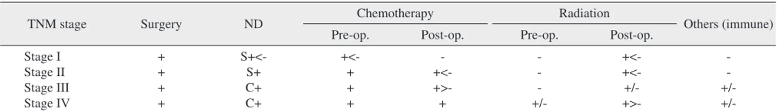
Table 1. Protocol for the treatment of head and neck malignancy in the Department of Oral and Maxillofacial Surgery at Pusan National University Hospital
TNM stage Surgery ND Chemotherapy Radiation
Others (immune)
Pre-op. Post-op. Pre-op. Post-op.
Stage I Stage II Stage III Stage IV
+ + + +
S+<- S+
C+
C+
+<- + + +
- +<- +>- +
- - - +/-
+<- +<- +/- +>-
- - +/- +/- (ND: neck dissection, op.: operation, S: selective, C: comprehensive, +: treatment, -: no treatment, >, <: treatment priority, +/-: treatment status according to disease severity)
Dong-Ho Geum et al: The impact factors on 5-year survival rate in patients operated with oral cancer. J Korean Assoc Oral Maxillofac Surg 2013
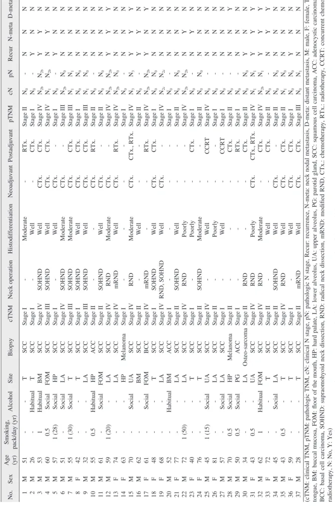
Table 2. Summary of patients No.SexAge (yr)Smoking, pack/day (yr)AlcoholSiteBiopsycTNMNeck operationHistodifferen tiationNeoadjuvantPostadjuvantpTNMcNpNRecurN-metaD-meta 1 2 3 4 5 6 7 8 9 10 11 12 13 14 15 16 17 18 19 20 21 22 23 24 25 26 27 28 29 30 31 32 33 34 35 36 37
M M M M M M M F M M M M F F M M F F F M F M F F M F M M M M F M F M M F M
51 26 53 60 57 51 55 42 32 55 61 59 74 63 70 62 61 48 68 52 77 72 40 76 45 81 57 70 50 34 43 62 72 45 43 59 28
- - 1 0.5 1 (28) - 1 (30) - - 0.5 - 1 (20) - - - - - - - - - 1 (50) - - 1 (15) - - 0.5 0.5 - 0.5 1 - - 0.5 - -
- Habitual Habitual Social Social Social Social - - Habitual Social - - - Social - Social - - Habitual - - - - Social - Social Social Social - - Habitual - Social - - -
T T BM FOM HP LA T T LA HP FOM LA LA HP UA BM FOM T LA BM LA LA T T UA LA LA HP PG LA UA FOM T LA T T T
SCC SCC SCC SCC SCC SCC SCC SCC SCC ACC SCC SCC SCC Melanoma SCC SCC BCC SCC SCC ACC SCC SCC SCC SCC SCC SCC SCC Melanoma ACC Osteo-sarcoma SCC SCC SCC SCC SCC SCC SCC
Stage I Stage I Stage IV Stage III Stage I Stage IV Stage III Stage I Stage III Stage II Stage II Stage IV Stage IV Stage I Stage IV Stage II Stage IV Stage I Stage IV Stage I Stage II Stage IV Stage I Stage II Stage IV Stage II Stage I Stage II Stage I Stage II Stage IV Stage IV Stage II Stage II Stage IV Stage I Stage II
- - SOHND SOHND - SOHND SOHND SOHND SOHND - SOHND RND mRND - RND - mRND SOHND RND, SOHND - SOHND RND - SOHND - - - - - RND RND RND - SOHND RND - mRND
Moderate Well Well Well Well Moderate Moderate Well Well - Well Moderate Well - Moderate Well - Well Well - Well Poorly Poorly Moderate Well Poorly Well - - - Poorly Moderate Well Well Well Well Well
- - CTx. CTx. CTx. CTx. CTx. CTx. CTx. CTx. CTx. CTx. CTx. - CTx. - - CTx. CTx. - - - - - - - - - - - CTx. - - CTx. CTx. CTx. CTx.
RTx. CTx. CTx. CTx. - - CTx. CTx. CTx. RTx. - - RTx. - CTx., RTx. - RTx. CTx. - - - - CTx. - CCRT - CCRT CTx. RTx. CTx. CTx., RTx. CTx. CTx. - CTx. CTx. CTx.
Stage II Stage I Stage IV Stage IV Stage I Stage III Stage III Stage I Stage III Stage II Stage II Stage IV Stage IV Stage I Stage IV Stage II Stage IV Stage I Stage IV Stage I Stage II Stage IV Stage I Stage II Stage IV Stage I Stage I Stage II Stage I Stage II Stage IV Stage IV Stage II Stage II Stage II Stage I Stage III
N0 N0 N2b N1 N0 N2b N1 N0 N1 N0 N0 N2b N2b N0 N3 N0 N2b N0 N2c N0 N0 N2b N0 N0 N0 N0 N0 N0 N0 N0 N1 N2b N0 N0 N1 N0 N0
- - N2b N2b - N0 N1 N0 N0 - N0 N2b N0 - N3 - N2b N0 N2c - N0 N2b - N0 - - - - - N0 N1 N1 - N0 N0 N0 N1
N Y Y N Y N N N N N N Y N N Y Y Y N N N N N N N N N N N N Y Y Y N N N Y N
N N Y Y N N N N N N N Y N N Y Y N N N N N Y Y N N N N N N N Y Y Y N N N N
N N N N N N N N N N N Y N N Y N Y N N N N N N N N N N Y N N Y Y Y N N N N (cTNM: clinical TNM, pTNM: pathologic TNM, cN: clinical N stage, pN: pathologic N stage, Recur: recurrence, N-meta: neck nodal metastasis, D-meta: distant metastasis, M: male, F: female, T: tongue, BM: buccal mucosa, FOM: floor of the mouth, HP: hard palate, LA: lower alveolus, UA: upper alveolus, PG: parotid gland, SCC: squamous cell carcinoma, ACC: adenocystic carcinoma, BCC: basal cell carcinoma, SOHND: supraomohyoid neck dissection, RND: radical neck dissection, mRND: modified RND, CTx.: chemotherapy, RTx.: radiotherapy, CCRT: concurrent chemo-
radiotherapy, N: No, Y: Yes) Dorperated with oral cancer. J Keantn Assoc Oral Maxillofac Surg 20s oieon fag-Ho Geum et al: The impactctators on 5-year survival rate in p13
followed by alveolar ridge of the upper jaw and palate with 7 (18.9%) cases each, floor of the mouth (FOM) with 4 (10.9%) cases, and buccal mucosa/retromolar trigone with 3 (8.1%) cases.(Table 4)
3) Histological types of cancer
For the distribution of tumor histology, there were 30 (81.1%) cases of squamous cell carcinoma (SCC), 3 (8.1%) cases of adenocystic carcinoma, 1 (2.7%) case of basal cell carcinoma, 2 (5.4%) cases of melanoma, and 1 (2.7%) case of osteosarcoma.(Table 5)
4) TNM stage
In the clinical TNM (cTNM) classification, 11 (29.7%) cases were found to belong to stage I, 11 (29.7%), to stage II, 3 (8.1%), to stage III, and 12 (32.5%), to stage IV. By analyz- ing the results of post-operative histopathologic specimen, in the pathologic TNM (pTNM) classification, 11 (29.7%) cases were for stage I, 11 (29.7%), for stage II, 4 (10.8%), for stage III, and 11 (29.8%), for stage IV.(Table 6)
5) Factors associated with cervical metastasis
The percentage of mutual concordance between cTNM and pTNM was 83.8%. Cases diagnosed as cervical lymph node metastasis (pN+) numbered 11 (29.7%), with 10 (27.1%) cases of recurrence of cervical lymph node metastasis within patients. For smoking, we recorded the average number of
cigarettes smoked each day; for drinking, drinking more than 0.5 bottles (Korean vodka, Soju) per day was considered ha- bitual. In the study, 13 patients were heavy smokers, and 17 were habitual drinkers.
All patients included in this study received wide excision on the primary site and neck dissection. The types of neck dissection conducted included supraomohyoid neck dissec- tion, modified radical neck dissection (mRND), and RND.
In this article, 19 cases were for pre-operation chemothera- py, 17, for post-operation chemotherapy, and 7, for radiation therapy after operation in those cases, including 2 patients who sequentially received both therapies as well as 2 patients who received concurrent chemo-radiotherapy.(Table 2)
1) Gender and age
Out of the 37 patients, 24 (64.8%) were male and 13 (35.2%) were female. Patients under 40 years accounted for 29.7%, and those over 50 years constituted 70.3%. The aver- age age was 55 years, ranging from 26 to 81.(Table 3)
2) Primary tumor sites
Areas most affected by oral cancer were the tongue and alveolar ridge of the lower jaw with 11 (29.7%) cases each,
Table 3. Gender and age Age
(yr)
No. of patient
Total
Male Female
20-29 30-39 40-49 50-59 60-69 70-79 80-89 Total
2 2 3 10 4 3 0 24 (64.8)
0 0 4 1 3 4 1 13 (35.2)
2 (5.4) 2 (5.4) 7 (18.9) 11 (29.8) 7 (18.9) 7 (18.9) 1 (2.7) 37 (100) Values are presented as number or number (%).
Dong-Ho Geum et al: The impact factors on 5-year survival rate in patients operated with oral cancer. J Korean Assoc Oral Maxillofac Surg 2013
Table 4. Primary tumor sites
Primary site n (%)
Tongue Lower alveolus Upper alveolus & palate Floor of the mouth
Buccal mucosa & retromolar trigone Etc. (parotid gland)
Total
11 (29.7) 11 (29.7) 7 (18.9) 4 (10.9) 3 (8.1) 1 (2.7) 37 (100) Dong-Ho Geum et al: The impact factors on 5-year survival rate in patients operated with oral cancer. J Korean Assoc Oral Maxillofac Surg 2013
Table 5. Histopathologic diagnosis
Type n (%)
SCC ACC BCC Melanoma Osteosarcoma Total
30 (81.1) 3 (8.1) 1 (2.7) 2 (5.4) 1 (2.7) 37 (100)
(SCC: squamous cell carcinoma, ACC: adenocystic carcinoma, BCC:
basal cell carcinoma)
Dong-Ho Geum et al: The impact factors on 5-year survival rate in patients operated with oral cancer. J Korean Assoc Oral Maxillofac Surg 2013
Table 6. Clinical and pathological TNM classification in patients
TNM stage Clinical TNM Pathologic TNM
Stage I Stage II Stage III Stage IV Total
11 (29.7) 11 (29.7) 3 (8.1) 12 (32.5) 37 (100)
11 (29.7) 11 (29.7) 4 (10.8) 11 (29.8) 37 (100) Values are presented as number (%).
Dong-Ho Geum et al: The impact factors on 5-year survival rate in patients operated with oral cancer. J Korean Assoc Oral Maxillofac Surg 2013
2) Survival rate according to stage
The survival rate according to pTNM is shown in Fig. 2 (stage I: 90.0%; stage II: 81.8%; stage III: 100.0%; stage IV:
45.5%).
3) Survival rate according to histological differentiation The survival rate according to histological differentiation among patients diagnosed with SCC was 94.7% for the well- differentiated type, 57.1% for the moderately differentiated type, and 25.0% for the poorly differentiated type.(Fig. 3)
4) Survival rate according to the primary site recurrence, cervical lymph node metastasis
After operation, there were 15 (40.5%) patients who expe- rienced recurrence in the primary site or cervical lymph node metastasis. Out of these 15 patients, 7 (46.7%) of them were 5 years. Cervical lymph node diagnosed as clinically nega-
tive (cN0) was positive in the pathological result (pN+) in patients who showed occult nodal metastasis (3 cases). There were 15 (40.5%) oral cancer patients who experienced recur- rence in the primary site or cervical lymph node metastasis, 11 of whom underwent resection or neck dissection on the re- currence area or affected cervical lesion. The other 7 (18.9%) patients were found to experience metastasis in the lung and base of the skull during 5 years.(Tables 2, 6)
6) Histological differentiation of squamous cell carcinoma The SCC patients were classified according to the histologi- cal differentiation of specimen. Out of all the patients, those diagnosed with SCC through biopsy (n=30) were analyzed by categories of prevalence and survival rate according to the differentiation. There were 19 (63.4%) cases for the well- differentiated type, 7 (23.3%) cases for the moderately differ- entiated type, and 4 (13.3%) cases for the poorly differentiated type according to histological differentiation.(Table 7)
7) Primary tumor resection, neck dissection, and recon- struction methods
All analyzed patients primarily underwent wide excision of the primary site. Of all patients who seemingly had no me- tastasis (cN0), including the patients who seem to have cervi- cal lymph node metastasis clinically, T2-4 and T1 tongue cancer patients had elective or therapeutic neck dissection performed. Various reconstruction methods were used in the reconstruction of the defect site. The reconstruction methods used in this study include cervical flap, pectoralis major myo- cutaneous flap (7 cases), radial forearm free flap (8 cases), fibular free flap (2 cases), latissimus dorsi flap (1 case), and deep circumflex iliac artery flap (1 case).
2. Analysis of survival rate
1) 5-year survival rate
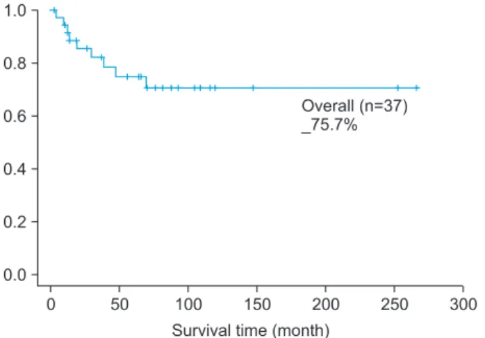
The survival rate of 37 oral cancer patients was 75.7%.(Fig. 1)
Table 7. Histopathologic differentiation distribution of SCC
Type n (%) Survival rate (%)
Well-differentiated Moderate-differentiated Poorly-differentiated Total
19 (63.4) 7 (23.3) 4 (13.3) 30 (100)
94.7 57.1 25.0
(SCC: squamous cell carcinoma)
Dong-Ho Geum et al: The impact factors on 5-year survival rate in patients operated with oral cancer. J Korean Assoc Oral Maxillofac Surg 2013
Fig. 1. Overall survival rate.
Dong-Ho Geum et al: The impact factors on 5-year survival rate in patients operated with oral cancer. J Korean Assoc Oral Maxillofac Surg 2013
Fig. 2. Stage-specific survival rate (Log rank test: P=0.027).
Dong-Ho Geum et al: The impact factors on 5-year survival rate in patients operated with oral cancer. J Korean Assoc Oral Maxillofac Surg 2013
palliative therapy7. Based on this, we statistically analyzed the other factors investigated in this study and used them as a scale to figure out how each of the factors affects the decreas- ing survival rate. We found a significant relationship with post-operative cervical lymph node metastasis.(Table 8)
IV. Discussion
Out of the 37 oral cancer patients included in this study, 23 (62.2%) were male and 14 (37.8%) were female. This represents a 1.6 : 1 ratio, showing a comparable result with the United States male oral cancer percentage ratio of 60.2%
and Funk et al.8 and Kim et al.9 reported percentage ratio of 65.7% males and 34.3% females. In this study, however, gen- alive. Regarding these factors as independent variables on the
5-year survival rate, the recurrence rate for the primary tumor and cervical lymph node metastasis after surgery was 60.0%
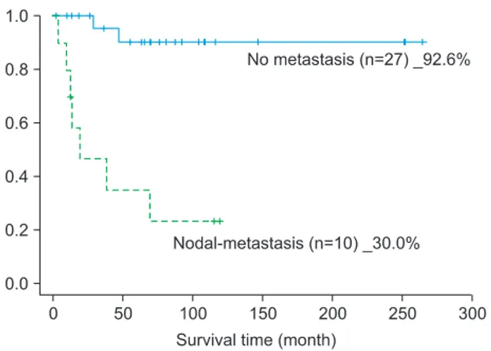
and 30.0%, respectively. These results showed significant lower percentage compared with the non-affected group’s 86.4% and 92.6%, respectively.(Figs. 4, 5)
Among the 10 patients who had recurrence of cervical lymph node, only 3 of them survived for 5 years.
5) Logistic regression test between factors and survival In this study, out of the patients experiencing metastasis to the other organs, no patients survived over 5 years.(Fig.
6) Currently, there are no radical treatments established for metastatic lesions derived from oral cancer, and the treatment choices of these terminally ill patients are clinical trials or
Fig. 3. Histopathologic differentiation-specific survival rate in squamous cell carcinoma (Log rank test: P=0.000).
Dong-Ho Geum et al: The impact factors on 5-year survival rate in patients operated with oral cancer. J Korean Assoc Oral Maxillofac Surg 2013
Fig. 4. Recurrence-specific survival rate (Log rank test: P=0.097).
Dong-Ho Geum et al: The impact factors on 5-year survival rate in patients operated with oral cancer. J Korean Assoc Oral Maxillofac Surg 2013
Fig. 5. Neck nodal metastasis-specific survival rate (Log rank test:
P=0.000).
Dong-Ho Geum et al: The impact factors on 5-year survival rate in patients operated with oral cancer. J Korean Assoc Oral Maxillofac Surg 2013
Fig. 6. Distant metastasis-specific survival graphs (Log rank test:
P=0.000).
Dong-Ho Geum et al: The impact factors on 5-year survival rate in patients operated with oral cancer. J Korean Assoc Oral Maxillofac Surg 2013
as a perilous factor that can affect survival.
According to Shah and Patel17, oral cancer’s TNM patient stage ratio was 37% (stage I), 36% (stage II), 18% (stage III), and 9% (stage IV). In domestic research, Kim et al.9 re- ported that, out of 180 oral cancer patients, 31 (17.2%) cases belonged to stage I, 24 (13%), to stage II, 14 (7.8%), to stage III, and 111 (61.7%), to stage IV. The difference in preva- lence between types of stage was deemed attributable to the patients’ residence, financial status, and cultural differences;
hence the need for additional epidemiological research on the patient’s economic activity or education level and oral health policy by location. In inferring the reason for the high preva- lence level of patients in early stages (stage I, II) and terminal stage (stage IV) in this study, patients diagnosed with oral cancer in the earlier stages were mostly transferred to our hospital because such can be found out by chance when they visit the local clinic due to different chief complaints, and others, by visiting the hospital doctors after suffering from edema and pain since the cancer has already progressed.
The concordance of cTNM and pTNM was 83.8%, re- lating to the reliability of the magnetic resonance imaging (MRI), computed tomography (CT), and positron emission tomography-CT (PET-CT) examinations before the operation to evaluate the primary site, cervical metastasis, and distant metastasis. In this study, PET-CT examination was mainly used for the evaluation of cervical lymph node metastasis or follow-up check before operation.
According to the reports of Goshen et al.18, PET-CT ex- amination has 88% accuracy, 100% sensitivity, and 77%
specificity. When it came to negative predictive value from patients who showed metastasis, it was found to be 100%.
In Nahmias et al.’s research19, 192 out of 1,678 lymph nodes had histopathologic metastasis, with sensitivity and specific- ity to N0 reported to be 79% and 82%, respectively; those to N+ were 95% and 25%, respectively. Thus, sensitivity and specificity for cervical metastasis were concluded to be 48%
and 99%, respectively. In addition, PET-CT ensures patients’
comfort when setting the irradiation site for radiation thera- py20.
In this study, there were 3 patients who showed signs of der was not related to the 5-year survival rate.
In this study, 13 (35.1%) patients were heavy smokers and 17 (46.0%) were habitual drinkers, showing lower percent- ages compared to the previous reports (57-65%)9-14. This seemed to be due to the bias of patients in the interview at the time of hospitalization; hence the difficulty in revealing that smoking and alcohol may have significant effects on the sur- vival rate in this report.
According to the research of Krolls and Hoffman15, oral cancer frequently strikes those in their 40s-70s. According to Kim et al.’s report9, males in their 70s and females in their 60s were affected mostly by oral cancer. In this study, out of all the age distributions of oral cancer patients, patients in their 50s (11 patients in their 50s with the average)--with average age of 55.5 years (±13.9)--were most affected. There were 11 (29.7%) patients under 40 years and 26 (70.3%) patients over 50 years, resulting in a higher number of older patients. Note, however, that 26- and 28-year-old patients with tongue cancer were observed in these cases as well. It is the form of oral cancer that can affect even a healthier and younger age group as well. Out of the 37 patients, the oldest patient was an 81-year-old male who did not show any recur- rence of the primary site or cervical lymph node metastasis after 1 year since operation but expired due to general weak- ness and respiratory disease; thus, this case was processed as censored data. There were no significant changes in the survival rate of oral cancer patients by age group; as factors that can have an effect on the 5-year survival rate among the elderly and patients who had systemic diseases, however, limitations in choosing the treatment method due to general condition, age, patient-related complications, and deaths caused by associated diseases may be considered16.
Based on the histopathologic examination of specimen after operation, SCC accounted for more than 80% of the re- sults, showing similar ratio of generally known oral tumors17. In this study, the survival rate of patients with cancer occur- ring on the FOM was lowest at 50.0% for 5 years. This seems to be due to the difficulty in terms of surgical approach and cervical lymph metastasis tendency on both sides; consider- ing these facts, the primary lesion’s location can be regarded
Table 8. Results of binary logistic regression analysis on the correlation between the factors and survival
Gender Age pTNM Recurrence Neck metastasis Distant metastasis
P-value 0.501 0.179 0.033 0.051 0.000* 0.000*
(pTNM: pathologic TNM)
*Significant value (coefficient) P<0.01.
Dong-Ho Geum et al: The impact factors on 5-year survival rate in patients operated with oral cancer. J Korean Assoc Oral Maxillofac Surg 2013
by Koo et al.25, similar results of 43% and 79% were reported.
Note, however, that the treatment of clinically negative cervi- cal lymph node is still controversial. Nonetheless, it is impor- tant to consider the potential for cervical metastasis before proceeding with surgery. According to Zbären et al.’s study26, 20% of patients with cervical lymph node metastasis--which is pN0 in the oral cancer and pathological examination-- have potential for metastasis or recurrence. On the other hand, O’Brien et al.27 stated that there may be a 30% possibility.
Similarly, Keski-Säntti et al.28 reported a low 5-year survival rate of 33% for patients confirmed to have cervical metastasis during observation and who underwent salvage surgery.
Thus, the 5-year survival rate of oral cancer patients with cervical lymph node metastasis is a very important factor in prognosis; bearing in mind the potential for cervical lymph node metastasis, and even if it is a case of cN0, planning ipsi- lateral or bilateral (tip of tongue, FOM, >T3, over the midline primary tumor) neck dissection must be considered25,29-31.
In this study, there were 15 (40.5%) patients with primary tumor recurrence or cervical lymph node metastasis, 7 of whom survived the second operation. The 5-year survival rate in case of recurrence was 46.7%; looking at cervical lymph node metastasis cases separately, however, the 5-year sur- vival rate was lower than 30%. After operation, if the recur- ring tumor grows deeper in the primary site or in the cervical region, it usually positions itself in a place that is difficult to excise; hence the difficulty of completing resection due to the surrounding anatomical structures. As a result, even with the salvage surgery option as mentioned previously, the survival rate is assumed to become even lower25,28,29. Moreover, cervi- cal lymph node metastasis itself implies the possibility of dis- tant metastasis, apparently causing the lower 5-year survival rate.
As found in this study, too, oral cancer has high possibil- ity of recurrence, in which case the prognosis is poor; hence the importance of continuous follow-up. The follow-up con- ducted in this study was monthly for up to 6 months after the operation and once every 3 months from 6 months to 1 year after operation; from 1 year to 5 years, only when there was recall and if required by the patient was the PET-CT exami- nation used to check closely for recurrence, cervical lymph node metastasis, and distant metastasis. According to the National Comprehensive Cancer Network guideline related to carcinoma of the head and neck, during follow-up, there is a need to consider the recurrence, risk of secondary primary tumor, treatment methods, and complications30,32. We need to examine carefully the clinical symptoms and findings of the occult neck nodal metastasis, with 4 patients testing positive
(cN+) in the clinical examination but pathologically negative (pN0) for cervical lymph node. Moreover, the sensitivity of the cervical lymph node examination before operation was 76.9%, and its specificity was 83.3%. From cN0, the poten- tial diagnostic methods for cervical lymph node metastasis (palpation, X-ray, fine needle aspiration cytology) are affect- ed by the number of lymph nodes removed and histological techniques for the examination of lymph nodes21. Therefore, when we perform surgery and treatment for the oral cancer patient, we should consider PET-CT or other examinations, cognizant of the need for neck dissection for occult lymph node metastasis as one of the complications of surgery. Like- wise, clinical follow-up and careful review of the clinical findings and diagnostic examination at an appropriate time after the operation are very important.
Cho and Kim22 reported a 54% 5-year survival rate among oral cancer patients during the period 1991 to 1996. Kim et al.9 found the 5-year survival rate of oral cancer patients from 1999 to 2006 to be 57.7%. From the report of Lee et al.23, however, the 5-year survival rate of oral cancer patients was 63.2%. This study took the records of patients who un- derwent operations during the period 1998 to 2008, and the 5-year survival rate of these patients was 75.7%, which was considerably higher than that of previous research studies.
Comparing the ratio of patients in stage IV, Lee et al.23 re- ported 57.1%, Cho and Kim22, 61.3%, and Kim et al.9, 61.7%.
Note, however, that the ratio of this study was lower (35.2%).
The etiology seems to be based on the other research studies for cases in stage IV and who did not undergo surgery but received palliative therapy. The same cannot be said for this study, though.
Our unusual stage III result (100%) for this study can be at- tributed to the small number of patients in stage III. The small number of individuals in the stage III results can also be seen in other research studies9,22-24. To guess the reason, the situ- ation wherein the primary carcinoma (T3) does not cause cervical lymph node metastasis and the situation wherein car- cinoma less than 4cm (T1, T2) causes cervical lymph node metastasis are unlikely to occur. Moreover, if there were T1 and T2 patients who did not show clinical cervical metastasis clinically, they can have cervical lymph node metastasis; if patients in other stages actually belonged to stage III, then such can explain the low ratio of stage III.
According to the 5-year survival rate in the case of cervical lymph node metastasis, in this study, a significant difference of 62.6% was noted between the two groups; in the research
correlation.
5. During the follow-up period, there were 15 (40.5%) patients with confirmed recurrence; the 5-year survival rate of these patients was 46.7%, which was lower than the total survival rate.
In summary, the factor wielding the biggest impact on long-term survival rate after operation was founded to be cervical lymph node metastasis after operation. Clinical and pathological TNM stage and local recurrence were found to be another strong factor impacting the 5-year survival rate.
Therefore, we can confirm the primary tumor and pathologi- cal findings of cervical lymph node after operation and po- tentially improve the long-term survival rate of the patients through close follow-up observation to detect any and all postoperative findings such as cervical metastasis.
References
1. Kademani D. Oral cancer. Mayo Clin Proc 2007;82:878-87.
2. Sun JR, Kim SM, Seo MH, Kim MJ, Lee JH, Myoung H. Oral can- cer incidence based on annual cancer statistics in Korea. J Korean Assoc Oral Maxillofac Surg 2012;38:20-8.
3. Woolgar JA, Rogers S, West CR, Errington RD, Brown JS, Vaughan ED. Survival and patterns of recurrence in 200 oral can- cer patients treated by radical surgery and neck dissection. Oral Oncol 1999;35:257-65.
4. Frierson HF Jr, Cooper PH. Prognostic factors in squamous cell carcinoma of the lower lip. Hum Pathol 1986;17:346-54.
5. Prieto I, Prieto A, Bravo M, Bascones A. Prognostic factors for cancer of the oral cavity. Quintessence Int 2005;36:711-7.
6. Rodriguez T, Altieri A, Chatenoud L, Gallus S, Bosetti C, Negri E, et al. Risk factors for oral and pharyngeal cancer in young adults.
Oral Oncol 2004;40:207-13.
7. Haigentz M Jr, Hartl DM, Silver CE, Langendijk JA, Strojan P, Paleri V, et al. Distant metastases from head and neck squamous cell carcinoma. Part III. Treatment. Oral Oncol 2012;48:787-93.
8. Funk GF, Karnell LH, Robinson RA, Zhen WK, Trask DK, Hoff- man HT. Presentation, treatment, and outcome of oral cavity can- cer: a National Cancer Data Base report. Head Neck 2002;24:165- 80.
9. Kim MY, Kim CS, Lee SH, Kim JW, Jang HJ. A clinicostatistical analysis of oral cancer patients for recent 8 years. J Korean Oral Maxillofac Surg 2007;33:660-8.
10. Napier SS, Speight PM. Natural history of potentially malignant oral lesions and conditions: an overview of the literature. J Oral Pathol Med 2008;37:1-10.
11. Chen JK, Katz RV, Krutchkoff DJ. Intraoral squamous cell carci- noma. Epidemiologic patterns in Connecticut from 1935 to 1985.
Cancer 1990;66:1288-96.
12. Blot WJ, McLaughlin JK, Winn DM, Austin DF, Greenberg RS, Preston-Martin S, et al. Smoking and drinking in relation to oral and pharyngeal cancer. Cancer Res 1988;48:3282-7.
13. Sandoval M, Font R, Mañós M, Dicenta M, Quintana MJ, Bosch FX, et al. The role of vegetable and fruit consumption and other habits on survival following the diagnosis of oral cancer: a pro- spective study in Spain. Int J Oral Maxillofac Surg 2009;38:31-9.
14. Kwon HK, Cha IH, Lim SJ, Choi CH, Kim BI. Risk factors for oral cancer; a case-control study. J Korean Oral Maxillofac Surg 2002;28:395-400.
15. Krolls SO, Hoffman S. Squamous cell carcinoma of the oral soft
patient and, if necessary, perform radiology imaging exami- nation. Likewise, the guideline suggests that CT, MRI, and PET-CT examinations--which use radiology and contrast-- be performed each year after operation for high-risk patients (1) with two or more lymph node metastasis, (2) wherein the affected size of a single lymph node is larger than 3 cm, (3) with extravasation spread of nodal lesion, and (4) with his- tory of previous recurrence30.
An advantage of this study is that it controlled variables by selecting oral cancer patients who received treatment from the same surgical team from a single lab. For its limitations, however, only a small number of target group were consid- ered for the epidemiological studies. In the future, there is a need to collect more patient data to determine the survival rate of each patient by treatment method and analyze how, given the same primary site and cervical lymph node metas- tasis status, the combined treatment method--such as che- motherapy and radiotherapy performed with primary tumor resection--affects the patient’s survival rate.
V. Conclusion
A total of 37 oral cancer patients were analyzed based on gender, age, drinking status, smoking status, primary site, type of carcinoma, TNM stage, neck dissection, combina- tion treatment and affected area, and cervical lymph node metastasis, including how these factors impacted the 5-year survival rate of each of the patients.
1. The 5-year survival rate of oral cancer patients was 75.68%; the pathological TNM stage-related, 5-year survival rate was as follows: 90.0% in stage I, 81.8% in stage II, 100%
in stage III, and 45.5% in stage IV. The observed difference in survival rate by stage was statistically significant.
2. In the case of cervical lymph node metastasis after op- eration, the 5-year survival rate was 30%; the patients who did not have such had a 92.6% 5-year survival rate, showing significant difference as well as the greatest impact on the survival rate.
3. Based on the histopathologic examination, the well- differentiated type accounted for the largest portion of SCC’s histological differentiation classification. As for the 5-year survival rate, the well-differentiated type recorded 94.7%, moderately differentiated type, 57.1%, and poorly differenti- ated type, 25.0%; thus showing a large difference in the sur- vival rate depending on the differentiation classification.
4. Two factors--postoperative cervical lymph node metas- tasis and distant metastasis--had positive moderate coefficient
Overall five-year survival rate in squamous cell carcinoma of oral cavity. J Korean Oral Maxillofac Surg 2009;35:83-8.
25. Koo BS, Lim YC, Lee JS, Choi EC. Management of contralat- eral N0 neck in oral cavity squamous cell carcinoma. Head Neck 2006;28:896-901.
26. Zbären P, Nuyens M, Caversaccio M, Stauffer E. Elective neck dissection for carcinomas of the oral cavity: occult metastases, neck recurrences, and adjuvant treatment of pathologically positive necks. Am J Surg 2006;191:756-60.
27. O'Brien CJ, Traynor SJ, McNeil E, McMahon JD, Chaplin JM.
The use of clinical criteria alone in the management of the clini- cally negative neck among patients with squamous cell carcinoma of the oral cavity and oropharynx. Arch Otolaryngol Head Neck Surg 2000;126:360-5.
28. Keski-Säntti H, Atula T, Törnwall J, Koivunen P, Mäkitie A. Elec- tive neck treatment versus observation in patients with T1/T2 N0 squamous cell carcinoma of oral tongue. Oral Oncol 2006;42:96- 101.
29. Lindberg R. Distribution of cervical lymph node metastases from squamous cell carcinoma of the upper respiratory and digestive tracts. Cancer 1972;29:1446-9.
30. Woolgar JA. Histological distribution of cervical lymph node me- tastases from intraoral/oropharyngeal squamous cell carcinomas.
Br J Oral Maxillofac Surg 1999;37:175-80.
31. Woolgar JA, Scott J. Prediction of cervical lymph node metastasis in squamous cell carcinoma of the tongue/floor of mouth. Head Neck 1995;17:463-72.
32. National Comprehensive Cancer Network (US). The complete library of NCCN clinical practice guidelines in oncology. Rock- ledge: National Comprehensive Cancer Network; 2013.
tissues: a statistical analysis of 14,253 cases by age, sex, and race of patients. J Am Dent Assoc 1976;92:571-4.
16. Syrigos KN, Karachalios D, Karapanagiotou EM, Nutting CM, Manolopoulos L, Harrington KJ. Head and neck cancer in the el- derly: an overview on the treatment modalities. Cancer Treat Rev 2009;35:237-45.
17. Shah JP, Patel SG. Head and neck surgery and oncology. 3rd ed.
Edinburgh: Mosby; 2003.
18. Goshen E, Davidson T, Yahalom R, Talmi YP, Zwas ST. PET/CT in the evaluation of patients with squamous cell cancer of the head and neck. Int J Oral Maxillofac Surg 2006;35:332-6.
19. Nahmias C, Carlson ER, Duncan LD, Blodgett TM, Kennedy J, Long MJ, et al. Positron emission tomography/computerized tomography (PET/CT) scanning for preoperative staging of pa- tients with oral/head and neck cancer. J Oral Maxillofac Surg 2007;65:2524-35.
20. Townsend DW, Beyer T, Kinahan PE, Charron M, Dachille M, Meltzer C, et al. Recent studies with a combined PET/CT scanner.
Amsterdam, New York: Excerpta Medica Foundation; 1999:229- 44.
21. Ferlito A, Devaney KO, Rinaldo A, Devaney SL, Carbone A. Mi- crometastases: have they an impact on prognosis? Ann Otol Rhinol Laryngol 1999;108:1185-9.
22. Cho JH, Kim CS. Clinical and statistical analysis of the oral cancer patients: a statistical analysis of 256 cases. J Korean Assoc Maxil- lofac Plast Reconstr Surg 1998;20:33-44.
23. Lee JW, Kim JW, Kim CS. A clinic-statistical study on cervical lymph node metastasis of oral squamous cell carcinoma. J Korean Oral Maxillofac Surg 2008;34:594-601.
24. Oh MS, Kang SH, Kim HJ, Zhenglin Z, Ryu JI, Nam W, et al.