MS 재건병원 정형외과, 고려대학교구로병원 정형외과*, 가톨릭대학교 의정부성모병원 정형외과†
Tension Band Wiring Technique for Distal Radius Fracture with a Volar Articular Marginal Fragment
- Technical Note -
Neunghan Jeon, M.D., Jong-Keon Oh, M.D.*, Jae-Woo Cho, M.D.*, Youngwoo Kim, M.D.†
Department of Orthopedic Surgery, MS Jaegeon Hospital, Daegu, Department of Orthopedic Surgery, Korea University Guro Hospital*, Seoul, Department of Orthopedic Surgery, The Catholic University of Korea, Uijeongbu St. Mary’s Hospital†, Uijeongbu, Korea
Received November 29, 2019 Revised December 3, 2019 Accepted December 3, 2019 Correspondence to:
Youngwoo Kim, M.D.
Department of Orthopedic Surgery, The Catholic University of Korea, Uijeongbu St. Mary’s Hospital, 271 Cheonbo-ro, Uijeongbu 11765, Korea Tel: +82-31-820-3690
Fax: +82-31-847-3671
E-mail: [email protected] Financial support: None.
Conflict of interests: None.
Most distal radius fractures are currently being treated with anterior plating using anatomical precon- toured locking compression plates via the anterior approach. However, it is difficult to fix the volar articular marginal fragment because these anatomical plates should be placed proximally to the water- shed line. There were just a few methods of fixation for this fragment on medical literature. Herein, we introduced a tension band wiring technique for fixation of a volar articular marginal fragment in the distal radius.
Key Words: Distal radius, Articular marginal fracture, Anterior approach, Tension band wiring
Copyright © 2020 The Korean Fracture Society. All rights reserved.
This is an Open Access article distributed under the terms of the Creative Commons Attribution Non-Commercial License (http://creativecommons.org/licenses/by-nc/4.0) which permits unrestricted non-commercial use, distribution, and reproduction in any medium, provided the original work is properly cited.
대부분의 원위 요골 골절은 일반적으로 전방접근법을 통 해 해부학적 모양에 맞게 설계된 잠김금속판을 이용한 고정 방법이 널리 사용된다. 하지만 수장부 관절면에 인접한 가장 자리 골절편을 동반한 원위 요골 골절에 대해서는 이러한 잠 김금속판으로는 고정이 용이하지 않다(Fig. 1). 그럼에도 현 재 이러한 관절 가장자리 골절편에 대한 고정 방법에 대해서
는 알려진 바가 거의 없는 실정이다. 이에 좋은 임상결과를 얻 었던 증례들을 바탕으로 장력대강선술식을 이용한 관절 가 장자리 골절편 고정 방법을 소개해 보고자 한다.
본 보고에 사용된 사진들은 모두 환자로부터 서면동의를 받았다.
증례 보고
환자를 일반 수술 테이블에 앙와위로 눕힌 후 환측 상지에 지혈대를 하고 일반적인 수술 소독을 시행하였다. 지혈대 압 박을 한 후 손목 전방에서 요수근굴곡건의 건막을 자르고 요 수근굴곡건을 척측으로 당겨 요수근굴곡건 구획의 바닥으 로 접근하는 변형된 Henry 접근법1)을 통해 골절 부위를 노출 시켰다. 골절 부위의 혈종 등을 씻어내고 겸자 등을 이용하여 골절편들을 정복하였다. 2개의 1.5 mm K-강선을 관절 가장 자리 골절편의 전방 원위 관절면 가장자리에서 시작하여 근 위 후방으로 전진시켜 고정시킨 후 거치시켜 놓는다. 이때 K- 강선의 끝이 원위 요골 후방 피질골에 살짝 걸치도록 하고 튀 어나오지 않게 하며 C-arm으로 위치를 확인한다(Fig. 2). 원 위 요골 잠김금속판을 watershed line의 원위부로 넘어가지
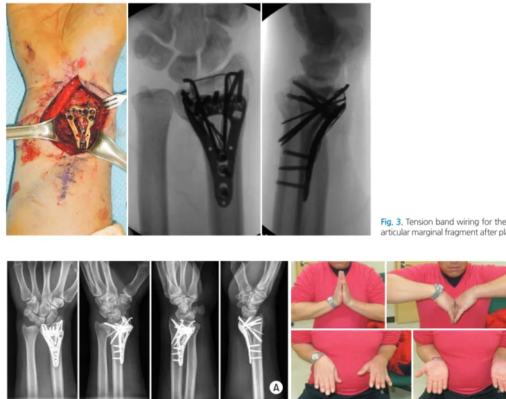
않도록 제 위치에 잘 위치시킨 후 금속나사를 삽입하여 고정 한다. 이때 골간부에 넣을 수 있는 금속나사 구멍 하나는 비 워 놓는다. 금속판 고정 후 강선을 관절 가장자리 골절편에 거치시켜 놓은 K-강선의 뒤로 통과시킨 후 금속판 전방 근위 부에서 교차시킨다. 앞서 비워 놓았던 골간부 금속나사 구멍 을 통해 피질골 나사를 삽입하고 완전히 조여 주기 전에 교차 시켜 놓았던 강선을 걸어 준다. 그 다음 강선을 매듭부에서 천천히 꼬아 압박력이 가해지도록 한 후 충분한 압박력이 걸 리면 피질골 나사를 완전히 조여서 고정해 준다. 고정이 끝나 면 K-강선을 최대한 골절편에 가깝게 구부리고 끝부분을 절 단한 다음 조금 더 전진시켜 최대한 돌출되지 않도록 해준다 (Fig. 3). 고정이 끝난 후 C-arm으로 고정상태를 확인한 후 창상 세척하고 절개 부위를 층별로 봉합하고 수술을 마친다.
A B
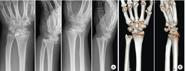
Fig. 1. (A) Volar articular marginal fragment (circles with the dotted line) with a comminuted metaphyseal fracture of the distal radius on plain radi- ography. (B) Three-dimensional computed tomography scan also shows the volar articular marginal fragment.
A B
Fig. 2. (A) Two K-wires were inserted from the distal edge of the volar articular marginal fragment to the posterior cortex proximally and posteriorly after reduction with pointed forceps through the modified Henry approach. (B) The position of two K-wires was checked on the anteroposterior and lateral view of the C-arm image.
1. 증례 1
추락 후 발생한 우측 원위 요골 골절로 응급실을 통해 내 원한 40세 남자 환자의 손목 단순방사선검사 및 컴퓨터 단층 촬영 결과 수장부에 골간단부 분쇄골절을 동반한 관절 가장 자리 골절이 관찰되었다(Fig. 1). 위에서 기술한 수술 술기대 로 장력대강선술식을 이용하여 고정한 후 별다른 합병증 없 이 수술 후 6주째 완전한 골유합을 얻었다(Fig. 4).
2. 증례 2
추락 후 발생한 우측 원위 요골 골절로 응급실을 통해 내 원한 46세 여자 환자의 손목 단순방사선검사 및 컴퓨터 단 층촬영 결과 골단부에 단순형의 관절 가장자리 골절이 관찰
되었다(Fig. 5). 위에서 기술한 수술 술기대로 장력대강선술 식을 이용하여 고정한 후 후방으로 돌출된 K-강선으로 손목 굴곡 시 약간의 자극증상이 있었으나 수술 후 2년 뒤 금속제 거를 하고나서 별다른 합병증 없이 완전히 기능 회복을 하였 다(Fig. 6).
고 찰
원위 요골 골절에 대해 전방접근법을 통해 해부학적 모양 에 맞게 설계된 잠김금속판을 이용한 고정 방법은 후방접근 법을 이용한 고정 방법에 비해 공간이 넓고 평평해서 금속판 을 대기 용이하고 혈류가 좋으며 대부분 원위 요골 골절 시 전방 피질골은 후방 피질골에 비해 분쇄가 심하지 않아 정복 이 쉬워 널리 사용된다.2) 하지만 전방 금속판 고정 시 금속판
Fig. 3. Tension band wiring for the volar articular marginal fragment after plating.
A B
Fig. 4. Radiologic union (A) and near full fuctional recovery (B) were achieved at postoperative 6 weeks.
이 watershed line의 원위부로 올 경우 굴곡건과 마찰을 일으 켜 합병증을 발생시킬 수 있으므로 반드시 watershed line의 근위부에 위치해야 하는데,2) watershed line의 원위부로 관절
가장자리 골절이 있을 경우 금속판을 통한 금속나사로는 이 골절편을 고정하기가 어렵다. 이러한 골절편에 대해서 손상이 없는 수장부 관절막 및 인대를 이용하여 강선 매듭만으로 고
A B
Fig. 5. (A) The simple volar articular marginal fragment of the distal radius on plain radiography. (B) Three-dimensional computed tomography scan also shows more clearly the volar articular marginal fragment.
A B
C D E
Fig. 6. (A) Immediate postoperative radiograph. Protrusion of the K-wire was seen on the oblique view (circle with the dotted line). (B) Complete union was seen on the postoperative 1-year X-ray. (C) Deficit of wrist flexion still remained due to protrusion of the K-wire at postoperative 1-year follow up. (D) Postoperative X-ray after implant removal. (E) The deficit of wrist flexion was full recovered after removal of the implant.
독으로 발생한 것이 아니라 원위 요골 골간단부의 주된 골절 편과 동반되어 수장부의 해부학적 잠김금속판과 같이 사용 을 해야 할 경우 동시에 고정을 하지 못할 가능성이 크다. 이 전에도 원위 요골 골절을 장력대강선술식으로 고정하여 치료 하는 술기가 소개된 적이 있으나5) 이는 원위 요골의 수장부 해부학적 잠김금속판이 개발되기 전에 주로 외고정기와 같이 사용하여 치료한 술기로 관절 가장자리 골절편에 사용된 술 기가 아니었다. 작은 피질골 나사를 이용하여 관절 가장자리 골절편을 따로 고정할 수도 있겠지만 후방부 피질골 분쇄가 동반된 원위 요골 골절의 경우는 반대편 피질골을 고정할 수 없어 고정력이 약해질 수 있으므로 손상 없이 골편에 붙어있 는 수장부 관절막과 인대를 같이 당겨서 고정할 수 있는 장력 대강선술식이 더 효과적일 것으로 생각된다.
하지만 원위 요골 골절에서 K-강선을 이용한 고정 방법은 많은 합병증을 초래할 수 있다.6) 특히 강선의 길이가 길어 후 방 피질골을 많이 넘어갈 경우 앞선 증례처럼 손목 후방에 자 극증상이 생겨 불편감을 호소할 수 있으며 심하면 신전건에 손상을 줄 수도 있다. 또한 K-강선 끝을 구부린 후 절단한 후 전전시켜서 전방피질골에 바짝 붙여 놓지 않으면 watershed line을 침범하여 정중신경이나 굴곡건을 자극할 수 있으므로 K-강선의 구부린 끝 부위가 피질골에 묻힐 정도로 확실하게 붙여주어야 한다. K-강선과 금속판의 간격이 좁을수록 역학 적으로 유리할 수 있으나 금속판의 위치는 watershed line의 근위부로 해부학적 모양에 따라 위치가 정해져 있으며 K-강 선의 경우는 watershed line의 원위부에서 삽입해야 하므로 간격이 조금 생길 수 밖에 없다. 또한 아직 임상 증례가 많지 않아 이 술기를 검증할 만한 생역학 연구나 더 많은 수의 증 례를 통한 임상결과 분석이 필요할 것으로 생각된다.
그럼에도 불구하고 수장측 관절 가장자리 골절을 동반한 원위 요골 골절에서 장력대강선을 이용한 고정 방법은 해부 학적 잠김금속판으로 고정이 어려운 골절편을 효과적으로 고 정하여 양호한 임상적 결과를 얻을 수 있는 유용한 방법이라
속판을 통한 고정이 어렵다. 지금까지의 문헌에서는 이러한 관절 가장자리 골절을 고정하기 위한 술기를 찾아 보기 어렵 다. 이에 관절 가장자리 골절편을 장력대강선술식을 이용하 여 고정하는 술기를 소개하고자 한다.
색인 단어: 원위 요골, 관절 가장자리 골절, 전방접근법, 장력
대강선술식ORCID
전능한, https://orcid.org/0000-0002-5509-8346 오종건, https://orcid.org/0000-0002-5184-6036 조재우, https://orcid.org/0000-0003-4962-3535 김영우, https://orcid.org/0000-0003-1851-7334
References
1. Conti Mica MA, Bindra R, Moran SL: Anatomic considerations when performing the modified Henry approach for exposure of distal radius fractures. J Orthop, 14: 104-107, 2017.
2. Orbay J: Volar plate fixation of distal radius fractures. Hand Clin, 21: 347-354, 2005.
3. Chin KR, Jupiter JB: Wire-loop fixation of volar displaced os- teochondral fractures of the distal radius. J Hand Surg Am, 24:
525-533, 1999.
4. Schumer ED, Leslie BM: Fragment-specific fixation of distal radius fractures using the Trimed device. Tech Hand Up Extrem Surg, 9: 74-83, 2005.
5. Katznelson A, Volpin G, Lin E: Tension band wiring for fixation of comminuted fractures of the distal radius. Injury, 12: 239- 242, 1980.
6. van Aaken J, Beaulieu JY, Della Santa D, Kibbel O, Fusetti C:
High rate of complications associated with extrafocal kirschner wire pinning for distal radius fractures. Chir Main, 27: 160-166, 2008.