DOI : 10.3341/jkos.2007.48.12.1711
안부속기에서 발생하는 림프구 증식성 질환(ocular adnexal lymphoproliferative lesion)은 대부분이 비호지킨성 B세포 림프종(non-Hodgkins B-cell lymphoma)으로 전체 결절외 림프종(extranodal lymphoma)의 약 2%를 차지하며 조직학적으로 양성 림 프구 과다증식(benign reactive lymphoid hyperplasia) 에서 악성 림프종(malignant lymphoma)까지 다양하 고 광범위한 연속선상의 질환군으로 여겨지고 있다.1
양성 림프구 과다증식은 항원의 자극에 의해 발생하 며 림프 조직이 가역적으로 커지는 질환으로 안부속기 에서 발생하는 림프구 증식성 질환의 13∼27% 정도를 차지하며 높게는 안와에 발생한 모든 림프구 증식성 질 환의 30%, 결막에 발생한 림프구 증식성 질환의 26%
까지 차지하는 것으로 보고된 바 있다.2-5 병리조직학적
으로는 작은 림프구로 이루어진 저등급의 악성 림프종 과 유사하나 림프구의 침윤 정도가 약하며 특징적으로 다클론성(polyclonality)을 보인다.6 스테로이드 사용 이나 방사선 치료에 잘 반응하며 림프구 증식성 질환 중에서 가장 예후가 좋은 것으로 알려져 있다.4,5,7
안부속기 림프구 증식성 질환에서 전신 림프종으로 이행하는 비율은 보고마다 다르나 Jakobiec et al8은 안부속기 악성 림프종의 경우 최대 60%까지 보고하고 있다. 일반적으로 악성도가 높은 경우(high grade)에 서 전신 림프종으로 이행하는 비율이 더 높은 것으로 알려져 있다.2,4,5,8 국내에도 안와 및 안부속기에 발생 한 악성 림프종으로 치료를 받고 전신 림프종으로 재발 한 환자는 있으나 양성 림프구 과다증식으로 진단 및 치료를 시행받고 관해가 이루어진 후 전신 악성 림프종 으로 재발한 보고가 없어 이를 보고하는 바이다.
증례보고
71세 여자 환자가 4년전부터 서서히 진행하는 우안 의 안검부종 및 안구돌출 증상을 주소로 내원하였다.
교정시력은 우안 0.8, 좌안 0.7이었으며 내원 당시 우 안 안검부종, 결막부종, 3 mm의 우안 안구돌출 소견 보 였다. 안운동 소견에서 제일안위에서 정위이나 동향운동 검사에서 우안의 경도의 외전 및 상전 장애를 보였다.
전신 악성 림프종으로 재발한 안부속기 양성 림프구 과다증식 1예
조영준1․박창준1․이성복1,2
충남대학교 의과대학 안과학교실1, 충남대학교병원 의과대학 의학연구소2
목적 : 안부속기 양성 림프구 과다증식 치료를 시행한 후 전신 악성 림프종으로 재발한 환자를 경험하였기에 이를 보 고하고자 한다.
증례요약 : 71세 여자 환자가 4년전부터 서서히 진행하는 우안의 상안검 부종 및 안구돌출을 주소로 내원하였다. 안 와 전산화 단층촬영에서 우안 외직근과 상직근의 종창이 보였으며 전신검사에서 이상 소견 보이지 않았다. 절개 생검을 시행하여 양성 림프구 과다증식으로 진단하였고 방사선 치료 후에 상안검 부종 및 안구돌출 증상은 사라졌다. 양성 림 프구 과다증식 치료 후 51개월이 지나 좌측 경부의 동통성 종괴를 주소로 다시 내원하여 경부 전산화 단층촬영을 시행 한 결과 좌측 경부의 림프절 증대와 좌측 인두벽이 비대칭적으로 두꺼워진 소견을 보였다. 절개 생검을 시행하였고 조 직검사에서 MALT 림프종으로 진단하였다. 경부 방사선 치료를 시행하였고 치료를 마친 4개월 후 시행한 경부 전산화 단층촬영에서 병소는 소실되었다.
결론 : 안부속기에서 발생한 양성 림프구 과다증식의 경우에도 타장기로의 재발 및 악성화 가능성이 있으므로 지속적 인 추적관찰 및 전신적인 평가가 필요하다고 생각된다.
<한안지 48(12):1711-1715, 2007>
<접수일 : 2007년 4월 29일, 심사통과일 : 2007년 10월 2일>
통신저자 : 이 성 복
대전시 중구 대사동 640 충남대학교병원 안과
Tel: 042-280-7608, Fax: 042-255-3745 E-mail: [email protected]
* 본 논문의 요지는 2005년 대한안과학회 제94회 추계학술대회 에서 포스터로 발표되었음.
= 증례보고 =
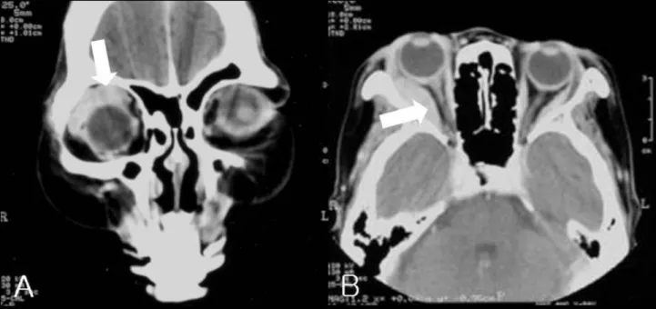
안와 전산화 단층 촬영상 우안 외직근과 상직근의 근 복(belly of muscle)과 건 부위까지 종창 소견이 나 타났다(Fig. 1). 내과에서 시행한 이학적 검사, 혈액 검사, 흉부, 복부 및 골반의 방사선 검사 및 전산화 단 층촬영 등에서 이상 소견은 보이지 않았다.
우안의 비대해진 외직근과 상직근을 포함한 안와주 위 조직을 절개 생검하였으며 절개 생검시 외직근과 상 직근의 주위 지방과의 경계는 깨끗하게 잘 유지되고 있 었고 안와주위 조직에는 이상소견 보이지는 않았다. 조
직 검사를 시행하였고 조직 검사상 소림프구의 증식 소 견이 보였다. CD3에 대한 조직면역염색에서 T림프구 의 증식소견을 보였고 CD20 조직면역염색에서는 B림 프구의 증식 소견을 보였다. 조직 검사에서 비정상적인 악성 림프구는 보이지 않아 B림프구와 T림프구가 혼재 된 양성 림프구 과다증식으로 진단하였다(Fig. 2). 스 테로이드 치료에 반응하지 않아 2000 cGy 용량의 방 사선 치료를 시행하였다. 치료를 마치고 2개월 후 우안 상안검 부종 및 안구돌출 증상은 사라졌으며 안와 전산 Figure 1. (A) There is enlargement of bellies of superior rectus muscle (arrow) and lateral rectus muscle on orbital computed tomography (CT) at the initial visit for the right eyelid swelling and proptosis. (B) Orbital CT shows the enlargement of belly of lateral rectus muscle (arrow).
Figure 2. Histopathologic findings of superior rectus muscle and lateral rectus muscle. (A) Photomicrograph of histopathologic specimen shows small lymphoid cells infiltration (hematoxylin-eosin stain, original magnification ×400). (B) Immunohistologic staining for CD3 shows hyperproliferation of T cells (immunoperoxidase stain, original magnification ×10). (C) Immunohistologic staining for CD20 shows hyperproliferation of B cell (immunoperoxidase stain, original magnification ×40). Consequently, benign reactive lymphoid hyperplasia was diagnosed.
화 단층촬영상 병소가 소실되었다.
양성 림프구 과다증식 치료 후 정기적인 추적관찰 중 재발로 의심되는 이상소견은 보이지 않았다. 51개월이 지났을 때 3개월 전부터 발생한 좌측 경부의 동통성 종 괴를 주소로 내원하였다. 경부 전산화 단층촬영에서 좌 측 경부의 림프절 증대와 좌측 인두벽이 비대칭적으로 두꺼워진 모습을 보였다(Fig. 3). 좌측 경부 림프절과 인두벽의 절개 생검을 시행하였고 조직 검사에서 소림프 구와 형질세포의 증식 소견을 보였고 kappa 및 lambda light chain에 대한 조직 면역 염색에서 lambda light
chain에 대한 단클론성(monoclonality)을 보여 경부 MALT 림프종으로 진단되었다(Fig. 4). 경부를 제외 한 다른 전신 부위에 대한 이학적 검사, 혈액 검사, 흉 부, 복부 및 골반의 방사선 검사 및 전산화 단층촬영 등에서 이상 소견은 보이지 않아 Ann Arbor classification상 stageⅡE로 분류되었다. 이후 3400 cGy의 경부 방사선 치료를 시행하였고 치료를 마친 4 개월 후 시행한 경부 전산화 단층촬영에서 병소는 소실 되었다. 이후 지속적인 추적관찰을 시행하였으며 경부 방사선 치료 후 30개월이 지난 현재 경부 및 안부속기
Figure 4. Histopathologic findings of left cervical lymph nodes and left oropharyngeal wall. (A) Photomicrograph of histopathologic specimen shows small lymphoid cells and plasma cells infiltration (hematoxylin-eosin stain, original magnification
×400). (B) Photomicrograph shows negative immunohistologic staining for kappa light chain (immunoperoxidase stain, original magnification ×200). (C) Photomicrograph shows strong and diffuse positive staining for lambda light chain (immunoperoxidase stain, original magnification ×200). Consequently, monoclonal MALT lymphoma was diagnosed.
Figure 3. Neck computed tomography scan shows enlargement of left cervical lymph nodes (arrows) (A) and asymmetrical thickening of left oropharyngeal wall (arrow) (B) at the visit for the left neck mass.
에서의 재발은 보이지 않고 있다.
고 찰
안와 및 안부속기에 발생하는 림프구 증식성 질환은 조직학적으로 악성 림프종이더라도 예후가 좋은 경우도 많아 양성과 악성의 구별이 쉽지 않으며 그 구분도 명 확하지 않다. 따라서 기존의 병리조직 검사 이외에 면역조직화학 검사가 반드시 필요하며 경우에 따라 southern blot이나 polymerase chain reaction 등을 이용하여 종합적으로 판별하고 있다.3,6
안부속기 림프구 증식성 질환에서 전신 림프종으로 재발을 예측하는 인자에 대해서는 아직까지 명확히 알 려져 있지 않다. Knowles et al4은 안부속기의 림프 구 증식성 질환에서 조직학적 분류나 면역학적 분류가 전신 림프종의 발생을 예측하는데 도움이 되지 않는다 고 보고하였고, Johnson et al9도 안와 림프종에서 면역학적 혹은 분자생물학적 분석이 전신 림프종 발생 을 예측하는 데에는 도움이 되지 않는다고 보고하였다.
안와 및 안부속기의 양성 림프구 과다증식의 재발에 대한 국외 보고를 보면 Knowles and Jakobiec2은 안와 림프구증식성 질환 환자 60명 중 양성 림프구 과 다증식으로 진단 받은 사람은 8명(13%)이었으며 이중 1명에서만 전신 림프종으로 진행하였다고 보고하였다.
Sullivan et al5은 안부속기에서 발생하는 림프구증식 성 질환 환자 69명에서 양성 림프구 과다증식은 9명 (13%)이었으며 그 중 1명에서는 20개월 후 전신 림프 종으로 진행하였으나 다른 8명의 환자에서는 재발이나 진행은 없었다고 보고하였다.
안와 및 안부속기의 악성 림프종 환자에 대한 국내 보고를 보면 Lee and Chung10은 안와 및 안부속기의 악성 림프종 환자 22안 중 안부속기에 림프종이 국한된 경우는 14안이며 이들 모두 치료 후 종양의 재발은 없 었다고 보고하였다. Jo et al11은 안부속기에 발생한 MALT 림프종 11명 13안을 조사한 결과 4안에서 원 발 부위에서 재발을 하였다고 보고하였다. Ji et al12 은 안와 및 안부속기에 악성 림프종으로 진단 받은 환 자 44명 54안에서 타장기로의 재발이 3안에서 관찰되 었으며 이들 3안 중 2안은 MALT 림프종, 1안은 diffuse large B-cell 림프종이었다.
국내에서 안와 및 안부속기의 양성 림프구 과다증식 은 몇 차례 보고되어 왔다.13,14 Lee et al13은 양성 림 프구 과다증식 4안을 포함한 안부속기의 림프구 증식성 질환 환자 55명 68안에서 원발 부위에서 재발은 7안, 전신 림프종으로 재발은 3안이었으며 모두 MALT 림 프종 환자에서 재발하였다고 보고하였다. 이들 55명중
양성 림프구 과다증식으로 진단받은 4안을 살펴보면 4 안 중 2안에서는 방사선 치료 후 평균 50.5개월의 추적 관찰동안 전신 림프종이 발생하지 않았고 2안에서는 치 료없이 평균 43개월 추적관찰한 결과 관해를 보이지는 않았으나 재발이나 진행은 없었다고 보고하였다. Lim et al14은 양성 림프구 과다증식 7명을 포함한 결막 림 프구 증식성 질환 환자 18명, 19안에서 전신 림프종이 동반된 환자는 2명으로 각각 small B 림프구성 림프 종이 한 명, MALT 림프종이 한 명이었다고 보고하였 다. 또한 결막에 발생한 양성 림프구 과다증식 환자 7 명 모두에서는 수술적 절제만으로도 재발이 없었다고 보고하였다.
지금까지 국내에서 안와 및 안부속기의 양성 림프구 과다증식으로 진단 및 치료를 시행 받고 관해가 이루어 진 후 전신 악성 림프종으로 재발한 증례는 아직까지 보고된 바 없다. 저자들은 안부속기 양성 림프구 과다 증식으로 치료 후 전신적으로 재발한 악성 림프종을 경 험하였기에 이를 보고하는 바이며 안부속기에서 발생한 양성 림프구 과다증식의 경우에도 타장기로의 재발 및 악성화 가능성이 있고 이를 예측할 수 없으므로 지속적 인 추적관찰 및 전신적인 평가가 필요하다고 생각된다.
참고문헌
1) Freeman C, Berg JW, Cutler SJ. Occurrence and prognosis of extranodal lymphomas. Cancer 1972;29:252-60.
2) Knowles DM, Jakobiec FA. Orbital lymphoid neoplasms: A clinicopathologic study of 60 cases. Cancer 1980;46:576-89.
3) Coupland SE, Krause L, Delecluse HJ, et al. Lympho- proliferative lesions of the ocular adnexa. Analysis of 112 cases. Ophthalmology 1998;105:1430-41.
4) Knowles DM, Jakobiec FA, McNally L, Burke JS. Lymphoid hyperplasia and malignant lymphoma occurring in the ocular adnexa (orbit, conjunctiva, and eyelids): a prospective multiparametric analysis of 108 cases during 1977 to 1987.
Hum Pathol 1990;21:959-73.
5) Sullivan TJ, Whitehead K, Williamson R, et al. Lympho- proliferative disease of the ocular adnexa: A clinical and pathologic study with statistical analysis of 69 patients.
Ophthalmic Plast Reconstr Surg 2005;21:3:177-88.
6) White WL, Ferry JA, Harris NL, Grove AS Jr. Ocular adnexal lymphoma: A clinicopathologic study with identification of lymphomas of mucosa associated lymphoid tissue type.
Ophthalmology 1995;102:1994-2006.
7) Bessell EM, Henk JM, Wright JE, Whitelocke RA. Orbital and conjunctival lymphoma treatment and prognosis. Radiother Oncol 1988;13:237-44.
8) Jakobiec FA, McLean I, Font RL. Clinicopathologic characteristics of orbital lymphoid hyperplasia. Ophthalmology
1979;86:948-66.
9) Johnson TE, Tse DT, Byrne GE Jr, et al. Ocular-adnexal lymphoid tumors: A clinicopathologic and molecular genetic study of 77 patients. Ophthalmic Plast Reconstr Surg 1999;15:171-9.
10) Lee JW, Chung WS. Treatment of orbital and adnexal malignant lymphoma. J Korean Ophthalmol Soc 2003;44:800-5.
11) Jo YJ, Yim JH, Park KS. MALT (mucosa associated lymphoid tissue) type lymphoma of the ocular adnexa. J Korean Ophthalmol Soc 2002;43:357-62.
12) Ji JY, Ahn YC, Kim YD. Radiotherapy for malignant lymphoma of orbit and ocular adnexa. J Korean Ophthalmol Soc 2005;46:201-14.
13) Lee YS, Lee MI, Park TS, Lee SY. The prognosis of ocular-adnexal lympholiferative lesions. J Korean Ophthalmol Soc 2003;44:1260-7.
14) Lim DW, Im SK, Yoon KC. Clinical features and treatment results of conjunctival lymphoproliferative lesions. J Korean Ophthalmol Soc 2004;45:1820-6.
=ABSTRACT=
A Case of Ocular Adnexal Benign Reactive Lymphoid Hyperplasia Recurred as Systemic Malignant Lymphoma
Young Joon Jo, M.D.1, Chang Jun Park, M.D.1, Sung Bok Lee, M.D.1,2
Department of Ophthalmology, College of Medicine, Chungnam National University1, Daejeon, Korea Research Institute for Medical Science, College of Medicine, Chungnam National University2, Daejeon, Korea
Purpose: We report a case of recurred systemic malignant lymphoma developed after the treatment for ocular adnexal benign reactive lymphoid hyperplasia.
Case summery: A 71-year-old female visited our hospital for right upper eyelid swelling and proptosis that had been progressing slowly for 4 years. Orbital computed tomography (CT) showed enlargement of the bellies of lateral and superior rectus muscles in the right orbit, but other abnormal findings were not detected in the systemic evaluation. Through incisional biopsy, benign reactive lymphoid hyperplasia was diagnosed on histopathologic examination. Upper eyelid swelling and proptosis resolved after radiation therapy. Fifty-one months after the treatment of benign reactive lymphoid hyperplasia, the patient visited our hospital again for a painful mass that had developed in the left neck. On neck CT, it showed enlargement of the left cervical lymph node and asymmetrical thickening of the left oropharyngeal wall. Systemic MALT lymphoma was confirmed, and radiation therapy was performed. According to neck CT, four months after radiation therapy, the enlargement and thickening resolved.
Conclusions: Because there is a possibility of systemic malignant lymphoma after benign reactive lymphoid hyperplasia, continuous follow-up and repeated systemic evaluation should be required after treatment of ocular adnexal benign reactive lymphoid hyperplasia.
J Korean Ophthalmol Soc 48(12):1711-1715, 2007
Key Words: Benign reactive lymphoid hyperplasia, Lymphoma, Orbital lymphoma
Address reprint requests to Sung Bok Lee, M.D.
Department of Ophthalmology, Chungnam National University Hospital
#640 Daesa-dong, Jung-gu, Daejeon 301-721, Korea
Tel: 82-42-280-7608, Fax: 82-42-255-3745, E-mail: [email protected]