pISSN: 0378-6471 eISSN: 2092-9374
DOI : 10.3341/jkos.2010.51.7.954
= 증례보고 =
당뇨황반부종 환자에서 백내장적출술과 유리체내 베바시주맙주입술 동시 수술의 효과
김부기⋅권의용⋅이동욱⋅안 민⋅조남천 전북대학교 의과대학 안과학교실
목적: 당뇨황반부종 환자에서 백내장적출술과 유리체내 베바시주맙주입술 동시 수술의 효과와 안전성을 알아보고자 하였다.
대상과 방법: 수정체초음파유화술과 인공수정체삽입술 시행 후 1.25 mg의 베바시주맙을 유리체 내로 주입하였고, 수술 전, 수술 후 1주일, 1개월, 3개월, 6개월에 최대교정시력과 중심황반두께를 측정하였고, 합병증 발생 여부를 조사하였다.
결과: 수술 전 평균 최대교정시력(LogMAR)은 0.84±0.50이었고, 평균 중심황반두께는 333.7±57.50 μm였다. 수술 후 평균 최대교정 시력은 1주째 0.52±0.40, 1개월째 0.51±0.42, 3개월째 0.52±0.34, 6개월째 0.46±0.37로 수술 전보다 유의하게 향상되었고, 평균 중심황반두께는 1주째 356.4±86.44 μm, 1개월째 338.8±138.4 μm, 3개월째 349.0±122.9 μm, 6개월째 334.2±100.4 μm이었다. 수 술 중과 수술 후에 합병증은 관찰되지 않았다.
결론: 백내장적출술과 유리체내 베바시주맙주입술의 동시 수술은 안전한 수술이고, 황반부종을 예방하는 데 있어서 1개월까지는 효과 가 좋으나, 3개월째부터는 황반부종의 예방효과가 미미하였다.
<대한안과학회지 2010;51(7):954-960>
■ 접 수 일: 2009년 4월 29일 ■ 심사통과일: 2010년 5월 18일
■ 책 임 저 자: 권 의 용
전북 전주시 덕진구 금암동 634-18 전북대학교병원 안과
Tel: 063-250-1965, Fax: 063-250-1965 E-mail: [email protected]
당뇨병은 백내장의 흔한 원인 중 하나이고, 65세 이하에 서 당뇨병이 있는 사람이 당뇨병이 없는 사람들에 비해 백 내장이 3~4배 정도 많이 발생한다고 알려져 있으며, 전체 백내장적출술 중에 당뇨병 환자에게 시행하는 백내장적출 술이 20%를 차지한다는 보고가 있다.1,2당뇨병망막병증 환 자에서 백내장적출술을 시행할 때는 포도막염, 후낭혼탁, 전낭수축, 황반부종, 당뇨망막병증의 악화 등의 합병증이 발생할 확률이 높다.3-6
황반부종은 백내장적출술 뒤 시력저하를 일으키는 중요 한 원인으로서,7백내장적출술 전에 황반부종이 없거나, 치 료가 잘 되던 환자에서 백내장적출술 후 황반부종이 발생 하였다면 저절로 빠른 시일 내에 없어지는 반면,8수술 전 치료에도 불구하고 지속적인 황반부종이 있거나, 발견하지 못했을 경우에는 황반부종이 잘 치료되지 않고, 시력은 저 하된다.9-11 수정체초음파유화술 후 발생하는 임상적으로 유의한 낭포황반부종은 0.1~12.0%로 알려져 있고, 형광안 저촬영에서는 3~70%까지 나타난다는 보고가 있다.12,13현 재까지 시도되고 있는 황반부종의 치료는 격자레이저광응
고술, 유리체절제술, 유리체내 스테로이드주입술(Intravitreal triamcinolone acetonide injection) 등이 있으나, 이같은 치 료에 반응하지 않고 영구적인 시력저하를 일으키기도 한 다.14-16
혈관내피성장인자(Vascular endothelial growth factor, VEGF)는 망막혈관의 밀착연접단백(Tight junction pro- tein)을 인산화시켜 망막혈관의 투과성을 증가시키는 작용 을 하는데, 당뇨망막병증과 같은 저산소증 상태에서 안구내 농도가 높아지고, 정상안에 혈관내피성장인자를 주입하였 을때 망막혈관의 투과성이 증가한다고 알려져 있다.17-19 그 리고 당뇨망막병증 환자에서 백내장적출술을 시행하였을 때 유리체내 혈관내피성장인자의 농도가 크게 증가한다는 보고가 있다.20 이같은 사실을 바탕으로 혈관내피성장인자 의 억제가 당뇨황반부종 및 백내장적출술 후 발생하는 황 반부종의 중요한 치료법으로 대두되고 있다. 혈관내피성장 인자의 단클론성 항체인 베바시주맙(Bevacizumab, Avastin®, Genentech Inc., San Francisco, CA)을 유리체 내로 주입 하여 당뇨황반부종과 백내장적출술 후 발생한 황반부종 치 료에 좋은 효과가 보고되었다.21-24
본 연구에서는 당뇨황반부종과 백내장으로 인해 시력이 저하된 환자들을 대상으로 백내장적출술을 시행하였고, 수 술 중 유리체내 베바시주맙을 주입하였다. 수술 뒤 경과관 찰하며 최대교정시력과 중심황반두께를 측정하였고, 수술 중, 수술 후 합병증 발생 여부를 관찰하여 백내장적출술과
유리체내 베바시주맙주입술 동반수술의 효과 및 안정성을 알아보고자 하였다.
대상과 방법
2007년 11월부터 2008년 11월까지 지속적인 당뇨황반 부종과 백내장으로 진단받은 환자들을 대상으로 수정체초 음파유화술, 인공수정체 삽입술, 유리체내 베바시주맙주입 술을 시행하였고, 이 중 6개월 이상 경과관찰 하였던 환자 14명, 14안을 대상으로 후향적으로 조사하였다. 수술 전 최 대교정시력과 안압 측정, 세극등현미경검사를 시행하였고, 안저검사, 형광안저촬영, 빛간섭단층촬영 등을 이용하여 당 뇨황반부종의 여부와 정도를 측정하였다.
1형 또는 2형 당뇨병 환자 중, 당뇨망막병증이 있으면서, 안저검사상 황반중심에서 1/2 유두지름의 원 안에 1유두 크기 이상으로 망막이 두꺼워지거나, 낭포성 변화 또는 확 산 부종이 있으며, 형광안저촬영상 황반 중심 1/2 유두 지 름 내에 형광 누출을 보이고, 빛간섭단층촬영상 황반부가 250 μm 이상으로 두꺼워져 있으면 당뇨황반부종으로 진단 하였고, 당뇨황반부종이 있으면서 Lens opacity classi- fication system Ⅲ (LOCS Ⅲ)의 분류에 의해 NO3, NC3 이상의 핵백내장을 가진 환자를 대상으로 수술을 시행하였 다. 녹내장이나 각막혼탁, 유리체출혈 외상 등의 시력저하 를 유발할 수 있는 다른 질병을 가지고 있거나, 유리체절제 술을 받은 기왕력이 있거나, 안압이 21 mmHg 이상 상승한 기왕력이 있거나, 수술 6개월 이내에 유리체내 스테로이드 주입술 또는 베바시주맙주입술을 받은 기왕력이 있는 경우 는 대상에서 제외하였다.
모든 대상안은 수술 전에 당뇨망막병증에 대한 치료를 받은 기왕력이 있었는데, 14안(100%)에서 범망막광응고 술, 11안(78.6%)에서 유리체내 베바시주맙주입술, 3안 (21.4%)에서 유리체내 스테로이드주입술, 3안(21.4%)에 서 격자레이저 광응고술을 시행받았다.
수술은 한 명의 숙련된 술자에 의하여 이루어졌으며, povidone iodine을 이용해 눈 주위와 결막낭을 소독한 뒤 12시 방향에 2.8 mm 크기의 투명각막절개를 시행하였고, 초음파유화술을 시행하였다. 연성접힘인공수정체를 삽입한 뒤 각막윤부로부터 2.5 mm의 섬모체 평면부에 30게이지 바늘을 이용하여 베바시주맙 1.25 mg (0.05 ml)을 유리체 내로 주입하였다. 수술 후 점안 항생제와 스테로이드 점안 액을 1일 4회 간격으로 점안하였다. 환자 중 수술 전에 안 압 상승의 기왕력은 없었으며, 경과관찰을 하면서 안압 측 정을 하여 안압이 21 mmHg 이상 상승하였을 경우에 안압 하강제를 점안하였다.
대조군은 당뇨망막병증이 있으면서 조사기간 동안 유리 체내 베바시주맙주입술 동반 시행 없이 수정체초음파유화 술과 인공수정체삽입술을 시행하고, 6개월 이상 경과관찰 시행한 환자로 정의 하였다. 대조군 중 시력 저하를 일으키 는 다른 질환이 있거나 6개월 이내에 유리체내 베바시주맙 또는 스테로이드주입술을 시행받은 눈은 제외하여서 대조 군은 12명, 12안이었다. 대조군 중 12안(100%)에서 범망 막광응고술, 5안(41.7%)에서 유리체내 베바시주맙주입술, 3안(25.0%)에서 격자레이저광응고술을 시행받은 기왕력 이 있었다.
수술 후 1주일, 1개월, 3개월, 6개월째 최대교정시력, 안 압검사, 세극등현미경검사, 안저검사 등을 시행하였고 빛간 섭단층촬영(Optical coherence tomography)을 이용하여 중심황반두께를 측정하여 수술 전과 비교하였으며 수술 후 발생할 수 있는 합병증 여부를 관찰하였다.
경과관찰 도중 중심황반두께가 수술 전에 비해 크게 상 승하고, 시력 저하가 발생하였을 때는 유리체내 스테로이드 주입술을 추가적으로 시행하였다.
시력은 용이한 분석을 위해 최대교정시력을 LogMAR (logarithm of the minimum angle of resolution)로 환산하 였고, 빛간섭단층촬영은 Stratus OCT version 3.0 (Carl Zeiss Meditec, Dublin, CA)를 사용하였다. 경과관찰 시 측정한 최대교정시력과 중심황반두께는 수술전과 paired T-test 를 이용하여 비교하였고, 통계적인 분석은 SPSS version 15.0을 사용하였으며, p-value가 0.05 미만인 경우를 통계 적으로 의의가 있는 것으로 정의하였다. 환자들에게 수술 전에 수술 과정과 가능한 합병증 등을 충분히 설명하였고 사전동의를 받았다.
결 과
수술안군의 평균 연령은 65.14±7.86세(범위: 52~80세) 였으며, 평균 경과관찰 기간은 297.12±84.14일(범위: 191
~342일)이었고, 남자가 8명, 여자가 6명이었다. 과거력상 대상안 모두에서 범망막광응고술을 시행받은 상태였고, 11 안(78.6%)에서 평균 1.55회의 유리체내 베바시주맙주입술 을 시행받은 기왕력이 있었으며, 유리체내 스테로이드주입 술은 3안(21.4%)에서 평균 1.67회 시행 받았다. 대조군의 평균 연령은 67.90±5.97세, 평균 관찰 기간은 316±90.57 일이었고, 남자가 4명, 여자가 8명이었다. 대조군의 과거 력상 모두에서 범망막광응고술을 시행받은 상태였고, 5 안(41.7%)에서 평균 1.8회의 유리체내 베바스주맙주입술 을 시행받은 기왕력이 있었다. 수술 전에 측정한 수술안군 의 평균 최대교정시력은 0.84±0.50, 평균 중심황반두께는
Table 1. Characteristics of patients
Characteristics Operated eyes Control Age (yrs)
Mean±SD* 65.14±7.86 67.90±5.97
Range 52~80 56~74
Gender (n)
Male 8 4
Female 6 8
Follow-up time (days)
Mean±SD 297.12±84.14 316±90.57
Range 191~342 205~498
Preoperative BCVA† (LogMAR‡)
Mean±SD 0.84±0.50 0.77±0.48
Range 0.3~2.3 0.4~1.0
p-value 0.37
Preoperative CMT§ (μm)
Mean±SD 337.1±57.50 302.1±56.65
Range 281~434 210~387
p-value 0.07
*SD=standard deviation; †BCVA=best corrected visual acuity;
‡LogMAR=logarithm of the minimum angle of resolution;
§CMT=central macular thickness.
Operative eyes Control
350
300 400
250
Preop 1 week 1 month 3 months 6 months
Figure 1. Changes in central macular thickness with OCT after cataract surgery combined with intravitreal bevacizumab injection. CMT=central macular thick- ness; OCT=optical coherence tomography.
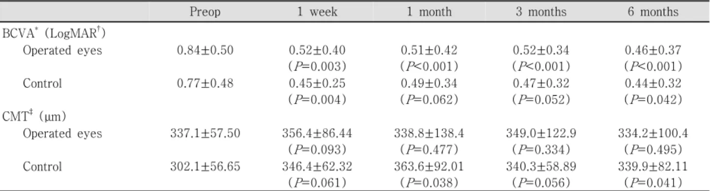
Table 2. Changes in BCVA and CMT after cataract surgery combined with intravitreal bevacizumab injection
Preop 1 week 1 month 3 months 6 months
BCVA* (LogMAR†)
Operated eyes 0.84±0.50 0.52±0.40
(P=0.003)
0.51±0.42 (P<0.001)
0.52±0.34 (P<0.001)
0.46±0.37 (P<0.001)
Control 0.77±0.48 0.45±0.25
(P=0.004)
0.49±0.34 (P=0.062)
0.47±0.32 (P=0.052)
0.44±0.32 (P=0.042) CMT‡ (μm)
Operated eyes 337.1±57.50 356.4±86.44
(P=0.093)
338.8±138.4 (P=0.477)
349.0±122.9 (P=0.334)
334.2±100.4 (P=0.495)
Control 302.1±56.65 346.4±62.32
(P=0.061)
363.6±92.01 (P=0.038)
340.3±58.89 (P=0.056)
339.9±82.11 (P=0.041)
*BCVA=best corrected visual acuity; †LogMAR=logarithm of the minimum angle of resolution; ‡CMT=central macular thickness.
337.1±57.50 μm였고, 대조군의 평균 최대교정시력은 0.77±
0.48, 평균 중심황반두께는 302.1±56.65 μm로 두 군 간에 통계적으로 유의한 차이를 보이지 않았다(P=0.373,0.066) (Table 1).
수술 후 수술안군의 평균 중심황반두께는 1주째 356.4±86.44 μm (P=0.093), 1개월째 338.8±138.4 μm (P=0.477), 3 개월째 349.0±122.9 μm (P=0.334)로 1주째 증가하는 경 향을 보이다가 1개월째는 감소하여 수술 전과 비슷한 정도 의 두께를 보였고, 3개월째는 다시 증가하였다. 수술 3개월 째의 중심황반두께는 1개월째에 비해 전반적으로 비슷하 였으나 3안에서 500 μm 이상으로 중심황반두께가 크게 증 가하여 유리체내 스테로이드주입술을 시행하였고, 6개월째 334.2±100.4 μm (P=0.495)로 다시 감소하는 소견을 보
였다. 대조군의 평균 중심황반두께는 수술 후 1주째 346.4±
62.32 μm (P=0.061), 1개월째 363.6±92.01 μm (P=0.038), 3개월째 340.3±58.89 μm (P=0.056), 6개월째 339.9±
82.11 μm (P=0.041)로 수술 1주째부터 증가하기 시작하 여 1개월째 가장 많이 증가하였고, 3개월째 감소하기 시작 하였으나 6개월째까지 수술 전에 비교하여 통계적으로 유 의한 상승을 보였다(Fig. 1, Table 2). 수술 후 평균 중심황 반두께를 수술 전과 비교하였을 때 수술안군은 1주째 28.21±
75.76 μm, 1개월째 1.64±88.54 μm, 3개월째 11.85±76.81 μm, 6개월째 -3±69.87 μm 였고, 대조군은 1주째 44.25±
72.98 μm, 61.5±104.35 μm, 38.17±77.07 μm, 37.58±
59.31 μm로 모든 조사기간 동안 수술안군이 대조군에 비 해 큰 변화를 보였다. 특히 대조군에서 1개월째 가장 큰 증 가를 보인 것에 반해 수술안군에서는 수술 전과 비교하였 을 때 1.64 μm의 작은 차이만을 보였다(Table 3).
Table 3. Difference in CMT after cataract operation from preoperative CMT
Operated eyes Control 1 week after operation
Mean±SD* (μm) 28.21±75.76 44.25±72.98
p-value 0.211
1 month after operation
Mean±SD (μm) 1.64±88.54 61.5±104.35
p-value 0.032
3 months after operation
Mean±SD (μm) 11.85±76.81 38.17±77.07
p-value 0.194
6 months after operation
Mean±SD (μm) -3±69.87 37.58±59.31
p-value 0.061
*SD=standard deviation.
Operative eyes Control 0.9
0.8 0.7 0.6 0.5 0.4 0.3 0.2 0.1
0
Preop 1 week 1 month 3 months 6 months
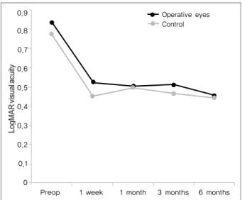
Figure 2. Changes in BCVA (LogMAR) after cataract surgery combined with intravitreal bevacizumab injection.
BCVA=best corrected visual acuity; LogMAR=loga- rithm of the minimum angle of resolution.
수술 후 수술안군의 평균 최대교정시력은 1주째 0.52±0.40 (P=0.003), 1개월째 0.51±0.42 (P<0.001), 3개월째 0.52±0.34 (P<0.001), 6개월째 0.46±0.37 (P<0.001)로 모두 수술 전보다 통계적으로 유의하게 향상되었다. 시력은 수술 후 3 개월째 중심황반두께가 증가함에 따라 약간 감소하는 소견 을 보였고, 3안에서 유리체내 스테로이드주입술을 시행한 후 다시 상승하였다. 대조군의 평균 최대교정시력은 1주째 0.45±0.25 (P=0.004), 1개월째 0.49±0.34 (P=0.062), 3개 월째 0.47±0.32 (P=0.052), 6개월째 0.44±0.32 (P=0.042)로 수술 전에 비해 1주째 통계적으로 유의한 시력의 상승이 있 었으나 중심황반두께가 가장 증가된 1개월째에 시력이 약 간 감소하는 경향을 보이다가 다시 6개월까지 안정되는 경 향을 보였다(Fig. 2, Table 2).
수술 중에 백내장적출술이나 유리체내 베바시주맙주입 술과 관련된 합병증은 발생하지 않았고, 수술 후 경과관찰 기간 동안에 안압상승, 안내염, 망막박리, 망막혈관폐쇄, 포 도막염 등의 합병증은 발견되지 않았다.
고 찰
황반부종은 백내장적출술 뒤 발생하는 시력저하의 중요 한 원인으로, 일반적으로 수술 뒤 6~8주 후에 임상적인 증 상이 나타나나 수개월 뒤 발생했다는 보고도 있다. 기전은 아직까지 명확하게 밝혀지지는 않았지만, 백내장수술 방 법,25 당뇨병,5 유리체탈출,12,13 포도막염,26 latanoprost 약 제의 사용,27광독성28등과 연관이 있는 것으로 생각되어지 고 있고 이 같은 인자들의 공통된 특징은 혈액-방수 장벽 손상과 연관된 안구내 염증을 일으키는 것이다.29Schalnus et al30은 백내장적출술에 의하여 염증 매개체들이 전방 내 에 유리되고, 이들이 유리체 내로 확산되어 망막혈관으로부
터 누출을 야기하여 황반부종을 일으킨다고 하였다. 특히 당뇨병 환자에서는 미세혈관장애가 있기 때문에 이미 혈액 -방수 장벽은 약화되어 있고, 백내장적출술에 의해 혈액- 방수 장벽의 손상이 심해져서 당뇨병이 없는 환자들에 비 해서 황반부종이 더 잘 나타난다고 한다.31,32
최근 백내장적출술 전후의 황반두께 변화를 빛간섭단층 촬영으로 측정하여 백내장적출술 후 황반부종의 발생율 및 정도를 평가하는 연구가 있었는데, von Jagow et al33은 당 뇨병이 없는 환자에서 수정체초음파유화술 후 중심황반두 께가 통계적으로 유의하게 증가하였다고 하였고, Kim et al34은 당뇨환자를 대상으로 수정체초음파유화술을 시행하 고 당뇨망막병증이 있는 군과 없는 군을 비교하였는데, 당 뇨망막병증이 있는 군이 없는 군에 비해 수술 후 중심황반 두께의 증가 정도가 심했고, 이는 시력 저하와 관련이 있었 다. 국내에서는 Wang and Choi35가 수정체초음파유화술 뒤 비증식성 당뇨망막병증 환자군과 당뇨병이 없는 환자군 모 두에서 수정체초음파유화술 후 중심황반두께가 증가한 것 을 보고하였다.
백내장적출술 뒤의 황반부종의 치료로는 격자레이저광 응고술을 시행하여 유의한 시력 상승과 형광안저촬영에서 누출의 감소 효과를 보았다는 보고가 있었으나,36 무작위 임상실험(randomized clinical trial)을 통한 효과가 보고된 적은 없었고, 안압상승, 망막박리 등의 합병증으로 한계성 이 있고, 유리체 내로 스테로이드를 주입하여 황반부종을 감소시키고 시력호전에 효과적이라고 보고되어 널리 사용 되고 있으나,37,38 안압상승, 안내염, 백내장, 망막박리 등의
합병증이 보고되어 주의를 요하고 있다.39-42
최근에는 당뇨황반부종 환자에서 백내장적출술을 할 때 유리체내 스테로이드주입술을 동시에 시행하여 좋은 효과 를 보였다는 보고가 있었는데, 통계학적으로 유의하게 시력 이 상승하였고 중심황반두께는 감소하였으며 시력에 위협 을 주는 합병증은 발생하지 않았다.43,44 그러나 Habib et al44은 백내장적출술과 함께 유리체내 스테로이드주입술을 시행받은 환자 중 33.3%가 안압이 21 mmHg 이상으로 상 승했고, 점안 안압하강제 치료가 필요하였다고 보고하였고, Lam et al43 역시 23.5%의 환자에서 일시적인 안압상승이 있었다고 하였다. 안압상승은 유리체내 스테로이드주입술 후 발생하는 주된 합병증 중 하나로써 약 40%에서 발생하 고 대부분은 7~9개월 후에 수술 전 안압으로 돌아오나 1~2%에서 안압하강제로 치료되지 않아서 섬유주절제술 등의 수술적 치료를 필요로 한다고 알려져 있다.45그리고 유리체내 스테로이드주입술 후 안압 상승이 발생하였다면 재주입술을 시행한 후 안압이 오를 가능성이 높다.46그러 므로 백내장적출술과 유리체내 스테로이드주입술의 동시 수술은 녹내장 환자, 특히 안압 조절이 불안정한 환자나 기 존에 유리체내 스테로이드주입술 후 안압상승이 있었던 환 자를 대상으로 하기에는 한계점이 있다.
Chen et al47은 당뇨황반부종 환자를 대상으로 백내장적 출술과 유리체내 베바시주맙주입술의 동시 수술을 보고하 였는데, 수술 전과 비교했을 때 통계적으로 유의한 시력상 승과 중심황반두께의 감소가 있었고, 백내장적출술만 시행 한 대조군에 비해 시력이 좋았으며 중심황반두께는 감소하 였다. 이는 백내장적출술과 유리체내 스테로이드주입술을 시행한 기존의 연구들과 비슷한 결과였고, 또한 안압 상승 을 포함하여 백내장적출술과 유리체내 베바시주맙주입술에 연관된 합병증은 발생하지 않았다.
본 연구에서 평균 중심황반두께는 1주째 증가하였고 1개 월째 감소하다가 3개월째는 다시 증가하였다. Wang and Choi35는 당뇨망막병증 환자에서 수정체초음파유화술 후 평균 중심황반두께가 1주째부터 증가하기 시작하여 1달째 가장 두꺼웠고, 6개월간 황반부종이 유지되었다고 보고하 였고, Seo and Park48은 유리체내 베바시주맙주입술 시행 후 당뇨망막부종이 감소하여 1개월째 가장 큰 효과를 보였 고 3개월째 중심황반두께가 증가하였다고 보고하였는데, 본 연구에서 유리체내 베바시주맙주입술로 백내장적출술 후 1개월째의 황반부종의 악화를 예방하였으나 3개월째에 는 베바시주맙의 효능의 감소로 인해 중심황반두께가 증가 했으리라 생각된다(Fig. 1, Table 2). 그리고 본 연구에서 수술 전의 중심황반두께와 비교하여 수술 1개월 후 중심황 반두께가 수술안군에서 1.64 μm만이 상승한 것에 비해 대
조군에서 61.5 μm가 상승한 것을 보았을 때, 백내장적출술 과 함께 시행한 유리체내 베바시주맙주입술이 수술 1개월 째 악화되는 황반부종을 예방하는 데 가장 큰 효과가 있음 을 알 수 있고, 수술 3개월 째 베바시주맙의 효능 감소로 인 해 수술안군에서 11.85±76.81 μm, 대조군에서 38.17±77.07 μm가 상승해서 두 군 간에 통계적으로 유의한 차이를 보이 지 않았다(P=0.194) (Table 3).
본 연구에서는 유리체내 스테로이드주입술 후에 빈번히 나타나는 안압 상승은 발생하지 않았으며, 수술 중과 수술 후 경과관찰 기간 동안에 망막박리, 안내염, 망막혈관폐쇄, 창상치유지연 등의 백내장적출술 및 유리체내 베바시주맙 주입술과 관계된 다른 합병증은 발생하지 않았다. 이는 Jonas et al49이 나이관련황반변성 환자를 대상으로 백내장 적출술과 유리체내 베바시주맙주입술을 동시에 시행하여 수술과 관련된 합병증이 발생하지 않았다는 보고와 일치한다.
당뇨병 환자는 당뇨병이 없는 사람들에 비해 면역력이 떨어지기 때문에 눈 수술 시 안내염에 대해 주의를 더욱 기 울여야 하는데,50 유리체내 베바시주맙주입술은 간단한 시 술로서 수술 시간에 거의 영향을 주지 않으면서 백내장적 출술에 추가적으로 시행이 가능하고, 흔하지는 않지만 심각 한 합병증인 안내염을 일으킬 수 있는 두 가지 수술을 동시 에 시행함으로써 안내염의 위험을 줄일 수 있으리라 생각 된다. 베바시주맙의 혈관형성억제효과로 수술 부위 치유 지 연이나 벌어짐이 나타날 가능성이 있지만 본 연구에서는 투명각막절개법을 사용했기 때문에 이 같은 합병증은 관찰 되지 않았다.49
결론적으로, 당뇨황반부종 환자에서 백내장적출술과 유 리체내 베바시주맙주입술의 동시 수술은 안전한 수술이고, 백내장적출술 후 빈번히 나타나는 황반부종의 악화를 예방 하는 데 있어서 1개월까지는 좋은 효과를 보였으나, 3개월 째부터는 베바시주맙의 효능 감소로 인해 황반부종의 예방 효과가 미미하였다.
참고문헌
1) Ederer F, Hiller R, Taylor HR. Senile lens changes and diabetes in two population studies. Am J Ophthalmol 1981;91:381-95.
2) Hamilton AM, Ulbig MVV, Polkinghorne P, et al. Epidemiology of di- abetic retinopathy. In management of diabetic retinopathy London:
BMJ Publishing Group. 1996:1-15.
3) Hykin PG, Gregson RM, Hamilton AM. Extracapsular cataract ex- traction in diabetics rubeosis iridis. Eye 1992;6:296-9.
4) Kato S, Oshika T, Numaga J, et al. Anterior capsular contraction after cataract surgery in eyes of diabetic patients. Br J Ophthalmol 2001;
85:21-3.
5) Dowler JG, Hykin PG, Lightman SL, Hamilton AM. Visual acuity fol-
lowing extracapsular cataract extraction in diabetes: a meta-analysis.
Eye 1995;9:313-7.
6) Chung J, Kim MY, Kim HS, et al. Effect of cataract surgery on the progression of diabetic retinopathy. J Cataract Refract Surg 2002;
28:626-30.
7) Rossetti L, Autelitano A. Cystoid macular edema following cataract surgery. Curr Opin Ophthalmol 2000;11:65-72.
8) Dowler JG, Sehmi KS, Hykin PG, Hamilton AM. The natural history of macular edema after cataract surgery in diabetes. Ophthalmology 1999;106:663-8.
9) Zaczek A, Olivestedt G, Zetterstrom C. Visual outcome after phacoe- mulsification and IOL implantation in diabetic patients. Br J Ophthalmol 1999;83:1036-41.
10) Krepler K, Biowski R, Schrey S, et al. Cataract surgery in patients with diabetic retinopathy: visual outcome, progression of diabetic retinop- athy, and incidence of diabetic macular oedema. Graefes Arch Clin Exp Ophthalomol 2002;240:735-8.
11) Chew EY, Benson WE, Remaley NA, et al. Results after lens extraction in patients with diabetic retinopathy: early treatment diabetic retinop- athy study report number 25. Arch Ophthalmol 1999;117:1600-6.
12) Flach AJ. The incidence, pathogenesis and treatment of cystoid mac- ular edema following cataract surgery. Trans Am Ophthalmol Soc 1998;96:557-634.
13) Milch FA, Yannuzzi LA. Medical and surgical treatment of aphakic cystoid macular edema. Int Ophthalmol Clin 1987;27:205-17.
14) Early Treatment Diabetic Retinopahty Study Research Group.
Photocoagulation for diabetic macular edema. Early Treatment Diabetic Retinopathy Study report number 1. Arch Ophthalmol 1985;
103:1796-806.
15) Pendergast SD, Hassan TS, Williams GA, et al. Vitrectomy for diffuse diabetic macular edema associated with a taut premacular posterior hyaloids. Am J Ophthalmol 2000;130:178-86.
16) Martidis A, Duker JS, Greenberg PB, et al. Intravitreal triamcinolone for refractory diabetic macular edema. Ophthalmology 2002;109:920-7.
17) Ferrara N. Vascular endothelial growth factor: Basic science and clin- ical progress. Endocr Rev 2004;25:581-611.
18) Aiello LP, Avery RL, Arrigg PG, et al. Vascular endothelial growth fac- tor in ocular fluid of patients with diabetic retinopathy and other reti- nal disorders. N Engl J Med 1994;331:1480-7.
19) Tolentino MJ, McLeod DS, Taomoto M, et al. Pathologic features of vascular endothelial growth factor-induced retinopathy in the nonhu- man primate. Am J Ophthalmol 2002;133:373-85.
20) Patel JI, Hykin PG, Cree IA. Diabetic cataract removal: postoperative progression of maculopathy-growth factor and clinical analysis. Br J Ophthalmol 2006;90:697-701.
21) Barone A, Prascina F, Russo V, et al. Successful treatment of pseudo- phakic cystoid macular edema with intravitreal bevacizumab. J Cataract Refract Surg 2008;34:1210-2.
22) Arevalo JF, Garcia-Amaris RA, Roca JA, et al. Primary intravitreal bev- acizumab for the management of pseudophakic cystoid macular ede- ma: pilot study of the Pan-American Collaborative Retina Study Group. J Cataract Refract Surg 2007;33:2098-105.
23) Kook D, Wolf A, Kreutzer T, et al. Long-term effect of intravitreal bevacizumab (Avastin) in patients with chronic diffuse diabetic mac- ular edema. Retina 2008;28:1053-60.
24) Haritoglou C, Kook D, Neubauer A, et al. Intravitreal bevacizumab (Avastin) therapy for persistent diffuse diabetic macular edema. Retina 2006;26:999-1005.
25) Ah-Fat FG, Sharma MK, Majid MA, Yang YC. Vitreous loss during conversion from conventional extracapsular cataract extraction to phacoemulsification. J Cataract Refract Surg 1998;24:801-5.
26) Fogla R, Biswas J, Ganesh SK, Ravishnkar K. Evaluation of cataract surgery in intermediate uveitis. Ophthalmic Surg Lasers 1999;30:191-8.
27) Warwar RE, Bullock JD, Ballal D. Cystoid macular edema and anterior uveitis associated with latanoprost use. Experience and incidence in a retrospective review of 94 patients. Ophthalmology 1998;105:263-8.
28) Henry MM, Henry LM. A possible cause of chronic cystic macu- lopathy. Ann Ophthalmol 1977;9:455-7.
29) Ursell PG, Spalton DJ, Whitcup SM, Nussenblatt RB. Cystoid macular edema after phacoemulsification: relationship to blood-aqueous bar- rier damage and visual acuity. J Cataract Refract Surg 1999;25:1492-7.
30) Schalnus RW, Ohrloff C, Magone T. The aqueous humor-vitreous body barrier and the blood-aqueous humor barrier after YAG laser capsulotomy in capsular sac vs ciliary sulcus fixation of the intraocular lens. Ophthalmologe 1995;92:289-92.
31) Miyak K. Indomethacin in the treatment of postoperative cystoid macular edema. Surv Ophthalmol 1984;28:554-68.
32) Yannauzzi LA, Landau AN, Turtz AI. Incidence of aphakic cystoid macular edema with the use of topical indomethacin. Ophthalmology 1981;88:947-54.
33) von Jagow B, Ohrloff C, Kohnen T. Macular thickness after un- eventful cataract surgery determined by optical coherence tomography.
Graefes Arch Clin Exp Ophthalmol 2007;245:1765-71.
34) Kim SJ, Equi R, Bressler NM. Analysis of macular edema after cata- ract surgery in patients with diabetes using optical coherence tomography. Ophthalmology 2007;114:881-9.
35) Wang SJ, Choi SH. The changes in macular thickness after phacoe- mulsification in patients with non-diabetes and nonproliferative dia- betic retinopathy. J Korean Ophthalmol Soc 2008;49:57-64.
36) Rosetti L, Autelitano A. Cystoid macular edema following cataract surgery. Curr Opin Ophthalmol 2000;11:65-72.
37) Tano Y, Sugita G, Abrams G, Machemer R. Inhibition of intraocular proliferations with intravitreal corticosteroids. Am J Ophthalmol 1980;
89:131-6.
38) Tano Y, Chandler D, Machemer R. Treatment of intraocular pro- liferation with intravitreal injection of triamcinolone acetonide. Am J Ophthalmol 1980;90:810-6.
39) Gillies MC, Simpson JM, Billson FA, et al. Safety of an intravitreal in- jection of Triamcinolone. Arch Ophthalmol 2004;122:336-40.
40) Jonas JB, Kreissig I, Degenring R. Intraocular pressure after intra- vitreal injection of triamcinolone acetonide. Br J Ophthalmol 2003;
87:24-7.
41) Moshfeghi DM, Kaiser PK, Scott IU, et al. Acute endophthalmitis fol- lowing intravitreal triamcinolone acetonide injection. Am J Ophthalmol 2003;136:791-6.
42) Islam MS, Vernon SA, Negi A. Intravitreal triamcinolone will cause posterior subcapsular cataract in most eyes with diabetic maculopathy within 2 years. Eye 2007;21:321-3.
43) Lam DS, Chan CK, Mohamed S, et al. Phacoemulsifi cation with intra- vitreal triamcinolone in patients with cataract and coexisting diabetic macular oedema: A 6-month prospective pilot study. Eye 2005;19:885-90.
44) Habib MS, Cannon PS, Steel DH. The combination of intravitreal tri- amcinolone and phacoemulsification surgery in patients with diabetic foveal oedema and cataract. BMC Ophthalmol 2005;5:15.
45) Jonas JB. Intravitreal triamcinolone acetonide for treatment of intra- ocular oedematous and neovascular diseases. Acta Ophthalmol Scand
=ABSTRACT=
The Results of a Combination of Cataract Surgery and Intravitreal Bevacizumab Injection for Diabetic Macular Edema
Bu Ki Kim, MD, Eui Yong Kweon, MD, Dong Wook Lee, MD, Min Ahn, MD, Nam Chun Cho, MD
Department of Ophthalmology, Chonbuk National University Medical School, Jeonju, Korea
Purpose: To evaluate the efficacy and safety of the combination of cataract surgery and intravitreal bevacizumab injection in patients with cataract and diabetic macular edema.
Methods: Patients received an intravitreal injection of bevacizumab(1.25 mg) combined with phacoemulsification and implantation of a posterior chamber intraocular lens. Best corrected visual acuity (BCVA, LogMAR) and, central macular thickness (CMT) were measured using OCT at baseline and at one week, one, three, and six months after surgery, and adverse events were recorded.
Results: The mean baseline LogMAR BCVA was 0.84±0.50 and mean CMT was 337.1±57.50 μm. At one week, one, three, and six months after surgery, the mean BCVAs were 0.52±0.40, 0.51±0.42, 0.52±0.34, and 0.46±0.37, and the mean CMTs were 356.4±86.44 μm, 338.8±138.4 μm, 349.0±122.9 μm, and 334.2±100.4 μm, respectively. No adverse events associated with cat- aract surgery or intravitreal bevacizumab injection were observed.
Conclusions: The short-term results from the present study suggest the combination of cataract surgery and intravitreal bev- acizumab injection are safe and effective for the prevention of macular edema aggravation for one month, but has little effect on prevention of macular edema aggravation three months after surgery for diabetic macular edema patients.
J Korean Ophthalmol Soc 2010;51(7):954-960
Key Words: Bevacizumab, Cataract surgery, Diabetic macular edema
Address reprint requests to Eui Yong Kweon, MD
Department of Ophthalmology, Chonbuk National University Hospital
#634-18 Geumam-dong, Deokjin-gu, Jeonju 561-712, Korea
Tel: 82-63-250-1965, Fax: 82-63-250-1965, E-mail: [email protected] 2005;83:645-63.
46) Jonas JB, Degenring R, Kreissig I, Akkoyun I. Safety of intravitreal high-dose reinjections of triamcinolone acetonide. Am J Ophthalmol 2004;138:1054-5.
47) Chen CH, Liu YC, Wu PC. The combination of intravitreal bev- acizumab and phacoemulsification surgery in patients with cataract and coexisting diabetic macular edema. J Ocul Pharmacol Ther 2009;25:83-9.
48) Seo JW, Park IW. Intravitreal bevacizumab for treatment of diabetic macular edema. Korean J Ophthalmol 2009;23:17-22.
49) Jonas JB, Spandau UH, Schlichtenbrede F, et al. Intravitreal bev- acizumab combined with cataract surgery for treatment of exudative macular degeneration. J Ocul Pharmacol Ther 2007;23:599-600.
50) Phillips WB 2nd, Tasman WS. Postoperative endophthalmitis in asso- ciation with diabetes mellitus. Ophthalmology 1994;101:508-18.