BIOMECHANICS OF ABUTMENTS SUPPORTING REMOVABLE PARTIAL DENTURES UNDER
UNILATERAL LOADING
Seong-Kyun Kim1, D.D.S., M.S.D., Ph.D., Seong-Joo Heo1, D.D.S., M.S.D., Ph.D., Jai-Young Koak1, D.D.S., M.S.D., Ph.D., Jeong-Taek Lee1, D.D.S., M.S.D., Ph.D., Hyun-Ki Roh1, D.D.S., M.S.D., Ph.D., Hyo-Jin Kim1, D.D.S., M.S.D., Ph.D., Seok-Hyung Lee2, D.D.S., D.M.D., Joo-Hee Lee3, D.D.S., M.S.D., Ph.D.
1Department of Prosthodontics and Dental Research Institute, College of Dentistry, Seoul National University, Korea
2Department of Prosthodontics, Samsung Medical Center, School of Medicine, Sungkyunkwan University, Korea
3Department of Prosthodontics, Asan Medical Center, College of Medicine, University of Ulsan, Korea
Statement of problem.In distal extension removable partial denture, the preservation of health of abutment teeth is very important, but abutment teeth are subjected to unfavorable stress under unilateral loading specially.
Purpose.The purpose of this study was to investigate the biomechanical effects of mandibu- lar removable partial dentures with various prosthetic designs under unilateral loading, using strain gauge analysis.
Material and methods. Artificial teeth of both canines were anchored bilaterally in a mandibular edentulous model made of resin. Bilateral distal extension removable partial den- tures with splinted and unsplinted abutments were fabricated.
Group 1: Clasp-retained mandibular removable partial denture with unsplinted abutments Group 2: Clasp-retained mandibular removable partial denture with splinted abutments by
6-unit bridge
Group 3: Bar-retained mandibular removable partial denture
Strain gauges were bonded on the labial plate of the mandibular resin model, approximately 2 mm close to the abutments. Two unilateral vertical experimental loadings (30N and 100N) were applied subsequently via miniature load cell that were placed at mandibular left first molar region. Strain measurements were performed and simultaneously monitored from a computer connected to data acquisition system. For within-group evaluations, t-test was used to com- pare the strain values and for between-group comparisons, a one-way analysis of variance (ANO- VA) was used and Tukey test was used as post hoc comparisons.
J Korean Acad Prosthodont : Volume 45, Number 6, 2007
※I acknowledge Seoul National University’s financial support for the new faculty.
A
s for the distal extension removable partial denture, the unilateral load to be given to the abutments was very important because most of strain was delivered in the off-vertical direction, which was disadvantageous to the abutments.There were several ways to judge whether a dis- tal extension removable partial denture was suc- cessful or not, but it was very important to keep the abutment and the residual alveolar ridge healthy. For successful remedy with the distal extension removable partial denture, efforts have been taken to disperse strain widely to keep abutments and other dental tissues healthy.1In order to disperse the strain that was given to the abutment, the abutment and its adjacent teeth were splinted for the distal extension remov- able partial denture. In the researches of move- ments of the splinted abutment, it was reported that mesio-distal force and movements of the bucco-lingual portion were reduced quite much,
but there was no change in the vertical movement of the abutment.2
Carlsson et al.3proposed abutment splinting for removable partial denture. And it was reported that how to splint the adjacent abutment was an important factor for distal extension removable partial denture when attachment retainer was used.4It was reported that the abutments of the distal extension partial denture, which were periodontally weak and moved, should be splint- ed.5However, there were many arguments over splinting natural teeth that were periodontally strong. Splinting was not applicable to the patients who took healthy chewing activities, having nor- mal periodontal ligament and little movements of teeth.
In comparison with fixed prosthesis, force act- ing on abutment supporting removable partial den- ture has claimed to increase the magnitude of moments. Particular interests, therefore, have been given to the influence of different pros- Results.The strain values of group 1 and 2 were tensile under loadings. In contrast, strain values of group 3 were compressive in nature. Strain values increased as the applied load increased from 30N to 100N (p<.05) except for right side in group 1. Under 30N loading, in left side, group 1 showed higher strain values than groups 2 and 3 in absolute quantity (p<.05). And group 2 showed higher strain values than group 1 (p<.05). In right side, group 1 and 2 showed high- er strain values than group 3 in absolute quantity (p<.05). Under 100N loading, in left side, group 1 showed higher strain values than groups 2 and 3 in absolute quantity (p<.05). And group 2 showed higher strain values than group 1 (p<.05). In right side, group 1 and 2 showed high- er strain values than group 3 in absolute quantity (p<.05). Under 30N loading, group 2 and 3 showed higher strain values in right side than in left side. Under 100N loading, right side strain values were higher than left side ones for all groups.
Conclusion.Splinting of two isolated abutments by bridge reduced the peri-abutment strain in comparison with unsplinted abutments under unilateral loading. Bar-retained removable partial denture showed the lowest strain of three groups, and compressive nature.
Key Words
Biomechanics, Strain, Abutment, Removable Partial Denture, Unilateral loading
thetic designs on strain magnitudes around abut- ment supporting removable partial denture. In vivo studies, unfortunately, were insufficient to derive constitutive descriptions concerning biome- chanical environment in bone tissue around abutments as measurements were carried not only above bone level, but also on prosthetic abutments, which do not represent the biome- chanical characterization of living bone.6Overall, it is noteworthy that controversy and lack of consensus still remains on intra osseous strain lev- els around splinted and unsplinted abutments sup- porting removable partial denture. Nevertheless, high success rates of mandibular removable par- tial denture supported by canines might sug- gest that high bone quality in the mandibular ante- rior region and decreased occlusal bite forces in elderly patients allow force distribution around abutments within physiologic levels.
This study intended to examine the influence of the methods of abutment splinting for the distal extension removable partial denture when uni- lateral loading was applied. For this, it has employed different designs of splinting and used strain gauge analysis to examine the pattern of strain that was shown on the labial plate around abutment. The purpose of this study was to investigate the biomechanical effects of mandibu- lar removable partial dentures with various pros- thetic designs under unilateral loading using strain gauge analysis.
MATERIAL AND METHODS
Fabrication of experimental mandibular model and removable partial dentures
An edentulous mandibular acrylic resin mod- el (Vertex SC, Dentimex, Zeist, Netherlands) was fabricated. On this model, anterior teeth were arranged according to the guidelines7estab-
lished for fabricating complete dentures to deter- mine canine locations. Indicator marks repre- senting canines were placed bilaterally. Following the removal of artificial teeth, the model was placed on the surveying table of a milling machine to prepare sockets. Artificial teeth of both canines (A5-500B, Nissin Dental Products Inc., Tokyo, Japan) were anchored into the sockets bilateral- ly. A 2-mm-thick layer was removed from the den- ture-supporting surface of the resin model and then replaced with polyvinylsiloxane impression material (Examix, GC corp., Tokyo, Japan) to simulate resilient edentulous ridge mucosa.
Canines were prepared according to prepara- tion guides. Surveyed two porcelain fused to metal crowns, a 6-unit porcelain fused to metal bridge and a bar (Hader bar, Cookson Co., La Chaux-de-Fonds, Switzerland) for abutments were fabricated. The removable partial dentures used in this study were classified as follows:
Group 1: Clasp-retained mandibular remov- able partial denture with unsplinted abutments
Group 2: Clasp-retained mandibular remov- able partial denture with splinted abutments by 6-unit bridge
Group 3: Bar-retained mandibular removable partial denture
Clasp- and bar-retained removable partial den- tures were fabricated and processed according to the principles established for processing remov- able partial dentures. RPA clasps were used for clasp-retained removable partial dentures. Three sets of removable partial dentures were fabri- cated for each case. Jig was fabricated at left first molar region to house miniature load cell (CSMN- 50L, Curiosity Technology, Seoul, Korea) for controlled experimental static loading. The load cell was connected to digital weight indicators (CTI- 1100A, Curiosity Technology, Seoul, Korea).
Quantification of strain
Linear strain gauges (120Ϊ; gauge length 1㎜;
KFG-1-120-C1-11, Kyowa, Japan) were bonded on the labial plate of the mandibular resin model, approximately 2 mm close to the abutments (Fig.
1).8The strain gauges were bonded with a special cyanoacrylate (M-Bond200, Vishay Micro- Measurements, Raleigh, NC, U.S.A.). The lead foils of the gauges were connected to bridge boxes (CTB- 100, Curiosity Technology, Seoul, Korea) via ter- minals. Each gauge was wired separately into a half Wheatstone bridge. The wires of the gauges were waterproofed by application of the air dry- ing polyurethane (MCoatA, Vishay Micro- Measurements, Raleigh, NC, U.S.A.). The strain gauges were then connected to cables. The cable led the signals to a dynamic signal conditioning strain amplifier (CTA-1000, Curiosity Technology,
Seoul, Korea) that was used to supply an excita- tion voltage in the Wheatstone bridge, thereby improving the signals. Two unilateral vertical experimental loadings (30N and 100N) were applied subsequently via miniature load cell that was placed at mandibular left first molar region.
The analog signals of electric resistance varia- tion were converted into digital signals via a 16 byte resolution converter (DAQCard-A1-16XE-50, National Instruments, Austin, U.S.A.) and processed by custom software (DA-1700B, Cas Korea, Seoul, Korea). Channel signals were orig- inally measured in millivolt and then converted to microstrain units (㎛/m). Measurement capa- bility was 1 ㎛/m. Ten measurements at each load were made under the same conditions, allowing at least 5 minutes for recovery. Each experiment was repeated on three sets of three different typed removable partial dentures.
Statistical analysis
For within-group evaluations, t-test was used to compare the strain values. For between-group com- parisons, a one-way analysis of variance (ANO- VA) was used to assess the differences and Tuchey test was used as post hoc comparisons.
RESULTS
Strain values around the abutments supporting removable partial denture under 30N and 100N Fig. 1. Measuring devices. Lined arrow indicates the loading
point and dotted arrows indicate the strain gauges.
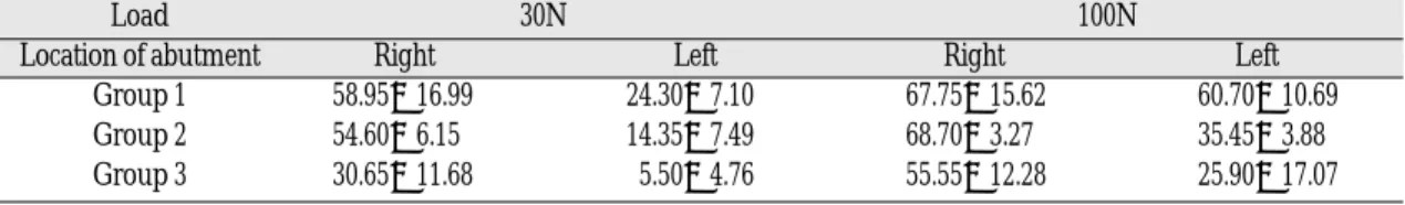
Table I. Microstrain on the labial plates around abutments supporting mandibular removable partial denture under unilateral left loadings
Load 30N 100N
Location of abutment Right Left Right Left
Group 1 58.95±16.99 24.30±7.10 67.75±15.62 60.70±10.69
Group 2 54.60±6.15 14.35±7.49 68.70±3.27 35.45±3.88
Group 3 30.65±11.68 5.50±4.76 55.55±12.28 25.90±17.07
loadings were presented in Table I. The strain val- ues of group 1 and 2 were tensile under 30N and 100N loadings. In contrast, strain values of group 3 were compressive in nature under 30N and 100N loadings. Within-group comparisons revealed that strain values increased as the applied load increased from 30N to 100N (p<.05) except for right side in group 1. Under 30N load- ing, in left side group, 1 showed higher strain val- ues than groups 2 and 3 in absolute quantity (p<.05). And group 2 showed higher strain values than group 1 (p<.05). In right side, group 1 and 2 showed higher strain values than group 3 in absolute quantity (p<.05). Under 100N loading, in left side, group 1 showed higher strain values than groups 2 and 3 in absolute quantity (p<.05).
And group 2 showed higher strain values than group 1 (p<.05). In right side, group 1 and 2 showed higher strain values than group 3 in absolute quantity (p<.05). Under 30N loading, group 2 and 3 showed higher strain values in right side than in left side. Under 100N loading, right side strain values were higher than left side ones for all groups. Splinting of two isolated canines by 6-unit bridge reduced the peri-abutment strain in comparison with unsplinted canines under uni- lateral loadings. Bar-retained removable partial denture showed the lowest strain of three groups, and compressive nature.
DISCUSSION
In the field of prosthodontics, when both canines of the lower jaw remained, crown designs were various and strain on the residual alveolar ridge and the abutments could be different. Designing an ideal crown, which minimize any burden to the residual alveolar ridge and the abutment, was nec- essary. And according to the status of the sup- porting tissue around canines, the quantity and quality of the residual alveolar ridge, and the
relationship with teeth, several treatment plans could be drawn up. The purpose of crown treat- ment, in case a small number of teeth remained, was to appropriately disperse strain of the abut- ment and the residual alveolar ridge in order to delay the edentulous status as possible as could and further, to preserve the function of the mouth as long as possible. Especially, canines were like- ly to remain to the end so to preserve canine teeth was very important in preventing the eden- tulous status and degenerative change of the lower jaw.
The load to be given to denture was delivered to abutment, surrounding alveolar bone and residual alveolar tissues of the denture muscle. Because of the difference in elasticity of alveolar periosteum and mucogingiva, distribution of strain happening in the denture muscle was various and analysis of strain by change in the denture design was very complicated.9By analyzing any change in strain dis- tribution on the denture supporting areas, an ide- al denture that performed its functions without giv- ing any harm to abutment, tissues around teeth and other oral structure should be considered. If exter- nal force was given to an object, the distribution type of internal strain became different, depending on the direction of the force, and the shape and mate- rial of the object receiving the external force. This internal strain could cause transformation in the object and in case the strain was big, it could cause permanent transformation or destruction.10 Especially, the lower jaw was very sensitive to this strain and was likely to cause degenerative change because its supporting areas were quite much reduced compared with that of the upper jaw, and the lower jaw in the edentulous status could not stand load better than the upper jaw in terms of its shape and structure. Therefore, in case both canine teeth remain in the lower jaw, it was sig- nificant to maintain and preserve the teeth.
Although the masticatory loads in mandibular removable partial denture are smaller than those in either the natural dentition or fixed restorations, removable partial denture is subject to both axi- al and transverse forces, the latter being smaller, but potentially more harmful. This study used the one-point concentration load, since it was almost impossible to reproduce crewing pattern by in vit- ro experiments. In this study, strain values of group 1 and 2 were tensile under 100N and 200N loadings. In contrast, strain values of group 3 were compressive in nature under 100N and 200N loadings. The difference of strain nature could be due to difference of retentive methods of removable partial denture. As for clasp-retained removable partial denture, pulling force hap- pened on abutment under loadings. In contrast, as for bar-retained removable partial denture, pressing force happened on abutment via cingulum rest under loading. In this case, pulling force could happen at center of bar on that retentive clip mounted. The tensile stress on abutment could be more harmful. Strain values on labial plate around unsplinted abutment were higher in compari- son with those of bridge-splinted abutment.
Increased strain values around unsplinted abut- ment could be critical because occurring defor- mations could be close or even pass the tolerance that would be deleterious to abutment.
CONCLUSION
The purpose of this study was to investigate the biomechanical effects of mandibular removable partial dentures with various prosthetic designs under unilateral loading, using strain gauge analysis. Artificial teeth of both canines were anchored bilaterally in a mandibular edentu- lous model made of resin. Bilateral distal exten- sion removable partial dentures with splinted and unsplinted abutments were fabricated.
Group 1: Clasp-retained mandibular remov- able partial denture with unsplint- ed abutments
Group 2: Clasp-retained mandibular remov- able partial denture with splinted abutments by 6-unit bridge
Group 3: Bar-retained mandibular removable partial denture
Strain gauges were bonded on the labial plate of the mandibular resin model, approximately 2 mm close to the abutments. Two unilateral ver- tical experimental loadings (30N and 100N) were applied subsequently via miniature load cell that were placed at mandibular left first molar region.
The strain values of group 1 and 2 were tensile under loadings. In contrast, strain values of group 3 were compressive in nature. Under 30N loading, in left side, group 1 showed higher strain values than groups 2 and 3 in absolute quantity (p < .05). In right side, group 1 and 2 showed higher strain values than group 3 in absolute quantity (p < .05). Under 100N loading, in left side, group 1 showed higher strain values than groups 2 and 3 in absolute quantity (p < .05).
In right side, group 1 and 2 showed higher strain values than group 3 in absolute quantity (p < .05).
Splinting of two isolated abutments by bridge reduced the peri-abutment strain in compari- son with unsplinted abutments under unilateral loading.
REFERENCES
1. Frechette A. Influence of partial denture design on distribution of forces on abutment teeth. J Prosthet Dent 1956;6:195-212.
2. Altay OT, Tsolka P, Preisket HW, Abutment teeth with extracoronal attachments: The effects of splinting on tooth movement. Int J Prosthodont 1990;3:441-8.
3. Carlsson GE, Hedegard B, Koivumaa KK. Studies in partial prosthesis IV, Final result of a 4-year lon- gitudinal investigation of dentogingivally sup-
ported partial dentures. Acta Odontal Scand 1965;23:443-69.
4. Kratochvil FJ, Thompson WD, Caputo AA.
Photoelastic analysis of stress patterns on teeth and bone with attachment retainers for removable partial dentures. J Prosthet Dent 1981;46:21-8.
5. Aydinlik E, Dayangac B, Celik E. Effect of splint- ing on abutment tooth movement. J Prosthet Dent 1983;49:477-80.
6. E1-Sheikh AM, Hobkirk JA. Force transmission in bar-retained implant-stabilized mandibular over- dentures: an in-vitro study. Eur J Prosthodont Restor Dent 2002;10:173-8.
7. Hickey JC, Zarb GA, Bolender CL. Boucher’s prosthodontic treatment for edentulous patients.
9th ed. St. Louis: CV Mosby; 1985. p. 352-66.
8. Asundi A, Kishen A. A strain gauge and photoe-
lastic analysis of in vivo strain and in vitro stress distribution in human dental supporting structures.
Arch Oral Biol 2000;45:543-50.
9. McCarteny JW. Motion vector analysis of an abut- ment for a distal-extension removable partial den- ture. A pilot study. J Prosthet Dent. 1980;43:15-21.
10. Loard JL and Teel S. Photoelasticity as a research technique for analyzing stress in dental struc- tures. J Dent Res 1955;34:831-7.
Reprint request to:
JOO-HEELEE, D.D.S., M.S.D., PH.D.
DEPARTMENT OFPROSTHODONTICS,ASANMEDICALCENTER, COLLEGE OFMEDICINE, UNIVERSITY OFULSAN,
388-1, PUNGNAP-DONG,SONGPA-GU,SEOUL, 138-736, KOREA [email protected]