J Korean Soc Coloproctol Vol. 20, No. 1, 2004
64
Mechanism of Genomic Instability and Its Clinical Applications
Suk-Hwan Lee, M.D., Ph.D.
Department of Surgery, Kyung Hee University College of Medicine, Seoul, Korea
Multiple genetic alterations are common prerequisite for carcinogenesis including colorectal cancers (CRCs). Recently, mutations within microsatellites have been described as a result of defective DNA mismatch repair (MMR) mechanisms, resulting in the phenomenon of microsatellite instability (MSI). This has been implicated in the etiology of hereditary non-polyposis colorectal cancer (HNPCC) and significant portions of sporadic colorectal cancers. However, the me- chanisms underlying the MSI are different from hereditary CRCs and sporadic CRCs. While the germline mutation of MMR genes is responsible for HNPCC, the hypermethylation of MLH1 gene promoter regions, an epigenetic, not in- herited alteration is responsible for most sporadic CRCs showing MSI. MSI tumors exhibit characteristic clinco- pathologic features, i.e, tumors are preferentially located to proximal to splenic flexure, poorly differentiated, mucinous cell type, frequently showing peritumoral lymphocytic infil- tration, and, of importance, showing better survival in stage- matched cases. In this article, the results of recent in- vestigations about MSI and its clinical applications are com- prehensively reviewed. Knowledge of these biochemical mechanisms are likely to lead to more effective diagno- sis and therapy of CRCs in the future. J Korean Soc Coloproctol 2004;20:64-73
Key Words: Genomic instability, Microsatellite instability, He- reditary non-polyposis colorectal cancer, Epige- netic alteration
유전자 불안정성, 현미부수체 불안정성, 가 족성 비용종증대장암, 에피제네틱 변화
서 론
인간은 외부의 환경과 상호 관계를 통해 끊임없는 진 화의 과정을 밟아 왔으며, 이러한 진화는 유전자의 자연 의 선택(evolution by natural selection)의 결과이다. 진화 과 정은 먼저 유전 정보의 무작위 변이가 일어나 후대로 전 해지며, 변이된 유전 정보가 생체의 생존과 번식을 위해 선택적으로 변화되는 것이다(clonal selection). 분자생물 학 연구 기법의 발달로 암 역시 이러한 유전자의 변화에 의해 일어나는 질환으로 인식되고 있다.
정상세포가 암세포로 되기까지는 유전자에 여러가지 변화를 일으켜야 한다. 즉 성장억제 또는 활성인자에 독 립적이어야 하며, 이상 클론의 선택과 확장(clonal selec- tion and expansion), 불멸화(immortalization), 세포고사 기전 의 불활성화, 혈관 신생, 세포 분화와 노화 기전의 회피 및 전이라는 다단계의 과정을 거쳐야 한다고 알려지고 있다.1
정상세포의 경우, 돌연변이원(mutagen)이 완전히 배제 된 환경하에서 우발적인 돌연변이의 발생 확률은 1.4×
10-10∼10-6 mutations/base pair/cell generation 정도로 매 우 낮다. 누드 마우스를 이용한 동물실험의 경우에는 2개 내지 3개의 돌연변이만으로도 암 발생이 가능하다. 그러 나 인간의 암세포에서 보이는 평균 100,000개 정도의 수 많은 돌연변이는 일반적인 산술 계산으로는 설명이 불가 능한 경우가 된다.2
Loeb는 인간 세포에서 일어나는 일상적인 돌연변이율 에 비추어 볼 때, 종양세포에서 발견되는 수많은 돌연변 이의 집적은 산술적으로 불가능한 일임을 지적하였으며, 이러한 불가능한 상황이 발생하는 원인을 ‘과돌연변이성 (hypermutability)’ ‘유전자 불안정성(genetic instability)’ 또는
‘돌연변이 표현형(mutator phenotype)’이라는 가설로 설명 하였다. 즉 정상 세포가 종양세포로 변하기 위해서는 우 선 세포 내의 유전자들이 쉽게 돌연변이를 일으키는 상
유전자 불안정성의 기전과 임상적 적용
경희대학교 의과대학 외과학교실
이 석 환
책임저자: 이석환, 서울시 동대문구 회기동 1번지 경희의료원 외과(우편번호: 130-702) Tel: 02-958-8266, Fax: 02-966-9366 E-mai: [email protected]
태, 즉 불안정한 상태로 변해야 한다는 것이다.3 본문에서는 유전자 불안정의 기전 중 현미부수체 불안 정성(microsatellite instability, 이하 MSI)과 이들의 임상적 적용에 대해 최근 연구들을 중심으로 기술하고자 한다.
본 론
1) 현미부수체(microsatellite), 현미부수체 불안정성 (microsatellite instability)의 정의
현미부수체는 유전체(genome)에 광범위하게 분포하고 있는 1∼5 bp의 DNA 조각들이다. 전체 유전체의 30%가 현미부수체로 이루어져 있다. 대표적인 mononucleotide microsatellite는 13개의 adenine이 반복된 형태이며 (A)13이 라고 표시한다. 사람에게서 가장 흔한 현미부수체는 dinucleotide인 cytosine과 adenine의 반복으로 (CA)n이라 고 표시한다. 또한 trinucleotide 반복은 감수분열시기에 주로 확장되는데, trinucleotide 반복에 이상이 초래될 경 우 Hungtington's disease나 fragile X syndrome과 같은 질병 을 유발한다.4 이러한 현미부수체 중 유전되며, 개개인에 따른 차이를 보이는 경우를 다형성 현미부수체(polymor- phic microsatellite)라 하며, 다형성 현미부수체는 매우 중 요한 유전적 표지자의 역할을 하는데, 법의학 분야에서 친자구별을 하거나, 암을 비롯한 다양한 질병의 원인 유 전자를 찾는 데 이용된다.
현미부수체 불안정성(MSI)이란 부정합 교정 유전자 (mismatch repair gene 이하 MMR gene)의 배선 돌연변이
(germline mutation)로 인해 유전자 전체에 걸쳐 존재하는
반복염기 서열인 현미부수체에 삽입, 탈락과 같은 체성 돌연변이(somatic mutation)가 일어나 DNA의 길이가 달라 지는 현상을 말한다. MSI는 replication error (RER)+, MIN (mutational instability) 등으로 혼동하여 사용되지만 본문에서는 MSI로 표기하도록 한다.
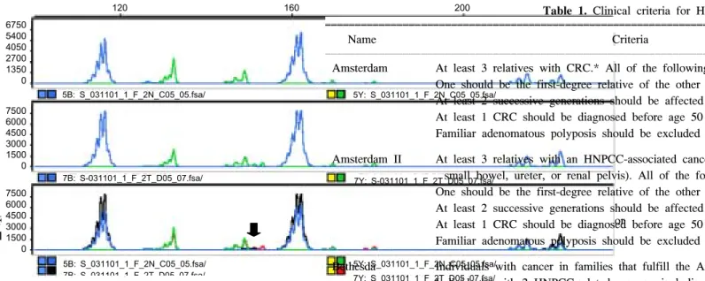
현미부수체 불안정성을 측정하기 위해서는 여러가지 종류의 현미부수체 표지자를 이용할 수 있으나 대장암의 경우는 National Cancer Institute Workshop on Microsatellite Instability에서는 5개의 현미부수체 표지자(BAT25, BAT26, D2S123, D5S346, D17S250)를 반드시 검사할 것을 권장하 고 있으며,5 5개의 표지자 중 2개 이상에서 불안정성을 보일 경우를 MSI-high (MSI- H), 5개 중 1개에서만 불안정 성을 보일 경우를 MSI-low (MSI-L), 불안정성이 없는 경 우를 MSS (microsatellite stable) 등으로 정의하고 있다 (Fig. 1∼3).
MMR gene의 이상으로 생기는 돌연변이는 점상 돌연 변이(point mutation), 염기쌍의 삽입 및 탈락, 염색체 분절 이나 전체 소실, 전위, 중복과 같이 매우 다양하며 유전자 검사는 비용과 시간이 매우 많이 소요되고 적절한 유전 학적 상담이 요구되므로 임상에서 유전자 검사를 받아야 하는 환자군을 선택하는 것은 매우 중요하다. 이를 위해 1991년 International Collaborative Group on HNPCC에서 가족성 비용종증 대장암(Herediatary Non-Polyposis Colorectal Cancer, 이하 HNPCC) 환자군의 선별을 위한 Amsterdam criteria를 발표하였다.6 그러나 이 기준 역시 환자군의 선 별에 너무 엄격하다는 비판이 있어 최근 International
Collaboration Group에서는 대장암 뿐 아니라 소장암, 자 Fig. 1. Example of microsatellite stable (MSS) tumor. All 5 markers from normal colonic mucosa (upper) and tumor tissue (lower) showed same DNA fragment pattern.
8000 6000 4000 2000 0
6400 4800 3200 1600 0
120 160 200
1B: S_031117_2_F_34T_A03_01.fsa/ 1Y: S_031117_2_F_34T_A03_01.fsa/
16B: S_031117_2_F_34N_H02_16.fsa/ 16Y: S_031117_2_F_34N_H02_16.fsa/
궁내막암, 요관암과 같은 extracolonic cancer를 추가한 Amsterdam criteria II를 발표하였으나,7 Amsterdam criteria I과 Amsterdam criteria II사이에 돌연변이 발견율은 각각 50%와 52%로 차이가 없는 것으로 알려지고 있다.8 위암 은 우리나라와 일본의 HNPCC가계에서 흔히 동반되지만 위암의 발병률이 서구에 비해 높아 extracolonic cancer에 포함되지 않았다. 또한 미국 국립 암 연구소에서는 HNPCC와 연관된 암의 90% 이상에서 MSI를 보이는 점에 착안하여 Bethesda guideline을 제시하였으며9 HNPCC 환 자 선별을 위해 MSI 검사가 필요한 환자군을 정의하였다 (Table 1).
2) 유전자 불안정성의 기전
대장암을 포함한 여러 종류의 암은 세포의 성장과 세 포 고사를 조절하는 유전자들에 일련의 돌연변이가 발생 하여 해당 유전자들의 기능이 증폭되거나 소실되어 일어 난다고 알려져 있다.10
유전자 불안정성은 두 가지 기전을 통하여 일어난다고 한다. 그 하나는 가족성 용종증의 경우로 adenomatous polyposis coli (APC) 유전자의 이상에 의해 여러가지 종류 의 oncogene과 종양억제 유전자에 돌연변이가 발생하여 염색체의 구조적, 수적 이상을 유발하는 상황으로 염색 체 불안정성(chromosomal instability)으로 정의하고, 또 다 른 하나는 HNPCC의 경우로, 유전자 복제과정에서 MMR
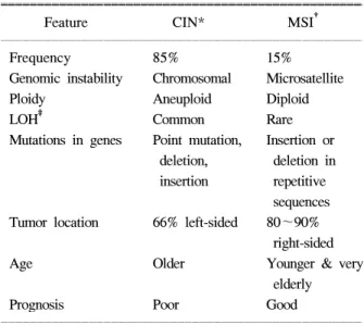
gene의 이상에 의해 DNA 복제 과정 중 실수로 만들어진 염기쌍이나 염기고리가 적절히 교정되지 못함으로써 많 은 유전자에서 점상 돌연변이나 작은 삽입, 탈락들이 발 생하게 된다. 특히 같은 종류의 염기가 반복되는 단순 반 복염기서열인 현미부수체(microsatellite) 구조에서는 자주 DNA 중합효소의 미끄럼 현상이 일어날 수 있다. 전자의 경우를 염색체 불안정성 경로(chromosomal instability pathway, CIN pathway)라 하며, 후자를 현미부수체 불안 정성 경로(microsatellite instability pathway, MSI pathway) 라 분류하며, 세포 유전학적, 임상적으로 상이한 특징을 나타낸다(Table 2).
대부부의 현미부수체는 intron에 존재하지만, 일부 유 전자들은 exon 부위에 현미부수체를 포함하고 있어서 MSI 형질을 갖고 있는 세포에서 흔히 돌연변이를 일으킨 다. 이러한 유전자들 중 세포의 성장, 분열, 세포 주기에 직접 또는 간접적으로 관여하거나, 세포고사를 조절하는 기능을 갖고 있는 경우는 그 돌연변이로 인하여 본연의 Fig. 2. Example of high-level microsatellite instability (MSI-H). Fragment pattern of amplified PCR products from a tumor that is unstable at four analyzed microsatellite loci except D5S346 (arrow).
6000 3000 0
2400 1600 800 0
12B: S_031116_1_F_3T_F01_11.fsa/ 12Y: S_031116_1_F_3T_F01_11.fsa/
10B: S_031116_1_F_3N_E01_09.fsa/ 10Y: S_031116_1_F_3N_E01_09.fsa/
120 160 200
Fig. 3. Example of low-level microsatellite instability (MSI-L). Fragment pattern of amplified PCR products from tumor that is unstable at one microsatellite locus (D2S123) (arrow).
6750 5400 4050 2700 1350 0
7500 6000 4500 3000 1500 0
7500 6000 4500 3000 1500 0
120 160 200
5B: S_031101_1_F_2N_C05_05.fsa/ 5Y: S_031101_1_F_2N_C05_05.fsa/
7Y: S-031101_1_F_2T_D05_07.fsa/
7B: S-031101_1_F_2T_D05_07.fsa/
5Y: S_031101_1_F_2N_C05_05.fsa/
7Y: S 031101 1 F 2T D05 07.fsa/
5B: S_031101_1_F_2N_C05_05.fsa/
7B: S 031101 1 F 2T D05 07 fsa/
Table 1. Clinical criteria for HN ꠚꠚꠚꠚꠚꠚꠚꠚꠚꠚꠚꠚꠚꠚꠚꠚꠚꠚꠚꠚꠚꠚꠚꠚꠚꠚꠚꠚꠚꠚꠚꠚꠚꠚꠚꠚꠚꠚꠚꠚꠚꠚꠚꠚꠚꠚꠚꠚꠚꠚꠚꠚꠚꠚꠚꠚꠚꠚꠚꠚꠚꠚꠚꠚ
Name Criteria
ꠏꠏꠏꠏꠏꠏꠏꠏꠏꠏꠏꠏꠏꠏꠏꠏꠏꠏꠏꠏꠏꠏꠏꠏꠏꠏꠏꠏꠏꠏꠏꠏꠏꠏꠏꠏꠏꠏꠏꠏꠏꠏꠏꠏꠏꠏꠏꠏꠏꠏꠏꠏꠏꠏꠏꠏꠏꠏꠏꠏꠏꠏꠏꠏ Amsterdam At least 3 relatives with CRC.* All of the following One should be the first-degree relative of the other At least 2 successive generations should be affected At least 1 CRC should be diagnosed before age 50 Familiar adenomatous polyposis should be excluded Amsterdam II At least 3 relatives with an HNPCC-associated cance
small bowel, ureter, or renal pelvis). All of the fo One should be the first-degree relative of the other At least 2 successive generations should be affected At least 1 CRC should be diagnosed before age 50 Familiar adenomatous polyposis should be excluded Bethesda Individuals with cancer in families that fulfill the Am
Individuals with 2 HNPCC-related cancers, including CRCs or associated extracolonic cancers
Individuals with CRC and a first-degree relative with extracolonic cancer and/or colorectal adenoma; 1 o <45 yr and the adenoma diagnosed at <40 yr Individuals with CRC or endometrial cancer diagnos Individuals with right-sided CRC with an undifferent histopathology diagnosed at <45 yr
Individuals with signet ring cell-type CRC diagnosed Individuals with adenomas diagnosed at <40 yr ꠏꠏꠏꠏꠏꠏꠏꠏꠏꠏꠏꠏꠏꠏꠏꠏꠏꠏꠏꠏꠏꠏꠏꠏꠏꠏꠏꠏꠏꠏꠏꠏꠏꠏꠏꠏꠏꠏꠏꠏꠏꠏꠏꠏꠏꠏꠏꠏꠏꠏꠏꠏꠏꠏꠏꠏꠏꠏꠏꠏꠏꠏꠏꠏ
*CRC = colorectal cancer.
단백질 기능이 변화되어 암이 발생하는 것이다. 가장 대 표적인 예가 BAX 유전자와 transforming growth factor II receptor (이하 TGFIIR) 유전자이다. BAX 유전자는 단백 질 해독 부위에(G)8의 현미부수체를 포함하고 있으며, MMR gene의 이상이 있는 경우 DNA 복제 과정 중 한 개 혹은 두 개의 guanine의 탈락이 일어나며 이로 인해 틀변 이(frame shift)가 일어나 BAX 유전자의 기능이 소실되며, 이로 인해 Bcl-2 매개의 세포 고사 경로가 억제되어 암이 발생한다. TGFIIR 의 경우에도(A)10 현미부수체를 포함하 고 있으며 불활성화될 경우 암화 과정 중 가장 중요한 암 억제 유전자의 기능 소실이 일어난다.
3) 현미부수체 불안정성의 기전
(1) 부정합 교정 유전자의 배선 돌연 변이: HNPCC는 가족성 대장암의 한 유형으로 일반 대장암에 비해 젊은 나이에 발생하며, 동시성 대장암(synchronous colorectal cancer)과 이시성 대장암(metachronous colorectal cancer)의 발생빈도가 높고, 우측 대장에 호발하며, 분화도가 나쁜 대장암의 비율이 높고, 예후가 좋으며, 자궁내막암, 난소 암, 위암, 간담도암 등 extracolonic tumor가 발생한다고 하 여 진단 및 치료에 매우 중요한 의미가 있다.1 그러나 진 단을 위해서는 가족성 용종증과 달리 질병의 특징적인 표현형인 용종증이 없으므로 전적으로 환자의 가족력에 의존하여 진단해야 하는 문제점이 있다. HNPCC 환자 의 50∼70%에서 hMLH1이나 hMSH2의 배선 돌연변이가 있다고 알려지고 있으며, 이와 같은 배선 돌연변이가 있
는 경우의 90%에서 MSI 형질을 획득하게 되고 상기한 바와 같이 exon 부위에 현미부수체 구조를 가진 일련의 유전자들이 체성 돌연변이를 일으키면서 빠른 속도로 암 이 발생하게 된다.5,11-14
HNPCC는 hMLH1, hMSH2, hPMS1, hPMS2, hMSH6와 같은 MMR gene의 배선 돌연 변이에 의해 유발되며, 90%
이상에서 MSI를 보인다고 하여 HNPCC 진단의 효율성을 높이기 위해 고위험군 환자에서 MSI assay를 권장하고 있 다.5,15
HNPCC 환자에서 발견되는 대부분의 배선 돌연변이는 hMLH1과 hMSH2 유전자라고 알려지고 있으나, 최근 들 어 MSI-L를 보이는 경우 MSH6 유전자의 배선 돌연변이 를 보인다는 보고도 있다.16-19 MSS tumor와 MSI-H tumor 는 상기한 바와 같이 서로 상이한 임상적 특징을 보이지 만 아직까지 MSI-L tumor의 역할에 대해서는 많이 알려 져 있지 않다. Jass 등은 대장암의 전암단계인 증식성용종 이나 serrated adenoma에서 MSI-L를 보이는 경우가 많으 며 이는 MSS tumor와 MSI tumor의 중간 정도인 ‘mild mutator pathway’의 증거라고 주장하였다.20-22 그러나 MSI assay에 사용된 현미부수체 표지자의 수가 증가함에 따라 MSI-L 표현형을 보이는 대장암의 비율이 증가하므로 아 직까지 MSI-L tumor의 생화학적, 임상적 의미에는 논란 이 많다.23
(2) CpG island mutator pathway: 대장암의 가족력이 없는 산발성 대장암의 15% 정도가 MSI를 보이지만 대부 분의 경우 MMR gene의 배선 돌연변이를 발견할 수 없다.
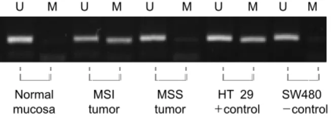
산발성 대장암 중 MSI tumor 역시 HNPCC와 유사한 임상 적 특성을 보인다. 즉 우측 대장암의 빈도가 높으며, 여자 에서 호발하고, 미분화암의 분포가 높으며, 병기에 따른 예후가 우수하다는 것이다. 산발성 대장암 중 MSI tumor 는 hMLH1 단백의 면역조직화학 염색에서 단백의 발현이 안 되는 경우가 많으며, 그 기전으로서 hMLH1 유전자 의 촉진자(promoter) 부위에 CpG island의 cytosine 메틸 화에 의한 것이라고 한다(Fig. 4).24-27 유전자의 기능 소 실이 단백질의 발현과 관련된 유전자의 변이 없이 촉진 자의 과메틸화에 의해 단백질의 기능이 소실되는 현상 을 epigenetic pathway라고 하며, 이러한 경우를 “CpG island methylator phenotype (CIMP) 또는 hypermethylator phenotype”이라고 칭한다.28,29 촉진자 부위의 메틸화는 5- aza-2'-dexoycytidine과 같은 methyl transferase inhibitor에 의 해 가역적으로 단백질이 재발현 된다고 하며 이를 치료 에 이용하려는 시도가 많이 있다.30
Table 2. Comparison of two pathways ꠚꠚꠚꠚꠚꠚꠚꠚꠚꠚꠚꠚꠚꠚꠚꠚꠚꠚꠚꠚꠚꠚꠚꠚꠚꠚꠚꠚꠚꠚꠚꠚꠚꠚꠚꠚꠚꠚꠚꠚꠚꠚꠚꠚꠚꠚꠚꠚꠚ
Feature CIN* MSI†
ꠏꠏꠏꠏꠏꠏꠏꠏꠏꠏꠏꠏꠏꠏꠏꠏꠏꠏꠏꠏꠏꠏꠏꠏꠏꠏꠏꠏꠏꠏꠏꠏꠏꠏꠏꠏꠏꠏꠏꠏꠏꠏꠏꠏꠏꠏꠏꠏꠏ
Frequency 85% 15%
Genomic instability Chromosomal Microsatellite
Ploidy Aneuploid Diploid
LOH‡ Common Rare
Mutations in genes Point mutation, Insertion or deletion, deletion in insertion repetitive
sequences Tumor location 66% left-sided 80∼90%
right-sided
Age Older Younger & very
elderly
Prognosis Poor Good
ꠏꠏꠏꠏꠏꠏꠏꠏꠏꠏꠏꠏꠏꠏꠏꠏꠏꠏꠏꠏꠏꠏꠏꠏꠏꠏꠏꠏꠏꠏꠏꠏꠏꠏꠏꠏꠏꠏꠏꠏꠏꠏꠏꠏꠏꠏꠏꠏꠏ
*CIN = chromosomal instability; †MSI = microsatellite instability; ‡LOH = loss of heterozygosity.
촉진자의 메틸화에 의한 단백질 발현의 억제는 주로 hMLH1 gene에서 일어나며, hMSH2 gene에서는 매우 드 문 것으로 알려지고 있다. Wheeler 등의 연구에 의하면, MSI tumor의 50%에서 hMLH1 유전자의 촉진자 과메틸화 가 있는 데 반해 MSS tumor 경우에는 단지 6%에서만 부 분적인 메칠화가 있으며, 이러한 경우는 단백질 발현의 소실은 없다고 한다.31
4) 유전자 불안정성의 임상적 적용
유전자 불안정성은 암발생의 첫번째 과정이며, 필수조 건이다. 유전자 불안정성은 현재 대장암을 포함한 많은 종류의 암에서 진단, 예후 예측, 치료의 표적 또는 치료 예측인자로 이용하기 위한 많은 시도가 이루어지고 있 다. 유전자 불안정성에 관여하는 기전들의 수많은 단백 질들의 상호 과정으로 이루어져 있으며, 이러한 단백질 중 상당수는 암 치료의 표적으로도 이용될 수 있다. 유전 자 불안정성의 임상 적용 예들을 진단, 예후 및 치료의 세 부분으로 나누어 기술하고자 한다.
(1) 진단: HNPCC는 전체 대장암의 13% 정도를 차지하 는 것으로 알려지고 있으며, 동시성 및 이시성 대장암의 발생 빈도가 높으므로 정확한 진단에 의거한 수술적 치 료 및 술 후 추적관찰이 필요하다. 그러나 상기한 바와 같이 용종증과 같은 임상에서 쉽게 찾을 수 있는 표현형 이 없으므로 진단에 많은 어려움이 있다. 모든 대장암 환 자에게 MMR gene의 배선돌연 변이 검사를 하는 것은 불 가능하며, 유전자 검사의 결과가 환자들의 일상생활에 불리하게 작용할 가능성도 있으므로 유전자 검사는 매우 신중하게 진행을 하여야 한다. HNPCC 환자군의 선별을
위한 진단 기준과 권고안에 의거할 경우에도 비용-효율 의 측면을 반드시 고려해야 한다. MSI assay의 경우, DNA 추출 및 증폭과정과 같은 여러 단계의 분자 생물학적 검 사가 동반되어야 하므로 임상적용에는 어려움이 많은 실 정이다. 면역조직화학 염색법은 임상에서 비교적 쉽게 적용할 수 있는 검사 방법으로 최근의 연구에 의하면 MSI assay와 일치율이 매우 높은 것으로 알려지고 있다 (Fig. 5).11,32 즉 고위험군 환자에 대한 면역조직화학 검사 후 MMR 단백의 발현이 없는 때 유전자 진단을 하는 것 이 효율적일 것이다.33
유전자 불안정성을 포함한 대장암에서 흔히 발견되는 분자생물학적 이상들은 대장암의 조기 검진에도 이용할 Fig. 4. Methylation of CpG islands in the MLH1 promoter
was determined by a methylation-specific PCR assay using bisulfate DNA modification. This reaction replaces cytosine with uracil in unmethylated DNA. Methylated DNA se- quences remain unaltered. The presence of a visible PCR product in those lanes marked M indicates the presence of methylated alleles. Normal colonic mucosa is unmethylated (U). DNA from HT 29 cells served as positive controls; from SW 480 cells, as negative controls.
U M U M U M U M U M
ꠐ ꠐ ꠐ ꠐ ꠐ ꠐ ꠐ ꠐ ꠐ ꠐ
ꠌꠏꠏꠏꠏꠎ ꠌꠏꠏꠏꠏꠎ ꠌꠏꠏꠏꠏꠎ ꠌꠏꠏꠏꠏꠎ ꠌꠏꠏꠏꠏꠎ
Normal MSI MSS HT 29 SW480
mucosa tumor tumor +control -control
Fig. 5. The expression of MLH1 (A and B) and MSH2 (C and D) was deter Avidin-Biotin-Peroxidase method. Sections were incubated overnight at 4oC 1:50, Clone G168-15, PharMigen, San Diego, CA) and MSH2 (dilution 1 Representative samples of tumors which expressed MLH1 (A) and MSH2 (D). In tumors did not express the MLH1 and MSH2, only the normal colo were stained as shown and considered as internal positive controls.
수 있는데, 최근 분변 DNA를 이용한 대장암의 선별검사 가 가능하게 되었으며 현재 대규모 인구 집단을 대상으 로 한 임상연구가 진행되고 있다.34,35 대장암에서 흔히 발 견되는 K-ras, p53, APC 유전자 및 MSI 표지자인 BAT 26 를 이용한 최근의 연구 결과에 의하면, 대장암 선별의 감 수성이 91%이며, 특이도는 93∼100%로 보고하고 있어 향후 연구 결과가 주목된다.34
(2) 예후 예측: HNPCC를 포함한 MSI tumor가 MSS tumor 보다 예후가 좋다는 것으로 알려지고 있다. 그러나 전통적인 예후인자들과 분자생물학적 예후인자 간의 상 관성에는 아직까지 설명할 수 없는 부분들이 많이 있다.
세포의 분화도가 나쁜 대장암 즉 미분화암이나 점액성 대장암은 일반적으로는 예후가 나쁜 것으로 알려지고 있 으나, MSI tumor들은 이러한 비분화암의 분포가 높으면 서도 좋은 예후를 보이고 있다.
예후에 관한 초기 연구들은 MSI tumor의 병기가 대개 낮으며, 원격전이가 적다고 보고하고 있으나, 병기별로 비교한 결과에서도 분명히 생존율이 높은 것으로 나타나 며,36 예후인자들의 다변량 분석 결과 MSI tumor의 상대 위험도가 0.67이라고 하는데 이는 진단 후 사망률이 MSS tumor에 비해 2/3 정도 수준이라는 것을 의미한다.37 MSI tumor의 예후가 좋은 생물학적 원인 규명은 아직 까지 불충분하지만, 크론병에서 보이는 양상과 유사한 종양 주변 림프구의 침윤이 면역체계의 방어 기전의 한 가지일 것이라는 주장이 있으며,38 Shibata 등은 이들 종양 이 덜 공격적이라고 하였다.39
(3) 치료: MSI와 관련된 in vitro 연구들은 MSI tumor가 alkylating agent에 잘 반응하지 않으며 예후도 나쁠 것으 로 보고하였으나, 최근의 대규모 임상 연구 결과들은 MSI tumor의 양호한 예후와 항암제 감수성과 관련이 있 다고 한다.40-43,45
여러 연구 결과에서 아직까지는 논란의 여지가 많지만 MSI 표현형을 보이는 암이 5-Fluorouracil (이하 5-FU) based chemotherapy에 반응성이 우수한 것으로 알려지고 있다.40,42,46
Watanabe 등의 보고에 의하면 5-FU based chemo- therapy를 받은 460명의 Dukes 병기 2기와 3기 환자의 생 존율 비교 연구 결과, MSI군의 5년 생존율이 64%, MSS군 의 5년 생존율이 49%로 통계학적으로 유의한 차이가 있 었으며, 다변량 분석을 통하여 MSI와 TGFIIR의 돌연변이 가 독립적인 예후인자라고 주장하였으며, 이러한 경우 5-FU based chemotherapy에 감수성이 높아져 향상된 예후
를 보일 것이라고 추정하였다.47
또한 Hemminki 등은 95명의 5-FU based chemotherapy를 받은 Dukes 병기 C 환자를 대상으로 한 연구에서 MSI군 의 3년 무병생존율이 90%로 MSS군의 43%에 비해 월등 히 좋은 예후를 보였다고 보고하면서 MSI군의 환자들이 5-FU based chemotherapy에 우수한 반응을 보인 결과라고 하였다.42
원격 전이가 있는 4기 대장암 환자들을 대상으로 한 고 용량 항암요법을 실시한 nonrandomized prospective study 에서 MSI군의 생존기간의 중앙치가 24개월로 MSS군의 13개월보다 통계학적으로 월등히 길었으며, 고용량 항암 제의 반응률 역시 65.7%와 35.1%로 유의한 차이가 있었 다고 한다. 다변량 분석 결과, 고용량 항암요법과 MSI 표 현형이 환자들의 생존율에 관계하는 독립적인 예후인자 였다고 보고하면서 MSI tumor의 예후가 좋은 원인은 암 종의 생물학적 공격성이 낮다기 보다는 항암제 감수성이 우수한 결과라고 주장하였다.46
그러나, 대장암 환자들 만을 대상으로 한 가장 최근 연 구에 의하면, MSI 표현형을 보이는 대장암 환자들이 오 히려 5-FU 항암요법에서 생존율이 낮았다고 보고하는 등 MSI 표현형을 포함한 분자 생물학적 표지자들과 항암제 감수성 간에는 아직까지 논란이 많은 실정이다.48 대부분 의 연구들이 후향적 연구이며 환자군의 선택에 편견이 개입될 가능성이 있으며, MSI 검사에 사용된 표지자들이 연구마다 상이하며, MSI의 정의에도 차이를 보이는 경 우도 있으므로 결과의 해석에는 주의를 기울여야 할 것 이다.
결 론
대장암 발암과정에 있어 유전자 불안정성은 가장 기본 적인 변화이다. 이러한 변화의 올바른 이해는 대장암 발 암과정에 대한 폭 넓은 연구를 통해 새로운 원인 유전자 의 규명을 가능케할 뿐 아니라 대장암의 조기 진단, 치료 및 예후 예측을 가능하게 할 것이며 머지않은 미래에 환 자 개개인에게 특성화된 치료(tailored treatment)에도 이용 될 전망된다.
REFERENCES
1. Kinzler KW, Vogelstein B. Lessons from hereditary colo- rectal cancer. Cell 1996;87:159-70.
2. Boland CR, Ricciardiello L. How many mutations does it take to make a tumor? Proc Natl Acad Sci USA 1999;
96:14675-7.
3. Loeb LA. Microsatellite instability: marker of a mutator phenotype in cancer. Cancer Res 1994;54:5059-63.
4. de la Chapelle A. Microsatellite instability. N Engl J Med 2003;349:209-10.
5. Boland CR, Thibodeau SN, Hamilton SR, Sidransky D, Eshleman JR, Burt RW, et al. A National Cancer Institute Workshop on Microsatellite Instability for cancer detection and familial predisposition: development of international criteria for the determination of microsatellite instability in colorectal cancer. Cancer Res 1998;58:5248-57.
6. Vasen HF, Mecklin JP, Khan PM, Lynch HT. The In- ternational Collaborative Group on Hereditary Non-Polyposis Colorectal Cancer (ICG-HNPCC). Dis Colon Rectum 1991;34:
424-5.
7. Vasen HF, Watson P, Mecklin JP, Lynch HT. New clinical criteria for hereditary nonpolyposis colorectal cancer (HNPCC, Lynch syndrome) proposed by the International Collaborative group on HNPCC. Gastroenterology 1999;116:1453-6.
8. Park JG, Vasen HF, Park YJ, Park KJ, Peltomaki P, de Leon MP, et al. Suspected HNPCC and Amsterdam criteria II:
evaluation of mutation detection rate, an international col- laborative study. Int J Colorectal Dis 2002;17:109-14.
9. Rodriguez-Bigas MA, Boland CR, Hamilton SR, Henson DE, Jass JR, Khan PM, et al. A National Cancer Institute Workshop on Hereditary Nonpolyposis Colorectal Cancer Syndrome: meeting highlights and Bethesda guidelines. J Natl Cancer Inst 1997;89:1758-62.
10. Vogelstein B, Fearon ER, Hamilton SR, Kern SE, Preisinger AC, Leppert M, et al. Genetic alterations during colorectal- tumor development. N Engl J Med 1988;319:525-32.
11. Dietmaier W, Wallinger S, Bocker T, Kullmann F, Fishel R, Ruschoff J. Diagnostic microsatellite instability: defi- nition and correlation with mismatch repair protein expres- sion. Cancer Res 1997;57:4749-56.
12. Calistri D, Presciuttini S, Buonsanti G, Radice P, Gazzoli I, Pensotti V, et al. Microsatellite instability in colorectal- cancer patients with suspected genetic predisposition. Int J Cancer 2000;89:87-91.
13. Liu B, Farrington SM, Petersen GM, Hamilton SR, Parsons R, Papadopoulos N, et al. Genetic instability occurs in the majority of young patients with colorectal cancer. Nat Med 1995;1:348-52.
14. Liu B, Parsons R, Papadopoulos N, Nicolaides NC, Lynch HT, Watson P, et al. Analysis of mismatch repair genes in he- reditary non-polyposis colorectal cancer patients. Nat Med 1996;2:169-74.
15. Aaltonen LA, Salovaara R, Kristo P, Canzian F, Hemminki A,
Peltomaki P, et al. Incidence of hereditary nonpolyposis colorectal cancer and the feasibility of molecular screening for the disease. N Engl J Med 1998;338:1481-7.
16. Wu Y, Berends MJ, Mensink RG, Kempinga C, Sijmons RH, van Der Zee AG, et al. Association of hereditary nonpolyposis colorectal cancer-related tumors displaying low microsatellite instability with MSH6 germline mu- tations. Am J Hum Genet 1999;65:1291-8.
17. Parc YR, Halling KC, Wang L, Christensen ER, Cunningham JM, French AJ, et al. HMSH6 alterations in patients with microsatellite instability-low colorectal cancer. Cancer Res 2000;60:2225-31.
18. Plaschke J, Kruppa C, Tischler R, Bocker T, Pistorius S, Dralle H, et al. Sequence analysis of the mismatch repair gene hMSH6 in the germline of patients with familial and sporadic colorectal cancer. Int J Cancer 2000;85:606-13.
19. Fishel R. The selection for mismatch repair defects in hereditary nonpolyposis colorectal cancer: revising the mutator hypothesis. Cancer Res 2001;61:7369-74.
20. Iino H, Jass JR, Simms LA, Young J, Leggett B, Ajioka Y, et al. DNA microsatellite instability in hyperplastic pol- yps, serrated adenomas, and mixed polyps: a mild mutator pathway for colorectal cancer? J Clin Pathol 1999;52:5-9.
21. Jass JR, Biden KG, Cummings MC, Simms LA, Walsh M, Schoch E, et al. Characterisation of a subtype of colorectal cancer combining features of the suppressor and mild mutator pathways. J Clin Pathol 1999;52:455-60.
22. Jass JR. Hyperplastic polyps of the colorectum-innocent or guilty? Dis Colon Rectum 2001;44:163-6.
23. Jass JR, Walsh MD, Barker M, Simms LA, Young J, Leggett BA. Distinction between familial and sporadic forms of colorectal cancer showing DNA microsatellite instability. Eur J Cancer 2002;38:858-66.
24. Herman JG, Umar A, Polyak K, Graff JR, Ahuja N, Issa JP, et al. Incidence and functional consequences of hMLH1 promoter hypermethylation in colorectal carcinoma. Proc Natl Acad Sci USA 1998;95:6870-5.
25. Ahuja N, Mohan AL, Li Q, Stolker JM, Herman JG, Hamilton SR, et al. Association between CpG island meth- ylation and microsatellite instability in colorectal cancer.
Cancer Res 1997;57:3370-4.
26. Lengauer C, Kinzler KW, Vogelstein B. DNA methylation and genetic instability in colorectal cancer cells. Proc Natl Acad Sci USA 1997;94:2545-50.
27. Kane MF, Loda M, Gaida GM, Lipman J, Mishra R, Goldman H, et al. Methylation of the hMLH1 promoter correlates with lack of expression of hMLH1 in sporadic colon tumors and mismatch repair-defective human tumor cell lines. Cancer Res 1997;57:808-11.
28. Toyota M, Ahuja N, Ohe-Toyota M, Herman JG, Baylin SB, Issa JP. CpG island methylator phenotype in colorectal cancer. Proc Natl Acad Sci USA 1999;96:8681-6.
29. Kuismanen SA, Holmberg MT, Salovaara R, Schweizer P, Aaltonen LA, de La Chapelle A, et al. Epigenetic phe- notypes distinguish microsatellite-stable and -unstable colo- rectal cancers. Proc Natl Acad Sci USA 1999;96:12661-6.
30. Deng G, Chen A, Hong J, Chae HS, Kim YS. Methylation of CpG in a small region of the hMLH1 promoter invariably correlates with the absence of gene expression. Cancer Res 1999;59:2029-33.
31. Wheeler JM, Beck NE, Kim HC, Tomlinson IP, Mortensen NJ, Bodmer WF. Mechanisms of inactivation of mismatch repair genes in human colorectal cancer cell lines: the predominant role of hMLH1. Proc Natl Acad Sci USA 1999;96:10296- 301.
32. Cawkwell L, Gray S, Murgatroyd H, Sutherland F, Haine L, Longfellow M, et al. Choice of management strategy for colorectal cancer based on a diagnostic immunohistoche- mical test for defective mismatch repair. Gut 1999;45:409- 15.
33. Terdiman JP, Gum JR, Jr., Conrad PG, Miller GA, Weinberg V, Crawley SC, et al. Efficient detection of hereditary non- polyposis colorectal cancer gene carriers by screening for tumor microsatellite instability before germline genetic testing.
Gastroenterology 2001;120:21-30.
34. Ahlquist DA, Skoletsky JE, Boynton KA, Harrington JJ, Mahoney DW, Pierceall WE, et al. Colorectal cancer screening by detection of altered human DNA in stool:
feasibility of a multitarget assay panel. Gastroenterology 2000;119:1219-27.
35. Dong SM, Traverso G, Johnson C, Geng L, Favis R, Boynton K, et al. Detecting colorectal cancer in stool with the use of multiple genetic targets. J Natl Cancer Inst 2001;
93:858-65.
36. Liang JT, Huang KC, Cheng AL, Jeng YM, Wu MS, Wang SM. Clinicopathological and molecular biological features of colorectal cancer in patients less than 40 years of age.
Br J Surg 2003;90:205-14.
37. Levin B. Why hereditary nonpolyposis colorectal carcinoma patients appear to have better survival than patients with sporadic colorectal carcinoma. Cancer 1998;83:201-3.
38. Graham DM, Appelman HD. Crohn's-like lymphoid reaction and colorectal carcinoma: a potential histologic prognosti- cator. Mod Pathol 1990;3:332-5.
39. Shibata D, Peinado MA, Ionov Y, Malkhosyan S, Perucho M.
Genomic instability in repeated sequences is an early so- matic event in colorectal tumorigenesis that persists after transformation. Nat Genet 1994;6:273-81.
40. Elsaleh H, Powell B, Soontrapornchai P, Joseph D, Goria F, Spry N, et al. p53 gene mutation, microsatellite in- stability and adjuvant chemotherapy: impact on survival of 388 patients with Dukes' C colon carcinoma. Oncology 2000;
58:52-9.
41. Elsaleh H, Powell B, McCaul K, Grieu F, Grant R, Joseph D, et al. P53 alteration and microsatellite instability have pre- dictive value for survival benefit from chemotherapy in stage III colorectal carcinoma. Clin Cancer Res 2001;7:
1343-9.
42. Hemminki A, Mecklin JP, Jarvinen H, Aaltonen LA, Joensuu H. Microsatellite instability is a favorable prognostic indi- cator in patients with colorectal cancer receiving chemo- therapy. Gastroenterology 2000;119:921-8.
43. Aebi S, Kurdi-Haidar B, Gordon R, Cenni B, Zheng H, Fink D, et al. Loss of DNA mismatch repair in acquired resis- tance to cisplatin. Cancer Res 1996;56:3087-90.
44. Fink D, Nebel S, Aebi S, Zheng H, Cenni B, Nehme A, et al. The role of DNA mismatch repair in platinum drug resistance. Cancer Res 1996;56:4881-6.
45. Fink D, Aebi S, Howell SB. The role of DNA mismatch repair in drug resistance. Clin Cancer Res 1998;4:1-6.
46. Liang JT, Huang KC, Lai HS, Lee PH, Cheng YM, Hsu HC, et al. High-frequency microsatellite instability predicts better chemosensitivity to high-dose 5-fluorouracil plus leucovorin chemotherapy for stage IV sporadic colorectal cancer after palliative bowel resection. Inter J Cancer 2002;
101:519-25.
47. Watanabe T, Wu TT, Catalano PJ, Ueki T, Satriano R, Haller DG, et al. Molecular predictors of survival after adjuvant chemotherapy for colon cancer. New England J of Med 2001;344:1196-206.
48. Ribic CM, Sargent DJ, Moore MJ, Thibodeau SN, French AJ,
Goldberg RM, et al. Tumor microsatellite-instability status as a predictor of benefit from fluorouracil-based adjuvant chemotherapy for colon cancer. N Engl J Med 2003;349:
247-57.