유방암의 병기를 결정할 때 종양의 범위와 다발성 유무를 정 확히 평가하는 것이 매우 중요하다. 특히 유방보존술(breast conserving operation)이 수술 방법으로 선호되고 있는 시점에 서 병변의 정확한 평가는 수술 방법을 변화시킬 수 있으므로 더욱 중요하다(1-4).
유방암 진단에서 유방촬영술의 민감도는 63-98%로 보고되 어 있다(5-8). 그러나 50세 미만의 환자나 이전에 수술이나 방사선치료를 받은 과거력이 있는 경우, 또는 치밀 유방은 진 단에 제한이 있어 민감도가 30-48%까지 감소한다(5, 9-15).
또한, 유방촬영술은 종양의 크기와 다발성(multicentric) 병변 을 과소평가하는 경향이 있다(1).
유방초음파는 유방촬영술에서 발견된 병변의 특성화와 종양
의 크기를 평가하는데 도움이 되나 다발성 혹은 예측되지 않 은 양측성 유방암 및 유방암의 관내 확산의 발견에는 제한이 있다(1, 16-18).
유방 자기공명영상은 유방암 진단에 있어 90% 이상의 민감 도를 보이며 유방촬영술보다는 높은 유방암 발견율을, 유방초 음파와는 비슷하거나 높은 유방암 발견율을 보이고, 침윤성 유 방암 크기의 진단에서는 유방촬영술이나 유방초음파보다 정확 하다고 보고된바 있다(18-22). 따라서 유방암 진단에서 자기 공명영상의 보완적 역할이 점차 중요해지고 있다.
이에 저자는 조직병리학적 결과를 표준으로 하여 유방암의 병변 범위 측정에서 유방초음파와 유방 자기공명영상의 정확 도를 비교하고 자기공명영상의 결과에 따른 수술 방법의 변화 여부를 조사하여 수술 전 자기공명영상의 유용성을 알아보고 자 하였다.
유방암 환자에서 수술 전 자기공명영상의 유용성:
유방초음파검사와 비교 연구
1이 지 은・최 혜 영・신 정 희1,2
목적: 유방암 환자에서 수술 전 유방 자기공명영상과 유방초음파 결과를 병리 결과와 비교하여 종양 범위 측정의 정확도를 평가하고 수술 방법의 변화 여부를 조사하여 자기공명영상의 유용 성을 알아보고자 하였다.
대상과 방법: 2004년 10월에서 2005년 8월까지 생검으로 유방암이 확진된 후 유방 자기공명
영상을 시행한 54명의 환자 중 병리 결과와 비교할 수 있었던 50명의 환자를 대상으로 하였 다. 유방 자기공명영상과 유방초음파를 분석하고 병리 결과를 표준으로 해서 병변의 개수와 최 대 직경을 비교한 후 과대측정, 동일, 과소측정으로 분류하여 자기공명영상과 유방초음파의 정 확도를 알아보았다. 또한, 자기공명영상 소견에 따른 수술적 치료의 변화 여부를 알아보았다.
결과: 총 50명 환자의 병리 결과에 따른 종양크기는 평균 2.2 cm(범위; 0.8-8.5 cm)이었다.
병변의 수는 1개인 경우가 30명, 2개인 경우가 10명, 3개인 경우가 1명, 4개 이상인 경우가 6 명이었다. 종양의 크기와 개수에서 자기공명영상은 전체 50명 중 38명(76%)에서 병리 소견 과 일치하였다. 일치하지 않는 경우는 7명(14%)에서 과대평가, 5명(10%)에서 과소평가되었 다. 유방초음파에서는 50명 중 28명(56%)에서 병리 소견과 일치하였으며 과대평가는 8명 (16%), 과소평가는 14명(28%)이었다. 자기공명영상의 결과에 의해 수술 치료 방법이 변한 경 우는 11명(22%)이었고 모두 변형근치유방절제술을 시행받았다. 11명 중 적절한 수술 방법을 시행한 경우는 9명(18%)이고 나머지 2명(4%)은 자기공명영상에서 과대평가되었던 경우이다.
결론: 유방암 환자에서 수술 전 유방 자기공명영상은 종양의 범위를 정확하게 평가할 수 있고 유방암의 국소적 병기 결정과 치료 방침 결정을 위해 유용한 영상기법이다.
1이화의대 목동병원 영상의학과
2성균관대학교 의과대학 삼성서울병원 영상의학과
이 논문은 2006년 2월 10일 접수하여 2006년 5월 19일에 채택되었음.
대상과 방법 환 자
2004년 10월부터 2005년 8월까지 조직 생검을 통해 유방 암으로 확진된 후 수술 시행 전 자기공명영상을 시행한 환자 는 54명이었다. 이 중 조직병리학적 결과를 얻지 못한 4명의 환자를 제외한 50명의 환자를 대상으로 하였다. 대상 환자는 모두 여자이며 나이는 29-69세, 평균 46.2세였다. 22명은 변 형근치유방절제술(modified radical mastectomy)을 시행 받았 고 28명은 유방보존술을 시행 받았다. 자기공명영상 시행 전 에 신보강화학요법(neoadjuvant chemotherapy)을 시행받은 환자가 11명이었으며 맘모톰절제술을 시행 받은 환자가 4명 이었다. 이전에 환측과 반대 측에 유방보존술을 시행 받은 환 자가 각각 1명이었다.
자기공명영상 촬영방법
조직 생검에서 유방암으로 확진된 환자에서 유방 초음파 시 행 후 수술 전 자기공명영상을 시행하였다.
유방 자기공명영상 촬영은 1.5T Avanto(Siemens, Erlangen, Germany)와 유방 전용 코일을 사용하였다. T2 TSE(T2- weighted turbo spin echo) 축상면 영상(axial image)과 지방 억제 T2 TSE 시상면영상(sagittal image)을 얻었다. 역동적 조영증강 검사를 시행했는데, 조영증강 전 지방억제 T1 3D FLASH(T1-weighted three dimensional fast low angle shot) 축상면영상(TR 4.42 ms, TE 1.4 ms, flip angle 12, field of view 280 mm, matrix 512×256), 영상획득시간 (acquisition time) 54초, 1.5 mm 두께)을 얻었으며, 0.2 mmoL/kg의 Gadolinium DTPA(Magnevist, Schering, Berlin, Germany)를 2 cc/sec로 주입 후 같은 조건으로 연속적인 9회 의 조영증강 영상을 얻었다. 후처리(post-processing) 영상으 로 표준감산(standard subtraction, 최대 조영증강 영상 — 조 영증강전 영상)영상, 역감산(reverse subtraction, 최대 조영증 강 영상 — 마지막 조영증강 영상)영상, 축상면과 관상면 (coronal image)의 MIP(maximum-intensity-projection)영 상을 얻었다. 각각의 조영증강 영상은 직전의 영상과 감산영상 을 얻어 신호강도 변화 곡선(Time-Signal Intensity Curve) 을 얻었다.
유방초음파 촬영방법
유방초음파는 두 명의 영상의학과 전문의가 검사하였으며 자 기공명영상과 유방초음파 사이의 간격은 0-23일, 평균 4.6일 이었다. 유방초음파는 HDI 5000(Advanced Technology Laboratories, Bothell, Wash.)으로 5-12 MHz 선형 탐촉자를 이용하여 시행하였다.
병변의 평가
유방자기공명영상은 2명의 영상의학전문의가 BIRADS (Breast Imaging and Reporting and Data System)-MRI
lexicon에 따라 판독하였으며 유방초음파도 BIRADS - US lexicon에 따라 판독하였다. 유방자기공명영상과 유방초음파의 판독지 결과를 환자의 수술결과와 비교하였다.
유방자기공명영상과 유방초음파에서 병변의 범위는 악성 종 양으로 생각되는 병변의 최대직경과 수를 측정하였다. 병변이 여러 개일 경우에는 가장 큰 병변의 직경을 측정하였다.
영상에서 보이는 병변과 조직병리학적 병변은 병변의 크기 와 위치가 비슷한 경우 동일한 병변으로 생각하였다. 영상에서 보이는 병변의 크기와 조직병리학적 병변의 크기의 비교는 세 계보건기구에서 이학적 검사와의 비교를 위해 제시한 기준을 이용하였다(23). 따라서 조직병리학적 결과를 표준으로 하여 자기공명영상과 유방초음파에서 발견된 병변의 최대 직경이 조 직병리학적 결과보다 30% 이상 크게 측정된 경우 과대 측정, 30% 이상 작게 측정된 경우 과소 측정으로 분류하였다. 30%
미만의 차이는 동일한 것으로 분류하였다.
조직병리학적 결과와 일치하는 경우를 0, 조직병리학적 결 과와 일치하지 않는 경우를 1로 하여 멕네마의 카이자승 검정 (McNemar Test)을 이용하여 유방 자기공명영상과 유방초음 파의 정확도를 비교하였다. SPSS 10.0.7 for window (Statistical Package for Social Sciences, SPSS, Chicago, Ill) 로 통계 분석을 하였으며 p 값이 0.05미만일 때 통계적 유의 성이 있는 것으로 정의하였다.
유방 초음파와 유방 자기공명영상의 결과에서 수술 방법을
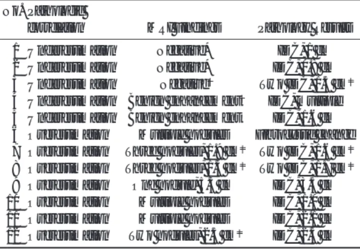
Table 1. The Results of MRI, US, and Pathologic Correlation in 50 Patients with Breast Cancer
Pathologic correlation MRI US
Underestimate 05 (10) 14 (28)
Equal 38 (76) 28 (56)
Overestimate 7 (14) 08 (16)
Note. Numbers in parentheses are percentages.
Table 2. MRI Findings with Underestimation or Overestimation Correlated with Pathologic Results
No. Pathologic
correlation MRI Findings Pathology Results 01 Underestimation Negative* IDC, 1 cm 02 Underestimation Negative* IDC, 0.8 cm 03 Underestimation Negative Two IDC, 1.5 cm 04 Underestimation Benign enhancement IDC, multiple 05 Underestimation Benign enhancement IDC, 1.6 cm 06 Overestimation Multiple nodules Fibrocystic change 07 Overestimation Three nodules, 1.9 cm Two IDC, 1.6 cm 08 Overestimation Three nodules, 1.3 cm Two IDC, 1.7 cm 09 Overestimation One nodule, 5.3 cm IDC, 3.5 cm 10 Overestimation Multiple nodules IDC, 1.0 cm 11 Overestimation Multiple nodules IDC, 2.1 cm 12 Overestimation Two nodules, 2.5 cm IDC, 2.5 cm IDC = Invasive ductal carcinoma
* : MRI after mammotome.
:MRI after neoadjuvant chemotherapy.
:Diameter of the largest nodule.
예측한 후 환자의 수술기록을 검토하여 유방 자기공명영상의 결과에 따라 수술방법이 바뀐 경우를 알아보고 자기공명영상 이 환자의 치료계획에 미치는 영향을 알아보았다.
연구 결과
총 50명 중 45명에서 조직병리학적으로 병변의 최대직경을 측정할 수 있었으며, 최대직경은 평균 2.2 cm(범위; 0.8-8.5 cm)이었다. 최대직경을 측정할 수 없었던 1명은 신보강화학요 법 후 잔류암이 없었고, 2명은 맘모톰절제술 후 잔류암이 없었 으며 다른 2명은 여러 개의 작은 병변이 산재하여 있어 정확 한 직경을 측정할 수 없었다. 병변의 최대직경이 2 cm 미만인 경우가 21명, 2 cm 이상이 24명이었다.
병변의 수는 1개인 경우가 30명, 2개인 경우가 10명, 3개인 경우가 1명, 4개 이상인 경우가 6명이었다.
자기공명영상에서는 과대측정이 7명(14%), 동일한 경우가 38명(76%), 과소측정이 5명(10%)이였다(Table 1). 과대측 정으로 보였던 7명 중 1명은 병변의 최대직경이 병리조직학적 결과보다 컸고, 나머지 6명은 모두 병변의 수가 병리조직학적 결과보다 많았다. 과소측정으로 보였던 5명 중 2명은 맘모톰 절제술을 시행한 경우로 조영증강을 보이는 병변이 없었으며 1명은 신보강화학요법으로 치료한 후 자기공명영상을 시행하 여 남아 있는 병변이 자기공명영상에서는 보이지 않았다. 다른 2명에서는 양성 조영증강을 보이는 부분만 있어 병변에서 제 외되었는데, 2명 중 1명은 신보강화학요법을 받은 환자였다 (Table 2, Fig. 1).
유방 자기공명영상은 수술 직전에 시행하는 경우가 대부분 이어서 유방초음파에서 보이지 않았으나 자기공명영상에서 보 였던 경우 조직 생검은 시행할 수 없었다. 그러나 4명에서 다
시 초음파를 시행하여 병변을 확인하였으며 4명 중 1명은 병 변이 있는 부위의 피부에 표시를 하여 수술 시 병변이 포함되 도록 했다.
유방초음파에서는 과대측정이 8명(16%), 같은 경우가 28명 (56%), 과소측정이 14명(28%)이었다(Table 1). 과대측정으 로 보였던 8명 중 4명에서는 병변의 수가 병리조직학적 결과 보다 많았고, 3명은 최대직경이 병리조직학적 결과보다 크게 측정되었으며, 나머지 1명은 신보강화학요법 시행 후 유방 초 음파에서는 남아 있는 병변이 있는 것으로 보았지만 병리조직 학적 결과에서 남아 있는 병변이 없었던 경우였다. 과소측정으 로 보였던 14명 중 6명은 병변의 수가 적었고, 4명은 최대직 경이 작게 측정되었으며 2명은 종양은 보이지 않고 석회화만 보였던 경우였다. 나머지 2명은 맘모톰 절제술을 시행한 경우 였다.
자기공명영상이 유방암의 평가에 있어 유방초음파보다 높은 정확도를 보였으며 이는 통계적으로 유의하였다(p = 0.041).
자기공명영상 결과에 따라 수술방법이 바뀐 경우는 11명 (22%)이었으며 11명 모두 변형근치유방절제술을 시행하였고 유방보존술로 바뀐 경우는 없었다. 이 중 9명은 자기공명영상 에서 병변의 범위를 정확히 평가하여 수술 방법이 적절히 바 뀌었고(Fig. 2), 2명은 자기공명영상에서 병변의 범위가 과대 평가되어 유방보존술을 시행하지 못했다(Fig. 3).
고 찰
유방촬영술과 유방초음파 결과가 유방암의 진단에 일차적으 로 사용되는 진단방법이나 민감도와 특이도에 제한이 있고 병 변의 범위를 평가하는데 한계가 있다. 유방 자기공명영상은 유 방촬영술이나 유방초음파 검사와는 달리 형태학적 기준과 역
A B
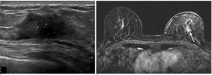
Fig. 1. 49-year-old woman with invasive ductal carcinoma in left breast.
A. After neoadjuvant chemotherapy, follow-up ultrasonography shows a 3.7×1.7 cm sized irregular and hypoechoic mass with calcification in upper center of left breast.
B. Early subtraction image (early enhancement image - pre-enhancement image) of MRI shows a focal enhancing area of left breast, which was considered as postchemotherapy fibrosis rather than residual tumor (arrow). Pathologic results showed 1.5 cm sized residual mass.
동적 조영증강의 결과를 조합하여 병변을 판단하기 때문에 유 방암을 더 정확하게 진단할 수 있다(19). 또한, 유방 자기공명 영상은 유방촬영술과 초음파검사로 유방암을 진단받았으면, 수 술 전 다초점성(multifocal), 다발성 혹은 양측성 유방암과 병 변의 범위를 결정하는데 있어서 중요한 역할을 한다. 즉, 정확 한 병변의 범위 평가가 가능하므로 한 번의 시술로 중심이 되 는 병변을 좀 더 완전히 제거할 수 있으며 또한 위성 병변을 제거할 수 있어 재수술 및 재발을 막을 수 있다(24). 그 외에 도 유방 자기공명영상은 종괴절제술(lumpectomy) 시행 후 잔 여종양의 존재 여부, 신보강화학요법 시행 후 반응률을 알아볼 때도 유용하게 이용될 수 있다(25-30).
Berg 등(5)은 양성병변을 포함한 유방의 병변 평가에서 유 방 자기공명영상이 72.9%, 유방초음파가 67.8%의 진단적 정 확도를 보인다고 보고하였다. 유방암 환자만을 대상으로 수술 전 유방 자기공명영상과 유방초음파의 병변 범위 측정의 정확 도를 비교한 보고가 없어 비교할 수 없으나 본 연구에서는 병 리결과와 비교했을 때 유방암 종괴의 크기와 개수의 평가에 있 어 유방 자기공명영상이 76%의 정확도를 보였고 유방초음파 는 56%의 정확도를 보여 유방 자기공명영상이 더 정확했다.
유방 자기공명영상에서는 잔류종양과 수술적 절제술 후에 생 긴 변화를 잘 구분할 수 없기 때문에 절제술 후에 시행한 자 기공명영상은 재수술 시행 여부를 결정하는데 별로 도움이 되
지 못한다(24). 본 연구에서는 수술적 절제술을 시행한 환자 는 없었지만 맘모톰 절제술을 시행한 환자가 포함되었다. 본 연구의 대상 환자군 중 맘모톰 절제술을 시행한 환자의 수가 적기는 하지만 맘모톰 절제술을 시행한 경우에는 반 수에서 자 기공명영상에서 병변이 보이지 않았으나 수술 후 조직병리결 과에서는 잔류종양이 확인되었다. 따라서 맘모톰 절제술 후 시 행하는 자기공명영상은 가음성 결과를 보일 수 있으므로 판독 할 때 주의를 요한다고 할 수 있다. 이에 대해서는 앞으로 좀 더 많은 수의 환자를 대상으로 하는 연구가 필요할 것으로 생 각된다.
유방 자기공명영상 결과에 따른 치료계획 변화는 환자군에 따라 8-30.6%까지 보고 되었으며(1), 본 연구에서도 이와 비 슷한 22%에서 치료계획의 변화를 보였다. Tillman 등(24)은 20%의 초기 유방암 환자에서 자기공명영상이 치료계획에 영 향을 미친다고 하였으며, 이 중 11%에서 치료 계획에 좋은 영 향을 주었고 6%에서 좋지 않은 영향을 주었으며 2%에서는 불 확실했다고 보고하였다. Schelfout 등(1)의 보고에서는 30.6%
인 64명에서 자기공명영상으로 인해 치료계획이 바뀌었다. 이 중 24명은 광범위 절제술(wider excision)을, 10명은 절개 생 검(open biopsy)을, 18명에서는 유방절제술(mastectomy)을 시행하였으며 12명에서는 추가적 양성 병변이 발견되거나 병 변의 크기를 과대측정하여 불필요한 광범위 절제술이나 조직
A B
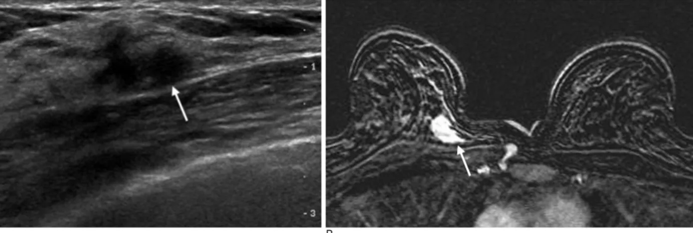
Fig. 2. 37-year-old woman with invasive ductal carcinoma in right breast.
A. Ultrasonography shows an ill-defined and hypoechoic mass with 1.3 cm in size at upper inner quadrant of right breast (ar- row).
B. Early subtraction image (early peak enhancement image - pre-enhancement image) of MRI shows an irregular marginated and early enhancing mass (arrow) with 1.8 cm in size, which is correlated with a sonographic lesion.
C. Nipple level image of MRI shows another 0.7 cm sized satel- lite nodule at inferior aspect of main mass (arrow). The surgical plan was changed from breast conserving surgery to modified radical mastectomy. A satellite nodule was confirmed as inva- sive ductal carcinoma.
C
생검(core biopsy)을 하였다고 보고하였다.
유방 자기공명영상은 약 3.5-7%에서 가양성을 보일 수 있 기 때문에 불필요한 치료계획 변화가 3-6%에서 생길 수 있 다고 한다(1, 18, 31). 본 연구에서도 4%에서 불필요한 치료 계획 변화를 보여 이와 비슷한 결과를 보였다. 또한, 유방 자 기공명영상은 특이도가 37-97%로 넓은 범위의 분포를 보이 기 때문에 자기공명영상에서 광범위한 병변을 보이는 경우 자 기공명영상에 전적으로 의존해 치료계획을 바꾸는 것은 주의 를 요한다(1, 18, 31). 자기공명영상의 이러한 단점을 보완할 수 있는 방법으로는 자기공명영상에서만 보인 병변에 대해 유 방초음파를 다시 시행하여 병변을 확인하는 방법이 있을 수 있 고 필요한 경우 초음파나 자기공명영상 유도하에 조직검사를 할 수 있다.
본 연구의 제한점은 대상 환자의 수가 적다는 점과 자기공 명영상에서 조영증강을 보인 병변에 대한 직접적인 조직병리 학적 확인을 시행하지 못했다는 점이다. 직접적인 확인을 시행 하지 못했기 때문에 가양성 병변으로 분류된 병변 중 실제 병 변이 포함되었을 가능성을 배제할 수 없다. 또한, 신보강화학 요법과 맘모톰 절제술 후에 시행한 자기공명영상이 포함되어 가음성 병변의 수가 실제보다 많은 것으로 측정되었을 가능성 이 있다.
결론적으로 유방 자기공명영상은 유방암의 범위를 유방초음
파보다 정확하게 평가할 수 있었으며 수술 전 시행한 유방 자 기공명영상의 결과에 따라 22%의 환자에서 치료계획의 변화 를 보였다. 따라서 유방암 환자에서 수술 전 유방 자기공명영 상은 유방암의 국소적 병기 결정과 치료 방침 결정을 위해 유 용한 영상기법이므로 수술 전 자기공명영상을 시행하는 것이 바람직하며, 맘모톰 절제술 후에 시행한 자기공명영상은 잔류 종양이 있더라도 음성으로 보일 수 있기 때문에 판독에 주의 를 요한다.
참 고 문 헌
1. Schelfout K, Van Goethem M, Kersschot E, Colpaert C, Schelfhout AM, Leyman P, et al. Contrast-enhanced MR imaging of breast le- sions and effect on treatment. Eur J Surg Oncol 2004;30:501-507 2. Yang WT, Lam WW, Cheung H, Suen M, King WW, Metreweli C.
Sonographic, magnetic resonance imaging, and mammographic as- sessments of preoperative size of breast cancer. J Ultrasound Med 1997;16:791-797
3. Boetes C, Mus RD, Holland R, Barentsz JO, Strijk SP, Wobbes T, et al. Breast tumors: comparative accuracy of MR imaging relative to mammography and US for demonstrating extent. Radiology 1995;197:743-747
4. Davis PL, Staiger MJ, Harris KB, Ganott MA, Klementaviciene J, McCarty KS Jr, et al. Breast cancer measurements with magnetic resonance imaging, ultrasonography, and mammography. Breast
A B
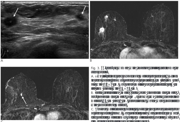
Fig. 3. 66-year-old woman with invasive ductal carcinoma in right breast.
A. Two ill-defined and hypoechoic masses are seen at 9 o’clock in right breast (arrows). Larger one is located 7 cm far from nip- ple with 1.2×1 cm. The other smaller one is identified at 3 cm far from nipple with 0.4×0.5 cm.
B. Early subtraction image (early peak enhancement image - pre-enhancement image) of MRI shows two early enhancing masses, 1.4 cm and 1 cm, respectively, which were correlated with sonographic masses.
C. Another multicentric nodule is noted in the upper center area of right breast (arrow). The surgical plan was changed to modi- fied radical mastectomy, but a multicentric nodule was con- firmed as benign lesion by histopathology.
C
Cancer Res Treat 1996;37:1-9
5. Berg WA, Gutierrez L, NessAiver MS, Carter WB, Bhargavan M, Lewis RS, et al. Diagnostic accuracy of mammography, clinical ex- amination, US and MR imaging in preoperative assessment of breast cancer. Radiology 2004;223:830-849
6. Burhenne HJ, Burhenne LW, Goldberg F, Hislop TG, Worth AJ, Rebbeck PM, et al. Interval breast cancers in the Screening Mammography Program of British Columbia: analysis and classifi- cation. AJR Am J Roentgenol 1994;162:1067-1075
7. Robertson CL. A private breast imaging practice: medical audit of 25,788 screening and 1,077 diagnostic examinations. Radiology 1993;187:75-79
8. Kerlikowske K, Grady D, Barclay J, Sickles EA, Ernster V. Effect of age, breast density, and family history on the sensitivity of first screening mammography. JAMA 1996;276:33-38
9. Howarth D, Slater S, Lau P, Booker J, Clark D, Sillar R.
Complementary role of adjunctive breast magnetic resonance imaging and scintimammograpy in patients of all ages undergoing breast cancer surgery. Australas Radiol 2005;49:289-297
10. Mann BD, Giuliano AE, Bassett LW, Barber MS, Hallauer W, Morton DL. Delayed diagnosis of breast cancer as a result of nor- mal mammograms. Arch Surg 1983;118:23-24
11. Niloff PH, Sheiner NM. False-negative mammograms in patients with breast cancer. Can J Surg 1981;24:50-52
12. Sickles EA. Findings at mammographic screening on only one standard projection: outcomes analysis. Radiology 1998;208:471- 475
13. Cornford EJ, Wilson AR, Athanassiou E, Galea M, Ellis IO, Elston CW, et al.Mammographic features of invasive lobular and invasive ductal carcinoma of the breast: a comparative analysis. Br J Radiol 1995;68:450-453
14. Mandelson MT, Oestreicher N, Porter PL, White D, Finder CA, Taplin SH, et al. Breast density as a predictor of mammographic detection: comparison of interval- and screen-detected cancers. J Natl Cancer Inst 2000;92:1081-1087
15. Kolb TM, Lichy J, Newhouse JH. Comparison of the performance of screening mammography, physical examination, and breast US and evaluation of factors that influence them: an analysis of 27,825 patient evaluation. Radiology 2002;225:165-175
16. Watson L. Breast cancer: diagnosis, treatment and prognosis.
Radiol Technol 2001;73:45-61
17. Fornage BD, Toubas O, Morel M. Clinical, mammographic and sonographic determination of preoperative breast cancer size.
Cancer 1987;60:765-771
18. Fischer U, Kopka L, Grabbe E. Breast carcinoma: effect of preop- erative contrast-enhanced MR imaging on the therapeutic ap-
proach. Radiology 1999;213:881-888
19. Hata T, Takahashi H, Watanabe K, Takahashi M, Taguchi K, Itoh T, et al. Magnetic resonance imaging for preoperative evaluation of breast cancer: a comparative study with mammography and ultra- sonography. J Am Coll Surg 2004;198:190-197
20. Gilles R, Guinebretiere JM, Lucidarme O, Cluzel P, Janaud G, Finet JF, et al. Nonpalpable breast tumors: diagnosis with contrast- enhanced subtraction dynamic MR imaging. Radiology 1994;191:
625-631
21. Bone B, Aspelin P, Bronge L, Isberg B, Perbeck L, Veress B.
Sensitivity and specificity of MR mammography with histopatho- logical correlation in 250 breasts. Acta Radiol 1996;37:208-213 22. Helbich TH, Becherer A, Trattnig S, Leitha T, Kelkar P, Seifert M,
et al. Differentiation of benign and malignant breast lesions: MR imaging versus Tc-99m sestamibi scintimammography. Radiology 1997;202:421-429
23. Yeh E, Slanetz P, Kopans DB, Rafferty E, Georgian-Smith D, Moy L, et al. Prospective comparison of mammography, sonography, and MRI in patients undergoing neoadjuvant chemotherapy for palpable breast cancer. AJR Am J Roentgenol 2005;184:868-877 24. Tillman GF, Orel SG, Schnall MD, Schultz DJ, Tan JE, Solin LJ.
Effect of breast magnetic resonance imaging on the clinical man- agement of women with early-stage breast carcinoma. J Clin Oncol 2002;20:3413-3423
25. 장윤우, 김동훈, 이민혁. 유방암 환자에서 수술 전 흉부 다검출 전산 화단층촬영을 통한 유방암 평가: 유방자기공명영상과의 비교. 대한 영상의학회지 2005;53:137-143
26. Inoue M, Sano T, Watai R, Ashikaga R, Ueda K, Watatani M, et al.
Dynamic multidetector CT of breast tumors: diagnostic features and comparison with conventional techniques. AJR Am J Roentgenol 2003;181:679-686
27. Sardanelli F, Calabrese M, Zandrino F, Melani E, Parodi R, Imperiale A, et al. Dynamic helical CT of breast tumor. J Comput Assist Tomogr 1998;22:398-407
28. LaTrenta LR, Menell JH, Morris EA, Abramson AF, Dershaw DD, Liberman L. Breast lesions detected with MR imaging: utility and histopathologic importance of identification with US. Radiology 2003;227:856-861
29. Orel SG, Schnall MD. MR imaging of the breast for the detection, diagnosis, and staging of breast cancer. Radiology 2001;220:13-30 30. Morris EA. Review of breast MRI: indications and limitations.
Semin Roentgenol 2001;36:226-237
31. Mumtaz H, Hall-Craggs MA, Davidson T, Walmsley K, Thurell W, Kissin MW, et al. Staging of symptomatic primary breast cancer with MR imaging. AJR Am J Roentgenol 1997;169:417-424
J Korean Radiol Soc 2006;55:411-417
Address reprint requests to : Hye-Young Choi, M.D., Department of Radiology, Ewha Womans University, College of Medicine, Seoul, Korea 911-1 Mokdong, Yangcheongu, Seoul 158-710, South Korea.
Tel. 82-2-2650-6179 Fax. 82-2-2650-5302 E-mail : choihy@ewha.ac.kr
Usefulness of preoperative breast MRI in breast cancer:
Comparison with breast US
1Jee Eun Lee, M.D., Hye-Young Choi, M.D., Jung Hee Shin, M.D.1, 2
1Department of Radiology, Ewha Womans University, College of Medicine
2Department of Radiology, Samsung Medical center, Sungkyunkwan University School of Medicine
Purpose: The purpose of this study was to evaluate the usefulness of preoperative breast MRI compared with breast US and pathologic finding in breast cancer patients
Materials and Methods: A total of 50 patients with breast cancer underwent surgery at our institute between October 2004 and August 2005. They were examined preoperatively with MRI and US. The maximum diame- ter and the number of the lesions on MRI and US were measured. These measurements were subsequently compared with the pathologic results. The results were divided into the equal, overestimated and underesti- mated groups. Changes of the therapeutic approach, based on MRI, were also evaluated.
Results: Breast cancer was correctly evaluated in 38 of 50 (76%) patients with MRI and in 28 of 50 (56%) pa- tients with US; the cancer was overestimated in 7 of 50 (14%) patients with MRI and in 8 of 50 (16%) patients with US; the cancer was underestimated in 5 of 50 (10%) patients with MRI and in 14 of 50 (28%) patients with US. The therapeutic approach was changed in 11 of 50 (22%) patients, and all the cases underwent modified radical mastectomy. The therapeutic approach was correctly changed in 9 (18%) patients. Unnecessary wider excision was performed in 2 (4%) patients.
Conclusion: In conclusion, preoperative breast MRI may be a useful modality for preoperative evaluation, es- pecially for the local staging of tumor and the treatment planning of patients with breast cancer.
Index words :Breast
Breast neoplasms, MR
Magnetic resonance (MR), comparative studies Breast, US