간세포암의 자발적인 전체괴사는 극히 드물며(1-4), 영상 진단에서는 특징적인 간세포암의 소견을 보이지 않아 양성종 양을 비롯한 다른 병변과 감별하기 어렵다. 저자들은 전체괴사 를 일으켜 매우 비전형적인 영상소견을 보였던 간세포암의 증 례를 보고하고자 한다.
증례 보고
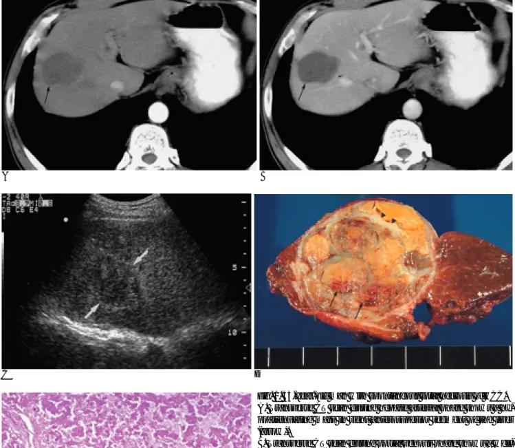
53세 남자 환자가 우연히 발견된 간종괴를 주소로 본원으로 전원되었다. 내원 당시 HbsAg와 Anti-HCV Ab는 음성이었 고, 혈중 알파태아단백(alpha fetoprotein, 이하 AFP)은 5 ng/ml이하로 정상이었으며, AST/ALT도 16/16 IU/L로 정상 이었다. 하루에 소주 1병의 음주력이 있었다. 간동맥기조영증 강전산화단층촬영(computed tomography, 이하 CT)에서는 8 번간분엽에 조영증강되지 않는 4.5 cm 크기의 저감쇠의 종괴 이며(Fig. 1A), 간문맥기CT에서도 종괴의 대부분은 조영증강 되지 않지만 변연부에는 미미한 조영증강이 있었다 (Fig. 1B).
CT소견으로 간농양 혹은 저혈관성 악성종양의 가능성을 제시 하였다. 초음파검사에서 이 종괴는 변연부에 고에코띠를 동반 하였으며 그 내부에는 낭성병변을 시사할 만한 소견 없이 비 균일한 저에코종괴로 보였다(Fig. 1C). 초음파소견과 CT소견 을 종합하여 악성 종양의 가능성이 높을 것으로 생각하였고, 종양절제술(tumorectomy)를 시행하였다. 육안병리소견에서, 종괴는 4.5×4 cm 크기의 피막을 가지는 연노랑색의 종괴였 다(Fig. 1D). 미세병리소견에서, 종괴의 모든 부분에서 괴사가 관찰되었고, 주변간조직에 경화는 없었다. 괴사된 종괴에서 섬
유주형배열을 보이는 간세포암세포의 잔재(ghost)가 보여서, 간세포암이 심한 괴사를 일으킨 것으로 진단하였다(Fig. 1E).
종괴의 모든 괴사부위는 응고괴사였으며 육안소견상 붉은색으 로 보인 부위는 현미경상 출혈에 의한 것이었다. 간문맥기CT 에서 보였던 종괴변연부의 미미한 조영증강부위의 현미경소견 은 종괴 가장자리 섬유화된 곳에 작은 혈관들(소동맥, 소정맥, 모세혈관)의 증식에 의한 가성피막이었으며 살아있는 종양세 포는 없었다. 염증세포의 침윤은 뚜렷하지 않았으며 종양부의 혈관들에 대한 현미경검사상 동맥 및 문맥내의 혈전이나 혈관 기형등의 소견은 보이지 않았다. 수술 후 환자는 별 문제 없이 회복 되었고 외래에서 12개월간 추적관찰 중이며 특이 증상 없이 AFP<5 ng/ml으로 유지되고 있다.
고 찰
자발적인 간세포암의 전체괴사는 극히 드물며, 영문문헌에 서는 지금까지 17예만이 보고되었다(1-4). 악성종양의 자발 성괴사의 정의는 항암제나 수술적 절제와 같은 특별한 치료가 없이 악성세포가 부분적 혹은 완전히 사라지는 것을 말한다(4, 5).
자발성괴사를 진단 하기 위해서는 수술전에 간세포암의 가 능성을 증명하는 것이 중요한데 혈중 AFP 혹은 PIVKA-ll (protein induced by vitamine K absence) 농도와 방사선학적 소견을 종합하여 간세포암을 결론지을 수 있다(4).
간세포암의 자발성괴사의 기전은 아직 알려지지 않았으나, 가능성 있는 기전은 종양의 급속한 성장이나 동맥내혈전에 의 한 허혈괴사이다(5, 6). 간세포암은 혈류공급에 매우 의존적인 종양이므로 종양의 급속한 성장은 상대적인 허혈괴사를 일으 킬 수 있다. 두번째 가설은, 면역기전으로 과도한 국소적 cytokine 생산으로 인해 일어나는 염증반응에 의한 종양의 괴 대한방사선의학회지 2003;48:177-179
─ 177 ─
자발성전체괴사간세포암
1김나라・김영준・정준용・이경호・김세형・김효철・한준구・최병인・이혜승2・이경규
자발성전체괴사를 일으킨 간세포암의 증례를 보고하고자 한다. 이 종괴는 간문맥기조영증강 CT에서 그 변연부에 미미한 조영증강소견이 있을 뿐, 종괴의 대부분이 저감쇠로 보였다. 초음 파에서는 변연부의 고에코띠를 동반한 저에코의 종괴로 보였다. 종양절제술을 시행하였고, 종 괴의 모든 부분에서 괴사가 관찰되었으며, 괴사된 조직에서 간세포암에 해당하는 세포의 잔재 (ghost)가 보여서, 간세포암이 심한 괴사를 일으킨 것으로 진단하였다.
1서울대학교 의과대학 방사선과학교실, 서울대학교 의학연구원 방사선의학연 구소
2서울대학교 의과대학 병리학교실
이 논문은 2002년 7월 23일 접수하여 2002년 10월 9일에 채택되었음.
사이다 (6, 7). 이 증례에서는 염증세포의 침윤은 뚜렷하지 않 았으며 종양부의 혈관들에 대한 현미경검사상 동맥 및 문맥내 의 혈전이나 혈관기형등의 소견은 보이지 않았다. 이 증례는
혈관이상이나 염증반응도 없으며 종괴가 작은데도 자발성전체 과사를 일으킨 드문 예로 다른 새로운 기전에 대한 향후 연구 가 필요할 것으로 생각된다.
김나라 외: 자발성전체괴사간세포암
─ 178 ─
A B
C D
E
Fig. 1. 53-year-old man with spontaneous total necrosis of HCC.
A. Transverse CT scan during hepatic arterial phase shows a hy- poattenuating mass in right anterosuperior segment of the liver (arrow).
B. Transverse CT scan during portal venous phase shows a well- defined hypoattenuating mass with subtle capsular enhancement (arrow). The curvilinear enhancing attenuation on posteromedial aspect is a branch of right anterior segmental portal vein (short arrow).
C. Ultrasonography shows heterogenously hypoechoic mass with peripheral echogenic rim (arrows).
D. Cut surface of surgical specimen shows a mass with internal necrosis and fibrous septum (arrowheads). All portion of necrosis is coagulation necrosis. The spotty areas of reddish color are due to hemorrhage (arrows).
E. Microscopic examination revealed that the mass is composed of totally necrotic material, without viable tumor cell. Note the polygonal necrotic tumor cells (ghost cells) arranged in trabecular pattern in varying thickness, indicating necrotic hepatocellular carcinoma (hematoxylin-eosin stain, ×200).
참 고 문 헌
1. Kaczynski J, Hansson G, Remotti H, Wallerstedt S. Spontaneous regression of hepatocellular carcinoma. Histopathology 1998;32:
147-150
2. Markovic S, Ferlan-Marolt V, Hlebanja Z. Spontaneous regression of hepatocellular carcinoma. Am J Gastroenterol 1996;91:392-393 3. Grossmann M, Hoermann R, Weiss M, et al. Spontaneous regres-
sion of hepatocellular carcinoma. Am J Gastroenterol 1995;90:1500- 1503
4. Matsuo R, Ogata H, Tsuji H, et al. Spontaneous regression of hepa- tocellular carcinoma: report of a case. Hepatogastroenterology 2001;
48:1740-1742
5. Lee SC, Chung HW, Chung JB, et al. Total necrosis of hepatocellu- lar carcinoma due to spontaneous occlusion of feeding artery.
Yonsei Med J 2002;43:123-127
6. Imaoka S, Sasaki Y, Masutani S, et al. Necrosis of hepatocellular carcinoma caused by spontaneously arising arterial thrombus.
Hepatogastroenterology 1994;41:359-362
7. Izuishi K, Ryu M, Hasebe T, Kinoshita T, Konishi M, Inoue K.
Spontaneous total necrosis of hepatocellular carcinoma: report of a case. Hepatogastroenterology 2000;47:1122-1124
대한방사선의학회지 2003;48:177-179
─ 179 ─
J Korean Radiol Soc 2003;48:177-179
Address reprint requests to : Byung Ihn Choi, M.D., Department of Radiology, Seoul National University College of Medicine and the Institute of Radiation Medicine, SNUMRC, 28, Yongon-dong, Chongno-gu, Seoul 110-744, Korea.
Tel. 82-2-760-2584 Fax. 82-2-743-6385 E-mail: [email protected]
Spontaneous Total Necrosis of Hepatocellular Carcinoma
1Na Ra Kim, M.D., Young Jun Kim, M.D., Jun Yong Jeong, M.D., Kyoung Ho Lee, M.D., Se Hyung Kim, M.D., Hyo-Cheol Kim, M.D., Joon Koo Han, M.D.,
Byung Ihn Choi, M.D., Hye-Seung Lee, M.D.2, Gyung Kyu Lee, M.D.
1Department of Radiology, Seoul National University College of Medicine and the Institute of Radiation Medicine, SNUMRC
2Department of Pathology, Seoul National University College of Medicine
We describe a case of spontaneous total necrosis of hepatocellular carcinoma (HCC). Contrast-enhanced CT scanning revealed a hypoattenuating mass during both the hepatic arterial and portal venous phase. During the latter, subtle capsular enhancement was noted. Ultrasonography demonstrated the presence of a hypoe- choic mass with a peripheral hyperechoic rim. The patient underwent tumorectomy, and a totally necrotic mass was found. Microscopic examination revealed necrotic tissue with HCC ghost cells, suggesting sponta- neous total necrosis of HCC.
Index words :Liver
Hepatocellular carcinoma Necrosis