ISSN 2234-3806 • eISSN 2234-3814
http://dx.doi.org/10.3343/alm.2013.33.6.431
Comparison of Methylation Profiling in Cancerous and Their Corresponding Normal Tissues from Korean
Patients with Breast Cancer
Eun-Jung Jung, M.D.1, In-Suk Kim, M.D.2, Eun Yup Lee, M.D.2, Jeong-Eun Kang, M.D.3, Sun-Min Lee, M.D.3, Dong Chul Kim, M.D.4, Ju-Yeon Kim, M.D.1, and Soon-Tae Park, M.D.1
Division of Surgical Oncology1, Department of Surgery, Gyeongsang National University Hospital, Jinju; Department of Laboratory Medicine2, Biomedical Research Institute, Pusan National University Hospital, Busan; Department of Laboratory Medicine3, Pusan National University Yangsan Hospital, Yangsan;
Department of Pathology4, Gyeongsang National University Hospital, Jinju, Korea
Background: Aberrant DNA hypermethylation plays a pivotal role in carcinogenesis and disease progression; therefore, accurate measurement of differential gene methylation patterns among many genes is likely to reveal biomarkers for improved risk assessment.
We evaluated the gene hypermethylation profiles of primary breast tumors and their corre- sponding normal tissues and investigated the association between major clinicopathologi- cal features and gene hypermethylation.
Methods: A single reaction using methylation-specific multiplex ligation-dependent probe amplification was used to analyze the DNA methylation status of 24 tumor suppressor genes in 60 cancerous tissues and their corresponding normal tissues from patients with primary breast cancer.
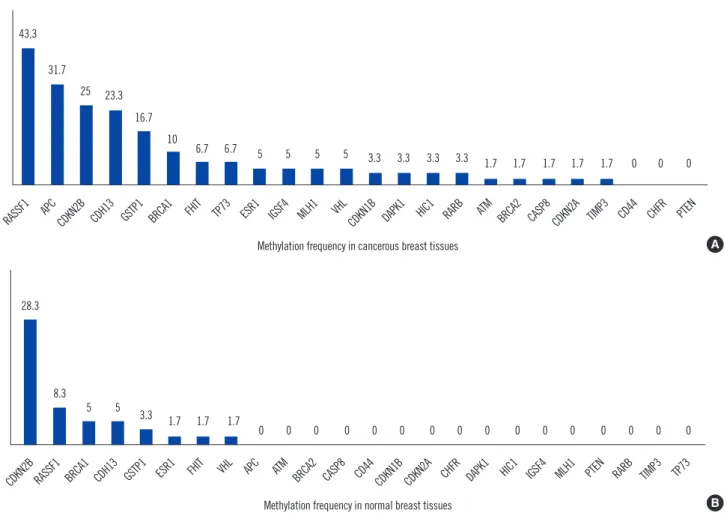
Results: In cancerous breast tissues, 21 of 24 genes displayed promoter methylation in one or more samples. The most frequently methylated genes included RASSF1 (43.3%), APC (31.7%), CDKN2B (25.0%), CDH13 (23.3%), GSTP1 (16.7%), and BRCA1 (10%).
APC was associated with lymph node metastasis, and BRCA1 was associated with nega- tive estrogen receptor and negative progesterone receptor expression. In normal breast tissues, 8 of 24 tumor suppressor genes displayed promoter hypermethylation; CDKN2B (28.3%) and RASSF1 (8.3%) hypermethylation were most frequently observed.
Conclusions: RASSF1 and CDKN2B hypermethylation in Korean breast cancer patients were the most frequent in cancerous tissue and corresponding normal tissue, respectively.
Our data indicates that methylation of specific genes is a frequent event in morphologi- cally normal breast tissues adjacent to breast tumors as well as the corresponding breast cancers. This study also suggests that gene methylation is linked to various pathological features of breast cancer; however, this requires confirmation in a larger study.
Key Words: Breast cancer, Epigenetics, Carcinogenesis, Methylation
Received: October 17, 2012 Revision received: June 20, 2013 Accepted: August 20, 2013 Corresponding author: In-Suk Kim Department of Laboratory Medicine, Pusan National University Hospital, 305 Gudeok-ro, Seo-gu, Busan 602-739, Korea
Tel: +82-51-240-7707 Fax: +82-51-240-7418 E-mail: [email protected]
© The Korean Society for Laboratory Medicine This is an Open Access article distributed under the terms of the Creative Commons Attribution Non-Commercial License (http://creativecom- mons.org/licenses/by-nc/3.0) which permits unrestricted non-commercial use, distribution, and reproduction in any medium, provided the original work is properly cited.
INTRODUCTION
Apart from thyroid cancer, breast cancer is the most common cancer among Korean women [1]. Genetic alterations associ-
ated with breast carcinogenesis, including specific gene amplifi- cations, deletions, point mutations, chromosome rearrange- ments, and aneuploidy, are well understood. In addition to these highly characterized mutations, epigenetic alterations are key
contributors to breast carcinogenesis [2-6]. The most widely studied epigenetic event in breast cancer is hypermethylation of CpG islands associated with the promoter regions of several genes [7, 8]. Hypermethylation of CpG islands in gene promoter regions is thought to be especially relevant for the silencing of important growth control genes. For breast cancer, some of the genes reported to undergo hypermethylation are involved in evasion of apoptosis (DAPK, TWIST1, and HOXA5), cell cycle regulation (CDKN2A and CCND2), cell invasion and metastasis (CDH1 and APC), DNA repair (BRCA1 and GSTP1), and cell signaling (ER and RARb2) [2, 9]. These epigenetic alterations occur at an early stage in breast carcinogenesis.
However, most epigenetic studies have been performed on women in western countries, and the reported frequencies and disease specificities vary across the studies [3, 10-14]. These discrepancies most likely relate to differences in the populations studied, the methods used, and the genes and regions where methylation has been studied. Most of the methods used to de- tect methylation are labor-intensive and/or allow the study of the methylation status of only one gene at a time. In this study, we used an approach that allows the simultaneous assessment of aberrant promoter methylation of 24 tumor suppressor genes in normal and cancerous tissues from 60 Korean patients with pri- mary breast cancer. The aims of this study were as follows: 1) to measure the frequency of gene hypermethylation in cancerous tissue and their corresponding normal tissue from Korean breast cancer patients and 2) to determine whether methylation changes in cancerous tissues from Korean breast cancer pa- tients are associated with major clinicopathological features.
METHODS
1. Patients and tissue samples
This study was approved by the Institutional Review Board of the Gyeongsang National University Hospital (Jinju, Korea), and written informed consent was obtained from all patients partici- pating in the study. We consecutively collected 60 pairs of can- cerous and the corresponding normal tissue samples from pa- tients with breast cancer. All samples were procured at the time of surgery, subjected to an initial gross pathological examination, frozen at -180˚C in liquid nitrogen and stored until use. Corre- sponding normal tissues were procured from the most distant site from the resected specimen. For each tumor and normal breast tissue sample, a section adjacent to the tissue used for DNA extraction was stained with hematoxylin and eosin for his- tological confirmation of the presence or absence of cancer
cells. However, tissue sizes were inadequate to perform tissue morphometry on these slides. Clinicopathologic characteristics of breast cancer were recorded by review of pathological files and electronic medical records (Table 1). Tumors were histologi- cally graded from 1 to 3 according to the Nottingham modifica- tion of the Bloom and Richardson histological grading scheme [15]. Estrogen receptor (ER), progesterone receptor (PR), and p53 status were determined by immunohistochemistry. HER2 status was determined according to the College of American Pa- thologists (CAP) and the American Society of Clinical Oncology (ASCO) joint guidelines [16]. The mean age of the patients was 50.5 yr (range, 29-77 yr), and the most common tumor type was invasive ductal carcinoma (78.3%). More than half (51.7%) of the patients had pathologically positive nodes. In terms of bio- logical markers, the ER and PR expression, Ki-67 proliferation index, HER2 status, and p53 overexpression were included in the analyses.
2. Tissue DNA extraction
Matched normal and tumor tissues were processed separately to avoid possible contamination. Each sample was frozen at -180°C and broken with a frozen mortar. The homogenate was collected and resuspended in TE buffer (10 mM Tris/HCl and 1 mM EDTA).
DNA was extracted by proteinase K digestion, followed by heat inactivation at 95°C and phenol-chloroform and ethanol precipi- tation. DNA was quantified using a Nanodrop 1000 spectropho- tometer (Thermo Scientific, Wilmington, DE, USA).
3. Methylation-specific, multiplex ligation-dependent probe amplification assay
Gene methylation status was evaluated by methylation-specific multiplex ligation-dependent probe amplification (MS-MLPA) [17] by using the ME001-C1 Tumor suppressor-1 kit (MRC-Hol- land, Amsterdam, Netherlands). In all, 24 tumor suppressor genes and 12 internal control genes were studied using 50-100 ng of sample DNA (Table 2). In brief, during the first step of the assay, DNA samples were denatured and hybridized with target- specific MLPA probes by overnight incubation. Then, the reac- tion was split into 2 tubes. One tube was processed as the stan- dard MLPA reaction, and the other tube was incubated with 10 U of HhaI at 49°C for 30 min. Digested probes are not amplified by PCR and, hence, do not generate a signal. In contrast, if the target DNA is methylated, the hemi-methylated probe/sample DNA hybrids are prevented from digestion by HhaI and the tar- get region is amplified, generating a signal. The amplified prod- ucts were analyzed by sequence-type capillary electrophoresis
(ABI 3100; Applied Biosystems, Foster City, CA, USA). The methylation status was subsequently quantified by comparing the relative signal peaks from the 2 tubes by using the Gene- Marker_V1.90 software (SoftGenetics, State College, PA, USA).
Methylation was scored as positive when the calculated methyl- ation levels were higher than 25%. Any value below that level was considered negative.
4. Sample size calculation and statistical analyses
Van der Auwera et al. [18] demonstrated a 58.9% difference in RASSF1A gene hypermethylation between cancerous and mor- phologically normal tissues (76.8% vs. 17.9%, respectively).
Thus, it was estimated that 41 patients per group would be re- quired to provide a power of 95% to detect a statistically signifi- cant difference between the cancerous and morphologically normal tissues in the Korean patients with breast cancer by us- ing a two-sided α-level of 0.05.
Continuous variables were presented as means±SD in base- line characteristics of patients with breast cancer (Table 1). For comparison of the methylation status of the 24 tumor suppres- sor genes in cancerous and corresponding normal tissues, data were presented as numbers and frequencies for categorical variables. The frequencies between groups were compared us- ing the χ2-test or, in the case of low frequency per variable, Fish- er’s exact method. The Kappa statistic was used to assess the agreement between dichotomous variables between cancerous and corresponding normal breast tissue samples. The clinico- pathologic parameters using categorical variables and the hy- permethylation status in cancerous tissues in patients with breast cancer were analyzed using the Pearson’s χ2-test or Fish- er’s exact method. A multivariate logistic regression analysis was used to identify independent predictors of clinicopathologic characteristics among the hypermethylated genes. Factors en- tered into the multivariate model were those with a P value less than 0.10 from the univariate analysis. A P value ≤0.05 of two- sided test was considered statistically significant. All statistical calculations were performed using SPSS, version 13.0 (SPSS Inc., Chicago, IL, USA).
RESULTS
1. Methylation pattern of the 24 tumor-suppressor genes in cancerous tissue and their corresponding, morphologically normal breast tissue from patients with breast cancer
The distribution of DNA methylation of 24 tumor-suppressor genes in normal and cancerous breast tissues is shown in Fig. 1.Table 1. Characteristics of patients with breast cancer (N=60) Clinicopathologic factors
Age at diagnosis (yr)
Mean±SD 50.5±12.1
Range 29-77
Body mass index
Mean±SD 24.3±3.1
Tumor type N of cases (%)
Invasive ductal carcinoma 47 (78.3)
Mucinous carcinoma 5 (8.3)
Ductal carcinoma in situ 5 (8.3)
Medullary carcinoma 3 (5.0)
Tumor stage N of cases (%)
T1 17 (28.3)
T2 36 (60.0)
T3 4 (6.7)
T4 3 (5.0)
Nodal involvement N of cases (%)
Negative 29 (48.3)
Positive 31 (51.7)
American Joint Committee on cancer stage N of cases (%)
I 14 (23.3)
II 39 (65.0)
III 7 (11.7)
IV 0 (0.0)
Histologic Grade N of cases (%)
1 11 (18.3)
2 28 (46.7)
3 21 (35.0)
ER status N of cases (%)
Negative 30 (50.0)
Positive 29 (48.3)
Not available 1 (1.7)
PR status N of cases (%)
Negative 30 (50.0)
Positive 29 (48.3)
Not available 1 (1.7)
BCL-2 N of cases (%)
Negative 25 (41.7)
Positive 33 (55.0)
Not available 2 (3.3)
Ki-67 proliferation index N of cases (%)
<20% 11 (18.3)
≥20% 24 (40.0)
Not available 25 (41.7)
P53 status N of cases (%)
Negative 11 (18.3)
Positive 47 (78.3)
Not available 2 (3.3)
HER2 status N of cases (%)
Negative 29 (48.3)
Positive 29 (48.3)
Not available 2 (3.3)
Abbreviations: ER, estrogen receptor; PR, progesterone receptor.
Twenty one of 24 genes (87.5%) displayed promoter hyper- methylation in one or more of the cancerous tissues (Fig. 1A).
However, only 8 of 24 tumor-suppressor genes (33.3%) dis- played promoter hypermethylation in the normal tissue samples (Fig. 1B). In the cancerous breast tissues, the most frequently hypermethylated genes were RASSF1 (43.3%) followed by APC (31.7%), CDKN2B (25.0%), CDH13 (23.3%), GSTP1 (16.7%), and BRCA1 (10%). Methylation was not observed in the CD44, CHFR, and PTEN gene promoters in cancerous tissues. In the normal breast tissues, the most frequently hypermethylated genes were CDKN2B (28.3%) and RASSF1 (8.3%). In their can- cerous tissues, the frequencies of RASSF1 (43.3% vs. 8.3%), APC (31.7% vs. 0%), and CDH13 (23.3% and 5.0%) were sig- nificantly higher than those of normal tissues (P <0.001, P <
0.001, and P =0.004, respectively; Table 2). However, the fre- quency of CDKN2B (25.0%) in cancerous tissues was similar to that (28.3%) in corresponding normal tissue (P =0.680).
In 65% of the cancerous tissues, the promoter regions were
hypermethylated in at least one of the 21 hypermethylated genes. The results are summarized in Table 3 . One tumor sam- ple was methylated at 14 sites. In 38.3% of the normal tissues, the promoter regions were hypermethylated in at least 1 of the 8 hypermethylated genes (Table 1). None of the normal tumor samples were methylated at more than 5 sites.
2. Association between DNA hypermethylation and clinicopathological factors
We investigated the association of clinicopathological features of the patients with the 7 most frequently hypermethylated genes (RASSF1, APC, CDKN2B, CDH13, GSTP1, BRCA1, and FHIT) in cancerous tissues. Table 4 shows the association between the hypermethylation of these 7 genes and various clinicopathologi- cal features of the patients. RASSF1 hypermethylation showed a statistically significant association with the invasive ductal carci- noma tumor type vs. other tumor types (53.2% [25/47] vs. 7.7%
[1/13], P =0.032); however, post-hoc comparisons among these Table 2. Distribution of DNA hypermethylation of 24 tumor-suppressor genes in cancerous and corresponding normal breast tissues (N=60)
Gene Gene full name Chromosome location Cancerous tissues, N (%) Normal tissues, N (%) P
APC Adenomatosis polyposis coli 5q22 19 (31.7) 0 (0) <0.001
ATM Ataxia teleangiectasia mutated 11q23 1 (1.7) 0 (0) 1.000
BRCA1 Breast cancer 1 17q21.31 6 (10.0) 3 (5.0) 0.488
BRCA2 Breast cancer 2 13q12.3 1 (1.7) 0 (0) 1.000
CASP8 Caspase 8 2q33.2 1 (1.7) 0 (0) 1.000
CD44 CD44 molecule 11p12 0 (0) 0 (0) 1.000
CDH13 Cadherin 13 16q23.3 14 (23.3) 3 (5.0) 0.004
CDKN1B Cyclin-dependent kinase inhibitor 1B 12p13.2 2 (3.3) 0 (0) 0.496
CDKN2A Cyclin-dependent kinase inhibitor 2A 9p21 1 (1.7) 0 (0) 1.000
CDKN2B Cyclin-dependent kinase inhibitor 2B 9p21 15 (25.0) 17 (28.3) 0.680
CHFR Checkpoint with forkhead and ring finger domains 12q24.33 0 (0) 0 (0) 1.000
DAPK1 Death-associated protein kinase I 9q22 2 (3.3) 0 (0) 0.496
ESR1 Estrogen receptor 6q25.1 3 (5.0) 1 (1.7) 0.619
FHIT Fragile histidine triad gene 3p14.2 4 (6.7) 1 (1.7) 0.364
GSTP1 Gluthathione S-transferase pi 11q13 10 (16.7) 2 (3.3) 0.015
HIC1 Hypermethylated in cancer 1 7p13.3 2 (3.3) 0 (0) 0.496
IGSF4 Immunglobulin superfamily member 4 11q23 3 (5.0) 0 (0) 0.244
MLH1 mutL homolog I 3p22.3 3 (5.0) 0 (0) 0.244
PTEN Phosphatase and tensin homolog 10q23.3 0 (0) 0 (0) 1.000
RARB Retinoic acid receptor beta 3p24.2 2 (3.3) 0 0.496
RASSF1 Ras association domain family 1A 3p21.3 26 (43.3) 5 (8.3) < 0.001
TIMP3 Tissue inhibitor of metalloproteinases 3 22q12.3 1 (1.7) 0 (0) 1.000
TP73 Tumor protein p73 1p36.32 4 (6.7) 0 (0) 0.119
VHL von Hippel Lindau 3p25.3 3 (5.0) 1 (1.7) 0.619
groups demonstrated no statistical significance between tumor types and RASSF1 hypermethylation. In addition, RASSF1 hy- permethylation was associated with lymph node involvement (55.2% [16/29] vs. 32.2% [10/31], P =0.073), but the associa- tion was statistically insignificant. APC hypermethylation was as- sociated with lymph node involvement (68.4% [13/29] vs. 19.3%
[6/31], P =0.034). CDKN2B, CDH13, and GSTP1 hypermethyl- ation were not associated with clinicopathological features.
BRCA1 hypermethylation was associated with histologic grade III (25.0% [5/20] vs. 2.5% [1/40], P =0.013), negative ER ex- pression (24.0% [6/25] vs. 0% [0/34], P =0.004), negative PR expression (20% [6/30] vs. 0% [0/29], P =0.014), and negative BCL-2 expression (20.8% [5/24] vs. 2.9% [1/34], P =0.046).
The difference between the clinicopathological characteristics and BRCA1 hypermethylation in cancerous tissue was statisti- cally significant as per the multivariate logistic regression analy- Table 3. Distribution of DNA methylation of 24 tumor-suppressor
genes in cancerous and corresponding normal breast tissues (N=
60)
Methylation Cancerous tissues,
N (%) Norma tissues,
N (%) P
Negative 21 (35.0) 37 (61.7) 0.003
Positive 39 (65.0) 23 (38.3)
N of methylation sites
1 9 (15.0) 15 (25.0)
2 14 (23.3) 6 (10.0)
3 7 (11.7) 1 (1.7)
4 2 (3.3) 0 (0.0)
5 3 (5.0) 1 (1.7)
>5 4 (6.8) 0 (0.0)
Fig. 1. Methylation frequencies of the tumor-suppressor genes in cancerous tissues and their corresponding normal tissues from the pa- tients with breast cancer. (A) Methylation frequency of the 24 analyzed cancer-related gene regions in cancerous breast tissues. (B) Meth- ylation frequency of the 24 analyzed cancer-related gene regions in the corresponding normal breast tissues. Numbers above each bar in- dicate the frequency of samples that were methylated in that region. The RASSF1 promoter was the most frequently methylated in cancer- ous tissues, and CDKN2B was the most frequently methylated in normal tissues. There were some gene regions whose methylation was not associated with breast tumor development (e.g., CD44, CHFR, and PTEN in tumors).
RASSF1 APC
CDKN2B CDH13 GSTP1 BRCA1 FHIT TP73 ESR1 IGSF4 MLH1 VHL
CDKN1B DAPK1 HIC1 RARB ATM
BRCA2 CASP8 CDKN2A TIMP3 CD44 CHFR PTEN 43.3
31.7
25 23.3 16.7
10 6.7 6.7 5 5 5 5 3.3 3.3 3.3 3.3 1.7 1.7 1.7 1.7 1.7 0 0 0
Methylation frequency in cancerous breast tissues
RASSF1 APC
CDKN2B BRCA1 CDH13 GSTP1 ESR1 FHIT VHL IGSF4 MLH1 TP73
CDKN1B DAPK1 HIC1 RARB
ATM BRCA2 CASP8 CD44 CDKN2A CHFR PTEN TIMP3
28.3
8.3
5 5
3.3 1.7 1.7 1.7
0 0 0 0 0 0 0 0 0 0 0 0 0 0 0 0
Methylation frequency in normal breast tissues
A
B
Table 4. Association between the methylation status in cancerous tissues and the clinicopathological characteristics in patients with breast cancer (N=60)
Gene
RASSF1 APC CDKN2B CDH13 GSTP1 BRCA1 FHIT
Positive (26) Negative
(34) Positive
(19) Negative (41) Positive
(15) Negative (45) Positive
(14) Negative (46) Positive
(10) Negative (50) Positive
(6) Negative
(54) Positive (4) Negative
(56) Tumor type
Invasive ductal
carcinoma 25 22 18 29 11 36 12 35 10 37 5 42 4 43
Ductal carcinoma
in situ 1 4 0 5 1 4 0 5 0 5 0 5 0 5
Medullary
carcinoma 0 3 0 3 0 3 1 2 0 5 0 5 0 5
Mucinous
carcinoma 0 5 1 4 3 2 1 4 0 3 1 2 0 3
P 0.032 0.303 0.207 0.781 0.527 0.588 0.892
Nodal involvement
Negative 10 21 6 25 5 26 6 25 3 28 3 28 0 31
Positive 16 13 13 16 10 19 8 21 7 22 3 26 4 25
P 0.073 0.034 0.101 0.451 0.175 1.000 0.049
Stage
I-II 22 31 17 36 12 41 12 41 10 43 5 48 3 50
III-IV 4 3 2 5 3 4 2 5 0 7 1 6 1 6
P 0.454 1.000 0.351 0.660 0.589 0.541 0.399
Histologic grade
1-2 18 22 13 27 10 30 11 28 6 34 1 39 3 37
3 8 12 6 14 5 15 3 17 4 16 5 15 1 19
P 0.713 0.844 1.000 0.347 0.718 0.013 1.000
ER status
Negative 8 17 6 19 5 20 8 17 2 23 6 19 2 23
Positive 18 16 13 21 10 24 6 28 8 26 0 34 2 32
Not available 0 1 0 1 0 1 0 1 0 1 0 1 0 1
P 0.321 0.438 0.777 0.244 0.321 0.004 1.000
PR status
Negative 10 20 7 23 7 23 9 21 4 26 6 24 2 28
Positive 16 13 12 17 8 21 5 24 6 23 0 29 2 27
Not available 0 1 0 1 0 1 0 1 0 1 0 1 0 1
P 0.232 0.304 1.000 0.258 0.747 0.014 1.000
BCL-2
Negative 8 16 6 18 4 20 6 18 3 21 5 19 0 24
Positive 17 17 12 22 11 23 7 27 7 27 1 33 4 30
Not available 1 1 1 1 0 2 1 1 0 2 0 2 0 2
P 0.247 0.449 0.588 1.000 0.757 0.046 0.341
P53 status
Negative 4 7 4 7 4 7 2 9 2 9 2 9 0 11
Positive 22 25 15 32 11 36 12 35 7 40 4 43 4 43
Not available 0 2 0 2 0 2 0 2 1 1 0 2 0 2
P 1.000 0.539 0.314 1.000 1.000 0.333 0.624
HER2/Neu status
Negative 15 14 12 17 7 22 9 20 6 23 5 54 3 26
Positive 10 19 6 23 8 21 5 24 4 25 1 28 1 28
Not available 1 1 1 1 0 2 0 2 0 2 0 2 0 2
P 0.358 0.325 1.000 0.180 0.539 0.126 0.381
Abbreviations: ER, estrogen receptor; PR, progesterone receptor.
sis. FHIT hypermethylation was associated with lymph node in- volvement (13.8% [4/29] vs. 0% [0/31], P =0.049).
We also investigated the 5 most frequently hypermethylated genes, CDKN2B, RASSF1, BRCA1, CDH13, and GSTP1, in the corresponding normal tissues and examined their association with clinicopathological features. CDKN2B hypermethylation was associated with positive ER expression (40.0% [12/30] vs.
13.8% [4/29], P =0.032) and positive PR expression (37.9%
[11/29] vs. 16.7% [5/30], P =0.055). Hypermethylation of RASSF1, BRCA1, CDH13, and GSTP1 in the corresponding normal tissues was not associated with clinicopathological fea- tures (data not shown).
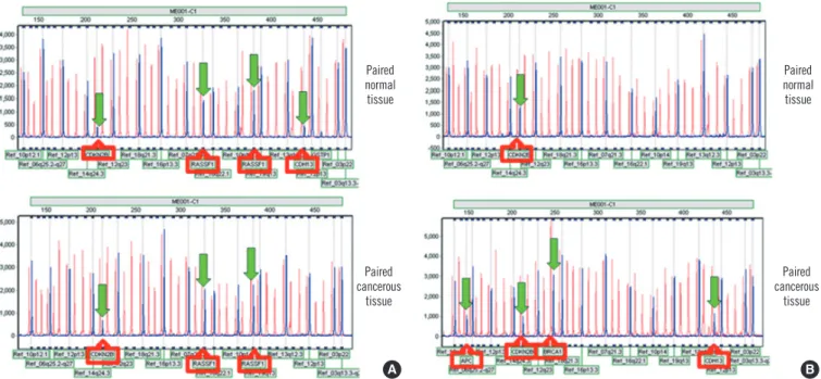
3. Concordant gene methylation in cancerous tissue and corresponding normal breast tissue
For most of the 24 tumor suppressor genes, there was a poor to slight agreement between methylation in cancerous and corre- sponding normal tissues. The results of the concordant methyl- ation pattern and disconcordant methylation pattern in the can- cerous and corresponding normal tissues are shown in Fig. 2A, B, respectively. When at least one gene(s) was found to be hy- permethylated in normal and cancerous tissues, poor agree- ment was noted (data not shown).
DISCUSSION
Hypermethylation of CpG islands is a common mechanism for silencing tumor suppressor genes and occurs frequently in breast cancer. Although there is a large body of literature and the PubMeth online database (www.pubmeth.org) reporting tu- mor suppressor gene methylation frequency in breast cancer, methylation research of breast cancer in the Korean patients is still lacking, except for a few reports [5, 19, 20]. Several tech- niques exist to study aberrant methylation, i.e., methyl-specific PCR, bisulfite-dependent sequencing, and methyl-sensitive re- striction enzyme-based assays. Most of these approaches are restricted to a limited number of genes. Although recent studies demonstrated that MS-MLPA analysis in breast cancer is rela- tively simple, sensitive, and highly specific to simultaneously de- tect the methylation status of multiple gene regions [17, 21-23], it is still necessary to confirm whether the MS-MLPA technique is an appropriate method to detect methylation.
In the present study, we investigated aberrant methylation of 24 genes in cancerous and their normal tissues from 60 Korean patients with breast cancer by using MS-MLPA. In the cancer- ous breast tissues, the most frequently hypermethylated genes were RASSF1 (43.3%), APC (31.7%), CDKN2B (25.0%), and CDH13 (23.3%). Using a PubMeth online database, which con-
A B
Fig. 2. Concordant vs. disconcordant methylation patterns in cancerous tissues and their corresponding normal tissues from the patients with breast cancer. (A) Concordant methylation pattern in normal and cancerous tissues from patients with breast cancer. Both tissues showed methylation changes for CDKN2B and RASSF1 genes. (B) Disconcordant methylation pattern in normal and cancerous tissues from patients with breast cancer. Morphologically normal tissue showed only CDKN2B methylation; however, the cancerous tissue showed methylation of multiple tumor suppressor genes, including APC, CDKN2B, BRCA1, and CDH13.
Paired normal tissue
Paired normal tissue
Paired cancerous
tissue
Paired cancerous
tissue
tains information and statistics about DNA hypermethylation in various cancers, and a review of the literature, the frequency of RASSF1 hypermethylation of breast cancer was determined to be 72.0% [22] and 71.4% [4], respectively. In this study, the frequency of RASSF1 hypermethylation was 43.34% (26/60) in cancerous tissues of all types of breast cancer and 53.2%
(25/47), in cancerous tissues of invasive ductal carcinoma. This value is somewhat lower than the frequencies reported in previ- ous studies and the online database [4, 24-26]. The discrep- ancy between these data might be the cut-off of positivity and analysis method rather than ethnic differences. However, these results are in accordance with the study by Buyru et al. [22], who reported that RASSF1 is the most frequently methylated gene and is methylated in 56.4% of invasive ductal carcinomas using the same MS-MLPA method as in this study. However, in a recent study based on the Korean ethnicity, the methylation frequency of ductal carcinoma in situ was 89% by using Methy- Light PCR analysis, which was determined as positive when the percentage of methylated reference was >4 [19]. Therefore, the method and cut-offs of positivity of methylation analyses are very important to reduce false positives due to inadequate con- version of non-methylated cytosine to uracil and mis-priming when high numbers of PCR cycles or nested primers are used [5, 6]. Optimal cut-off determination can result in over-fitting and false-discovery. In the future, we will validate our findings by using a larger sample set from clinical trials and develop bio- markers for clinical use. RASSF1 hypermethylation has sug- gested lymph node involvement, but the association between hypermethylation and nodal involvement was statistically insig- nificant (P =0.073). RASSF1 methylation has been previously shown to be associated with nodal metastasis in patients with breast cancer [27]. Further, the frequency of RASSF1 methyla- tion was shown to be higher in various metastatic sites as com- pared to the primary tumors [28], suggesting the potential of RASSF1 hypermethylation as a predictor for lymph node metas- tasis in breast cancer.
CDKN2B was the most frequently hypermethylated gene in corresponding normal breast tissues and the third most, in can- cerous tissues. The CDKN2B hypermethylation has been fre- quently noted in various cancers, such as leukemia, lymphoma, colorectal cancer, brain cancer, liver cancer, gastric cancer, multiple myeloma, ovarian cancer, and lung cancer [29-32]. In cancerous, para-cancerous, and non-cancerous tissues in he- patocellular carcinoma, CDKN2B hypermethylation occurred at frequencies of 50%, 40%, and 25%, respectively [33]. The fre- quencies of CDKN2B promoter methylation in corresponding
normal and cancerous tissues in colorectal cancer were re- ported to be 6.8% (6/88) and 26.1% (23/88), respectively [34].
Both studies suggested that CDKN2B promoter methylation might be an early event in hepatocarcinogenesis or colorectal cancer [33, 34]. This study demonstrates that CDKN2B pro- moter methylation in corresponding normal tissues in breast cancer is a frequent event and CDKN2B hypermethylation may be an early event of breast carcinogenesis in Korean patients with breast cancer.
The APC gene is the second most hypermethylated gene in cancerous tissues and the frequency was 31.7% (19/60); hy- permethylation of this gene was not observed in corresponding normal tissues. Similar reports indicated that hypermethylation of the APC promoter was detected in 18 of 50 (36%) primary breast cancers and in none of the 21 non-cancerous breast tis- sue samples [35]. These results indicate that APC hypermethyl- ation is a cancer-specific change. In addition, APC hypermeth- ylation was associated with lymph node involvement in this study (P =0.034). Therefore, APC hypermethylation might result in more aggressive behavior, which can potentially become a useful prognostic indicator warranting a more aggressive thera- peutic approach.
BRCA1 hypermethylation was the third most hypermethyl- ated gene (5%) in the corresponding normal breast tissue and the fifth most (10%) in cancerous tissues in this study. Although there was no statistical significance between the clinicopatho- logic characteristics and BRCA1 hypermethylation using multi- variate logistic regression analysis, BRCA1 hypermethylation was associated with negative ER expression (P =0.004), nega- tive PR expression (P =0.014), negative BCL-2 expression, and high histologic grade (P =0.013). Methylation in breast cancer has long been linked to hormone regulation, but this correlation has not been established yet. The status of both ER and PR are very important to help determine patients who would benefit the most from hormone therapy. BRCA1 hypermethylation is more frequent among breast cancer patients with negative ER and negative PR expression, and this study suggests that hyper- methylation of the gene promoter in cancerous tissues may be associated with hormone expression.
Detection of promoter CpG island hypermethylation offers several advantages compared to other DNA alterations in can- cer. These events may provide ideal biomarkers for molecular diagnosis and early detection of cancer.
In conclusion, we have clearly demonstrated that normal-ap- pearing breast tissue from the Korean patients with breast can- cer exhibited frequent aberrant DNA methylation changes. This
study also suggests that gene methylation may be linked to vari- ous pathological features of breast cancer; however, this re- quires confirmation in a larger study group.
Authors’ Disclosures of Potential Conflicts of Interest
No potential conflicts of interest relevant to this article were re- ported.
Acknowledgements
This study was supported by a Biomedical Research Institute Grant (2010-02), Pusan National University Hospital, and grants from the Research Foundation of Gyeongsang National University Hospital, GNUHCRF-2010-005.
REFERENCES
1. The Korea Central Cancer Registry. Annual report of cancer statistics in Korea in 2009. Seoul: The Korean Ministry of Health and Welfare, 2011.
2. Li S, Rong M, Iacopetta B. DNA hypermethylation in breast cancer and its association with clinicopathological features. Cancer Lett 2006;237:
272-80.
3. Van De Voorde L, Speeckaert R, Van Gestel D, Bracke M, De Neve W, Delanghe J, et al. DNA methylation-based biomarkers in serum of pa- tients with breast cancer. Mutat Res 2012;751:304-25.
4. Marzese DM, Hoon DS, Chong KK, Gago FE, Orozco JI, Tello OM, et al.
DNA methylation index and methylation profile of invasive ductal breast tumors. J Mol Diagn 2012;14:613-22.
5. Cheol Kim D, Thorat MA, Lee MR, Cho SH, Vasiljevíc N, Scibior-Bent- kowska D, et al. Quantitative DNA methylation and recurrence of breast cancer: a study of 30 candidate genes. Cancer Biomark 2012;11:75-88.
6. Zhu W, Qin W, Hewett JE, Sauter ER. Quantitative evaluation of DNA hypermethylation in malignant and benign breast tissue and fluids. Int J Cancer 2010;126:474-82.
7. Esteller M. CpG island hypermethylation and tumor suppressor genes:
a booming present, a brighter future. Oncogene 2002;21:5427-40.
8. Esteller M. Cancer epigenetics: DNA methylation and chromatin altera- tions in human cancer. Adv Exp Med Biol 2003;532:39-49.
9. Widschwendter M and Jones PA. DNA methylation and breast carcino- genesis. Oncogene 2002;21:5462-82.
10. Xu J, Shetty PB, Feng W, Chenault C, Bast RC Jr, Issa JP, et al. Methyl- ation of HIN-1, RASSF1A, RIL and CDH13 in breast cancer is associat- ed with clinical characteristics, but only RASSF1A methylation is associ- ated with outcome. BMC Cancer 2012;12:243.
11. Wang S, Dorsey TH, Terunuma A, Kittles RA, Ambs S, Kwabi-Addo B.
Relationship between tumor DNA methylation status and patient char- acteristics in African-American and European-American women with breast cancer. PLoS One 2012;7:e37928.
12. van Hoesel AQ, van de Velde CJ, Kuppen PJ, Putter H, de Kruijf EM, van Nes JG, et al. Primary tumor classification according to methylation pattern is prognostic in patients with early stage ER-negative breast cancer. Breast Cancer Res Treat 2012;131:859-69.
13. Tserga A, Michalopoulos NV, Levidou G, Korkolopoulou P, Zografos G, Patsouris E, et al. Association of aberrant DNA methylation with clinico- pathological features in breast cancer. Oncol Rep 2012;27:1630-8.
14. Szyf M. DNA methylation signatures for breast cancer classification and prognosis. Genome Med 2012;4:26.
15. Elston CW and Ellis IO. Pathological prognostic factors in breast cancer.
I. The value of histological grade in breast cancer: experience from a large study with long-term follow-up. Histopathology 2002;41:154-61.
16. Wolff AC, Hammond ME, Schwartz JN, Hagerty KL, Allred DC, Cote RJ, et al. American Society of Clinical Oncology/College of American Pathol- ogists guideline recommendations for human epidermal growth factor receptor 2 testing in breast cancer. J Clin Oncol 2007;25:118-45.
17. Nygren AO, Ameziane N, Duarte HM, Vijzelaar RN, Waisfisz Q, Hess CJ, et al. Methylation-specific MLPA (MS-MLPA): simultaneous detec- tion of CpG methylation and copy number changes of up to 40 se- quences. Nucleic Acids Res 2005;33:e128.
18. Van der Auwera I, Bovie C, Svensson C, Trinh XB, Limame R, van Dam P, et al. Quantitative methylation profiling in tumor and matched morpho- logically normal tissues from breast cancer patients. BMC Cancer 2010;
10:97.
19. Park SY, Kwon HJ, Lee HE, Ryu HS, Kim SW, Kim JH, et al. Promoter CpG island hypermethylation during breast cancer progression. Vir- chows Arch 2011;458:73-84.
20. Kim JH, Shin MH, Kweon SS, Park MH, Yoon JH, Lee JS, et al. Evalua- tion of promoter hypermethylation detection in serum as a diagnostic tool for breast carcinoma in Korean women. Gynecol Oncol 2010;118:
176-81.
21. Marzese DM, Gago FE, Vargas-Roig LM, Roqué M. Simultaneous analy- sis of the methylation profile of 26 cancer related regions in invasive breast carcinomas by MS-MLPA and drMS-MLPA. Mol Cell Probes 2010;24:271-80.
22. Buyru N, Altinisik J, Ozdemir F, Demokan S, Dalay N. Methylation pro- files in breast cancer. Cancer Invest 2009;27:307-12.
23. Marzese DM, Gago FE, Vargas-Roig LM, Roque M. Simultaneous analy- sis of the methylation profile of 26 cancer related regions in invasive breast carcinomas by MS-MLPA and drMS-MLPA. Mol Cell Probes 2010;24:271-80.
24. Dammann R, Yang G, Pfeifer GP. Hypermethylation of the cpG island of Ras association domain family 1A (RASSF1A), a putative tumor sup- pressor gene from the 3p21.3 locus, occurs in a large percentage of human breast cancers. Cancer Res 2001;61:3105-9.
25. Honorio S, Agathanggelou A, Schuermann M, Pankow W, Viacava P, Maher ER, et al. Detection of RASSF1A aberrant promoter hypermeth- ylation in sputum from chronic smokers and ductal carcinoma in situ from breast cancer patients. Oncogene 2003;22:147-50.
26. Yeo W, Wong WL, Wong N, Law BK, Tse GM, Zhong S. High frequency of promoter hypermethylation of RASSF1A in tumorous and non-tu- mourous tissue of breast cancer. Pathology 2005;37:125-30.
27. Müller HM, Widschwendter A, Fiegl H, Ivarsson L, Goebel G, Perkmann E, et al. DNA methylation in serum of breast cancer patients: an inde- pendent prognostic marker. Cancer Res 2003;63:7641-5.
28. Mehrotra J, Vali M, McVeigh M, Kominsky SL, Fackler MJ, Lahti-Do- menici J, et al. Very high frequency of hypermethylated genes in breast cancer metastasis to the bone, brain, and lung. Clin Cancer Res 2004;
10:3104-9.
29. Dodge JE, Munson C, List AF. KG-1 and KG-1a model the p15 CpG is- land methylation observed in acute myeloid leukemia patients. Leuk Res 2001;25:917-25.
30. Aggerholm A, Guldberg P, Hokland M, Hokland P. Extensive intra- and interindividual heterogeneity of p15INK4B methylation in acute myeloid
leukemia. Cancer Res 1999;59:436-41.
31. Herman JG, Jen J, Merlo A, Baylin SB. Hypermethylation-associated inactivation indicates a tumor suppressor role for p15INK4B. Cancer Res 1996;56:722-7.
32. Christiansen DH, Andersen MK, Pedersen-Bjergaard J. Methylation of p15INK4B is common, is associated with deletion of genes on chromo- some arm 7q and predicts a poor prognosis in therapy-related myelo- dysplasia and acute myeloid leukemia. Leukemia 2003;17:1813-9.
33. Qin Y, Liu JY, Li B, Sun ZL, Sun ZF. Association of low p16INK4a and
p15INK4b mRNAs expression with their CpG islands methylation with human hepatocellular carcinogenesis. World J Gastroenterol 2004;10:
1276-80.
34. Ishiguro A, Takahata T, Saito M, Yoshiya G, Tamura Y, Sasaki M, et al.
Influence of methylated p15 and p16 genes on clinicopathological fea- tures in colorectal cancer. J Gastroenterol Hepatol 2006;21:1334-9.
35. Jin Z, Tamura G, Tsuchiya T, Sakata K, Kashiwaba M, Osakabe M, et al.
Adenomatous polyposis coli (APC) gene promoter hypermethylation in primary breast cancers. Br J Cancer 2001;85:69-73.