Primary cutaneous B cell lymphomas (PCB- CLs) constitute 20-25 % of all cutaneous lym- phomas in Western literatures and those with multiple skin involvement are less frequent1. Though various classification systems for PCB- CLs are still debatable between Western and Oriental series in many aspects, PCBCLs are di- vided into primary cutaneous follicular center cell lymphoma (PCFCCL), primary cutaneous im- munocytoma (PCI) and primary cutaneous large B-cell lymphoma (PCLBCL) of the leg, according to the European Organization for Research and Treatment of Cancer (EORTC) classification2. Among them, PCLBCL of the
leg is a very unique entity in that it predomi- nantly affects elderly patients and its extent at presentation is fairly evenly distributed between stages and also it presents on and confined to legs2. Most of the patients could have addition- al nodules and/or tumors at adjacent sites such as flank, back and pubic areas or at dis- tant sites like arms and forehead besides the legs, and the prognosis of this lymphoma is known as less favorable compared with morphologically similar large follicular center cell lymphoma (FCCL) on the head and trunk2,3. However, many investigators recently prefer to use the uni- fying term ‘large B-cell lymphoma of the leg’ at a clinical point of view and the subset of pa- tients with PCLBCL of the leg did not have a worse outcome than in other locations and one could argue that the poor prognosis of this subset is age related1.
However, there have been few reports of Ko- rean patients with PCBCLs. A series of PCBCL cases from Japan recently reported Japanese
Primary Cutaneous Diffuse Large B-cell Lymphoma with Multifocal Subcutaneous Lesions
Min-Ja Jung, M.D., Young-Hoon Kim, M.D., Jeong-Joon Eim, M.D., Baek-Yeol Ryoo, M.D.*, Seung-Sook Lee, M.D.**, Ki-Ho, Kim, M.D.
Department of Dermatology, Dong-A University College of Medicine, Pusan, Korea Departments of Internal Medicine (Hemato-oncology)* and Pathology**
Korea Cancer Center Hospital, Seoul, Korea
We report herein a case of primary cutaneous diffuse large B-cell lymphoma with multiple skin le- sions in a Korean woman. A 56-year-old woman presented with rapidly growing multiple subcutaneous nodules in her right flank and right upper arm. Microscopic examination of skin biopsy specimen showed diffuse infiltrates of large atypical lymphocytes with vesicular nuclei, prominent nucleoli and moderate degree of mitotic figures in deep dermis and subcutis. Immunophenotypic studies revealed the lymphoid infiltrates reacted with CD45, CD20 and bcl-2 protein, but none of the sections expressed CD3, bcl-6 protein and CD30. In physical examination and staging work-up, we could not find any other extracutaneous or systemic involvement. She was treated with 2 cycles of high-dose multiagent chemotherapy with the Vanderbilt and the BEAM regimen combined with the autologous peripheral blood stem cell transplantation. Until now, 10 months after termination of treatment, she has shown improvement of all skin lesions and no development of extracutaneous disease.
(Ann Dermatol 14(1) 51-55, 2002).
Key Words : Primary cutaneous diffuse large B cell lymphoma, Multifocal skin lesions
Received April 30, 2001.
Accepted for publication October 20, 2001.
Reprint request to : Ki-Ho Kim, M.D. Department of Dermatology, College of Medicine, Dong-A University
#1, 3Ga, Dongdaeshin-Dong, Seo-Ku, Pusan, 602-103, Korea
Tel. (051) 240-5435, Fax. (051) 241-6726 E-mail. [email protected]
patients seemed to be different from those in EORTC series in aspects of frequency, histoar- chitectural growth pattern, and prognosis4,5.
Despite the EORTC classification defines a primary cutaneous lymphoma rather strictly as a non-Hodgkin lymphoma presenting in the skin
without any evidence of extracutaneous disease at the time of diagnosis and within the first 6 months after diagnosis, PCBCLs including our case can present as multifocal skin lesions and even as extracutaneous diseases1.
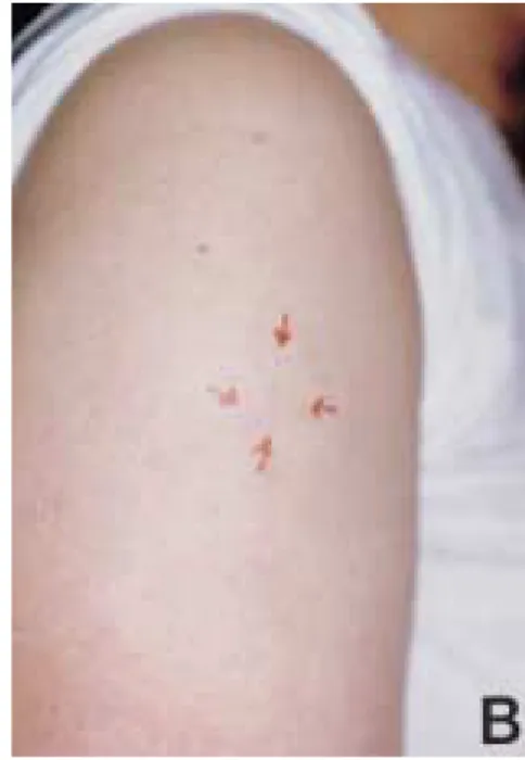
Fig. 1. Multiple subcutaneous nodules in the right flank (A) and right upper arm (B).
Fig. 2. These slides show diffuse cellular infiltration in the deep dermis and subcutis (A: H&E stain, ×1). The infiltrative cells are large atypical lymphoid cells with vesicular nuclei, prominent nucleoli and mitotic figures (B:
H&E stain, ×400).
CASE REPORT
A 56-year-old Korean woman visited our de- partment in March, 1999 with multiple subcuta- neous nodules in her right flank and right upper arm. Those lesions were rapidly growing since she had recognized them only 7 days before. She noted 4 subcutaneous nodules of firm consisten- cy without any symptoms or signs like pain, tenderness, generalized fever, night sweating or weight loss (Fig. 1). A complete physical exam- ination, laboratory findings including blood chemistry, peripheral blood smear, serum protein electrophoresis, and bone marrow biopsy and aspiration, and radiologic evaluation including chest and abdominal computed tomography (CT) and bone scan revealed no abnormal findings. The skin biopsy specimen from the right upper arm showed the diffuse infiltration of bottom-heavy pattern in deep dermis and sub- cutis without epidermal and subepidermal changes. It was composed of large atypical lymphoid cells with vesicular nuclei, prominent nucleoli and moderate degree of mitotic figures, seemingly, mixtures of immunoblasts and cen- troblasts (Fig. 2). Immunophenotypic studies revealed the lymphoid infiltrates reacted with CD45 ( LCA ; leukocyte common antigen ), CD20 ( L26 ; a pan B cell marker ) and bcl-2 protein, but none of the sections expressed CD3 ( a pan T cell marker ), bcl-6 protein and CD30 ( Ki-1 ) (Table 1)(Fig. 3).
We concluded that the patient had a primary cutaneous diffuse large B-cell lymphoma (DLB-
CL) with multifocal skin lesions and then treated her with 2 cycles of high-dose multiagent chemotherapy with the Vanderbilt regimen (VP-16, cyclophosphamide, vincristine, bleomycin, metho trexate, pred- nisolone) and the BEAM regimen (BCNU, VP-16, Ara-C, melphalan) combined with the autologous peripheral blood stem cell trans- plantation at Department of Hemato-oncology, Korea Cancer Center Hospital. Until now, 10 months after termination of treatment, she has shown the disappearance of all skin lesions and no further development of extracutaneous disease.
DISCUSSION
Cutaneous B-cell lymphomas (CBCLs) con- tribute 10% to 25% of all cutaneous lym- phomas in Western literatures and may be PCBCLs or non-Hodgkin lymphomas with secondary cutaneous involvement. CBCLs occur as a monomorphous picture of solitary or mul- tiple deep-red colored nodules or tumors without surface change, arising from normal-looking skin within less than 1 year, and the lesions are sometimes disseminated all over the body or lo- cated in aggregates in the preferential localiza- tions, trunk or head and neck1.
Primary CBCLs are recently recognized as a distinctive disease entity from extracutaneous or nodal B-cell lymphomas with the advent of improved immunophenotyping and im- munogenotyping2,3. Even though most cases of Fig. 3. Immunohistochemical stains of paraffin section show positive reaction to CD20 (A) and bcl-2 oncoprotein (B) (×200).
CBCLs have an indolent course and tend to re- main localized, CBCL lesions occurring any- where may metastasize to the extracutaneous sites including lymph node, bone, and bone marrow in 3% to 18% of all cases2,6. Conversely, 25% of non-Hodgkin’s lymphoma cases occur in extranodal sites including the skin, the next most common site of extranodal involvement to the gastrointestinal tract3 and a systemic B cell lymphoma can involve the skin in 6 to 20% of cases7. Usually, secondary CBCLs substantially show the same but a little different clinical features, e.g. disseminated or multiple nodules, a poorer overall prognosis, more frequent relapses in nodal and cutaneous lesions, compared with primary CBCLs6.
Due to such difficulties in discerning primary CBCLs from secondary CBCLs, we have to adopt the classification system proposed by the EORTC. According to this classification system, primary cutaneous lymphoma is defined as a non-Hodgkin lymphoma presenting in the skin, with no evidence of extracutaneous disease at the time of diagnosis and within the first 6 months after diagnosis, as assessed by appropriate staging procedures6.
Diffuse large B cell lymphoma (DLBCL) comprise a histogenetically heterogenous group.
According to a model proposed by Dalla-Favera et al and Karmer et al, at least two distinct ge- netic pathways, bcl-2 and bcl-6, may lead to DLBCL development; the bcl-2 rearrangement consorts with the DLBCL transformed from a clinically undetectable follicular phase and the bcl-6 pathway would be responsible for ‘de novo’
DLBCL8,9.
Bcl-2 is a proto-oncogene located in the 18q21 band. By karyotyping, t(14;18) has been identified in 85%-90% of follicular lymphoma
bcl-2 expression was found in 40%-45% of DLBCL, more often in extensive and primary nodal lymphomas than in extranodal cases10,11. And bcl-2 protein expression seemed to be re- lated to a reduced disease-free survival and a worse prognosis9,11. However, there seems to be almost no correlation between bcl-2 rearrange- ment and bcl-2 expression especially in DLBCL9. Recent studies suggest that amplification of the bcl-2 gene at chromosome 18q21 is an important mechanism for bcl-2 protein overexpression in diffuse large B cell lymphomas12. And so it would be better to consider bcl-2 rearrange- ment as a more meaningful prognostic factor rather than bcl-2 protein expression itself.
Bcl-6 is a novel proto-oncogene located in the 3q27 region and acts as a transcriptional re- pressor. Several studies found that the bcl-6 rearrangements occur in 30%-35% of DLBCL and bcl-6 rearrangements are the most fre- quent genetic lesion in nodal DLBCL13,14. But any possible correlation between the bcl-6 re- arrangement and clinical prognosis remained to be clarified in future9,14.
A series about the patients with multifocal PCLBCL of the leg reported to have a more unfavorable prognosis than those with localized PCLBCL of the leg or multifocal and localized PCFCCL or PCI. And so, they suggest that pri- mary cutaneous DLBCL with multifocal skin le- sions should always be treated with the more aggressive treatment including multiagent chemotherapy15.
To our knowledge, 6 cases of cutaneous B cell lymphoma16, 2 cases of primary CBCL17,18 and 3 cases of primary cutaneous DLBCL19-21 have been reported in Korean dermatologic litera- tures. Primary cutaneous DLBCL with multifo- cal subcutaneous nodules was not yet reported in Korea, but we expect a rather more primary cu- taneous DLBCL cases should be underreported regardless of its multifocality.
According to Japanese series recently reported4,5,
Bcl-6 proto-oncogene negative
CD 30 lymphocyte activation antigen negative
DLBCL was the most frequent subtype of PCBCL in Japanese patients and the prognosis of Japanese patients with DLBCL was worse than that of reported European cases. Also, the unfavorable clinical course was at least partly re- lated to high expression of bcl-2, and the site-re- lated difference in clinical prognosis in Japanese patients was not as great as that observed in European patients.
The presenting case showed that microscopi- cally, large atypical cells infiltrated in the sub- cutaneous fat layer and adjacent deep dermis.
And, the lymphoid infiltrates reacted with pan B cell marker and bcl-2 protein. Clinically, there presented with multiple subcutaneous nodules at noncontiguous anatomic sites without evidence of extracutaneous disease at the time of diagno- sis and within the first 6 months after diagnosis, as assessed by appropriate staging procedures.
And so, we think that this case is a primary cu- taneous diffuse large B-cell lymphoma presented as multifocal subcutaneous nodules.
REFERENCES
1. Burg G : Cutaneous B cell lymphoma, multiple myelo- ma, and myelomonocytic leukemia. In Moschella SL, Hurley HJ : Dermatology, 3rd ed., WB Saunder Co, Philadelphia, 1992, pp1829-1844.
2. Willemze R, Kerl H, Sterry W et al : EORTC classifica- tion for primary cutaneous lymphomas: A proposal from the cutaneous lymphoma study group of the Euro- pean Organization for Research and Treatment of Can- cer. Blood 90:354-371, 1997.
3. Santucci M, Pimpinelli N, Arganini L : Primary cuta- neous B-cell lymphoma: A unique type of low-grade lymphoma: Clinicopathologic and immunologic study of 83 cases. Cancer 67:2311-2326, 1991.
4. Tanaka M, Ichinohasama R, Iwasaki M, Sato M, Tasami M : Primary cutaneous B-cell lymphomas in Japan: a re- port of three cases and a comparison of Japanese and white patients. J Am Acad Dermatol 31:54-60, 1994.
5. Liu Q, Ohshima K, Kikuchi M : Primary cutaneous B- cell lymphoma in Japanese patients. Patho Int 50:960- 966, 2000.
6. Pandolfino TL, Siegel RS, Kuzel TM, Rosen ST, Guitart J : Primary cutaneous B-cell lymphoma: Review and current concepts. J Clin Oncol 18:2152-2168, 2000.
7. Sterry W, Kruger GRE, Steigleder GK : Skin invol- vement of malignant B-cell lymphomas. J Dermatol
Surg Oncol 10:276-277, 1984.
8. Dalla-Favera R, Yye BH, Lo Coco F et al : Identification of genetic lesions associated with diffuse large-cell lym- phoma. Ann Oncol 5:55-60, 1994 (suppl).
9. Karmer MHH, Hermans J, Wijburg E et al : Clinical rel- evance of BCL2, BCL6 and MYC rearrangements in diffuse large B-cell lymphoma. Blood 92:3152-3162, 1998.
10. Karmer MHH, Hermans J, Parker J et al : Clinical signif- icance of bcl2 and p53 protein expression in diffuse large B-cell lymphoma: A population-based study. J Clin Oncol 14:2131-2138, 1996.
11. Hill ME, MacLennan KA, Cunningham DC et al : Prog- nostic significance of BCL-2 expression and bcl-2 major breakpoint region rearrangement in diffuse large cell Non-Hodgkin’s lymphoma: A British National Lym- phoma Investigation Study. Blood 88:1064-1051, 1996.
12. Monni O, Joensuu H, Franssila K et al : Bcl-2 overex- pression associated with chromosomal amplification in diffuse large B-cell lymphoma. Blood 90: 1168-1174, 1997.
13. Pescarmona E, De Sanctis V, Pistilli A et al : Patho- genetic and clinical implications of Bcl-6 and Bcl-2 gene configuration in nodal diffuse large B-cell lymphomas. J Pathol 183:281-286, 1997.
14. Ofitt K, Lo Coco F, Louie DC et al : Rearrangement of the bcl-6 gene as a prognostic marker in diffuse large- cell lymphoma. N Engl J Med 331:74-80, 1994.
15. Bekkenk MW, Vermeer MH, Geerts ML et al : Treat- ment of multifocal primary cutaneous B-cell lymphoma:
a clinical follow-up study of 29 patients. J Clin Oncol 17:2471-2478, 1999.
16. Cho KH, Kim YG, Kim CW, Lee YS : A clinicopatho- logic study of cutaneous lymphoma. Kor J Dermatol 29:782-794, 1991.
17. Kim KH, Nam JT, Joh GY, et al : Primary cutaneous B- cell lymphoma. Ann Dermatol 6:249-255, 1994.
18. Lee HK, Cho YW, Song KY, Yoo BH, Ro BI : Primary cutaneous B-cell lymphoma. Ann Dermatol 7:58-61, 1995.
19. Kim JW, Yoon YM, Kim DS, Kim SW : A case of cuta- neous B-cell lymphoma. Kor J Dermatol 35: 312-316, 1997.
20. Chun TJ, Seo SJ, Song KY, Hong CK, Ro BI : A case of cutaneous diffuse large B-cell lymphoma. Kor J Derma- tol 37:1330-1334, 1999.
21. Koh GJ, Kim KJ, Chang SE et al : A case of primary cu- taneous diffuse large B-cell lymphoma. Kor J Dermatol 38:1651-1655, 2000.