ISSN 2288-0356(Online) Original Article
Comparative Study of Extracellular Proteomes for Bacillus subtilis and Bacillus amyloliquefaciens
Maria Claret Lauan
1,2, IlynLyzette Santos
1,3, Jinkyu Lim
1*1Major inFood Biomaterials, Kyungpook National University, Daegu, South Korea
2Department of Biology, University of the Philippines, Las Baňos, Philippines
3Dx Assays, Singapore
Abstract
Bacillus subtilisandBacillus amyloliquefaciensare closely related species that share a similar genomic background, and are both known to secrete large amounts of proteins directly into a medium. The extracellular proteomes of two strains ofBacillus subtilisand two strains ofBacillus amyloliquefaciens were compared by 2-D gel electrophoresis during the late exponential growth phase. The relative abundance of some minor protein spots varied among the four strains ofBacillus. Over 123 spots of extracellular proteins were visualized on the gel forB. subtilisCH 97, 68 spots forB. subtilis3-5, 230 spots forB. amyloliquefaciensCH 51, and 60 spotsfor B. amyloliquefaciens86-1. 2D gel electrophoresis images of the fourBacillusstrains showed significantly different protein profiles. Consistent with the 2D gel electrophoretic analysis, most of theB. subtilisproteins differed from the proteases secreted by theB. amyloliquefaciensstrains. Among the proteins identified fromB. subtilis, approximately 50% were cytoplasmic and 30%
were canonically extracellular proteins. The secreted protein profiles forB. subtilisCH 97 andB. subtilis3-5 were quite different, as were the profiles forB. amyloliquefaciensCH 51 and 86-1. The four proteomes also differed in the major protein composition. TheB. subtilisCH 97 andB. amyloliquefaciens CH 51 proteomes both contained large amounts of secreted hydrolytic enzymes. Among the four strains,B. subtilis3-5 secreted the least number of proteins. Therefore, even closely related bacteria in terms of genomic sequences can still have significant differences in their physiology and proteome layout.
Keywords:Bacillus, Secreted proteome, MALDI-TOF, 2-D gel
Introduction1)
Bacillus subtilisis
regarded as a representative model organism of Gram positive bacteria, and its genome sequence, published in 1997 by Kunst et al. provides its “blue-print of life”. Plus, due to the availability of its genome sequence and advent of recent technologies, this organism is also regarded as a model of functional genomics (transcriptomics, proteomics, metabolomics etc.). The growing field of “omics” has also broughtthe genome sequence to cell physiology.While about 4100 different open reading frames (ORFs) have already been predicted to express proteins from the genome of
Bacillus subtilis,
the genome sequence information has extended the proteomic analysis ofBacillus
in various areas.Moreover, the advances and availability of technology for identifying proteomes, such as high-resolution 2-DE with a high reproducibility, high-throughput mass spectrometry with a high sensitivity, and efficient database searching techniques and
software using sophisticated bioinformatics algorithms, have resulted in significant progress in functional proteomics.
The ability of
Bacillus
cells to secrete large amounts of proteins has long been of interest to the fermentation industry. AlthoughBacillus amyloliquefaciens
andBacillus
subtilis are closely related in terms of their genome sequences, there are significant differences between these two species as regards their growth characteristics and secreted protein profiles.Accordingly, this study compared the extracellular proteins of
Bacillus subtilis
andBacillus amyloliquefaciens
during the late exponential growth phase using their respective protein profile patterns. The proteomic view of these extracellular proteins can provide a comprehensive understanding of metabolism and growth processes, while the secreted enzymes offer practical industrial applications, such as fermentation monitoring and developing fermentation products. Among the 4100 proteins expressed inB. subtilis
, the number of extracellular proteinsReceived: March 12, 2013 / Revised: March 27, 2013 / Accept: March 28, 2013
*Corresponding Author: Jinkyu Lim, Tel. 82-53-950-5755, Fax. 82-53-950-6750, Email. [email protected]
The authors would like to thank Dr. Jeong Hwan Kim from Gyeongsang National University for providing the Bacillus strains used in this study.
is not yet known or how the secretion machinery proteins facilitate their extracellular secretion. Therefore, this study identified the secretory proteins separated on 2-D gels to demonstrate the secretory protein profiles from
B. subtilis
andB. amyloliquefaciens
and compare the functional proteomes quantitatively and qualitatively. Hence, the secreted proteins were identified using a proteomic approach based on two-dimensional gel electrophoresis and a peptide mass fingerprinting analysis using matrix assisted laser desorption ionization-time of flight (MALDI-TOF) mass spectrometry.2)
Materials and Methods Strains and culture conditions
Four strains of
Bacillus
, namelyBacillus subtilis
CH97,Bacillus subtilis
3-5,Bacillus amyloliquefaciens
CH51, andBacillus amyloliquefaciens
86-1, were each grown in 500 mL of a Luria-Bertani (LB) broth in 2 L Erlenmeyer flasks agitated at 200 rpm at 42℃, and taken after 12 and 24 h.Extracellular sample preparation for proteome analysis The supernatant fractions from each strain were collected after high-speed centrifugation of the cultures at 10000 rpm for 20 min. The supernatants were then dialyzed at 4℃with 4 buffer changes over 36 h, lyophilized to powders, dissolved in 0.1X PBS with protease inhibitors, and desalted using a PD-10 column (GE Healthcare, Uppsala, Switzerland). The proteins in the void volume from the desalting column were precipitated using 10%
tri-chloroacetic acid. Thereafter, the precipitated proteins were washed with 100% ice-cold ethanol, washed with 70% ethanol, and dried under a vacuum. Finally, the dried proteins were dissolved in a rehydration buffer containing 7 M urea, 2 M thiourea, 4% CHAPS, 0.1% dithiothreitol, and 0.2% ampholyte with a pH range of 3 to 10.
2D protein gel electrophoresis
Two-dimensional (2-D) sodium dodecyl sulfate-polyacrylamide gel electrophoresis (SDS-PAGE) was conducted to separate the proteins. First, dimensional isoelectric focusing was carried out using the passive-loading method with an immobilized pH gradient (IPG) strip at pH 4-7 (Amersham Biosciences, Freiburg, Germany). The IPG strips were initially equilibrated in an equilibration buffer containing 6M urea, 2% SDS, 0.375 M Tris-HCl, pH 8.8, 20% glycerol, and 130 mM dithiothreitol for the first equilibration and then in an equilibration buffer containing 135 mM iodoacetamide. Next, the strips were overlaid on a 12% separating gel for the2-D SDS-PAGE. The
resulting 2-D gels were fixed with 40% v/v ethanol and 10%
v/v acidic acid for 1-2 h and then stained with colloidal CBB (Amersham Biosciences, Freiburg, USA),
Preparation of peptide mixtures for MALDI-MS All the visible spots were excised from the gel, destained by washing three times in deionized water and two times in 50 mM NH4HCO3 and acetonitrile (60:40) with gentle shaking, and freeze-dried. The gel pieces were then digested for 18h using 10 ng trypsin/μL of 50 mM NH4HCO3and the peptides extracted using 0.1% trifluoroacetic acid in 50 mM NH4HCO3. Analysis of peptides by MALDI-TOF MS and identification of proteins
The MALDI-TOF measurement of the spotted peptide solutions was carried out using a Voyager DE-STR MALDI Biospectrometry Workstation Proteome-Analyzer 4700 (Applied Biosystems, USA). The spectra were recorded in the reflector mode within a mass range from800to 3500 Da. The peak lists were created using the “peak to mascot” script of the 4700 Explorer™Software and calibrated using
M-over-Z
(Genomic Solutions, USA). The database searches used the search engines Ms-Fit (http://prospector.ucsf.edu) and MASCOT (Matrix Science).Results and Discussion
Comparison of extracellular proteomes for
B. subtilis
CH97 andB. subtilis
3-5Several studies have already shown that the highest levels of protein secretion are usually during the late exponential phase until the onset of the stationary phase. Therefore, in this set of experiments, the
B. subtilis
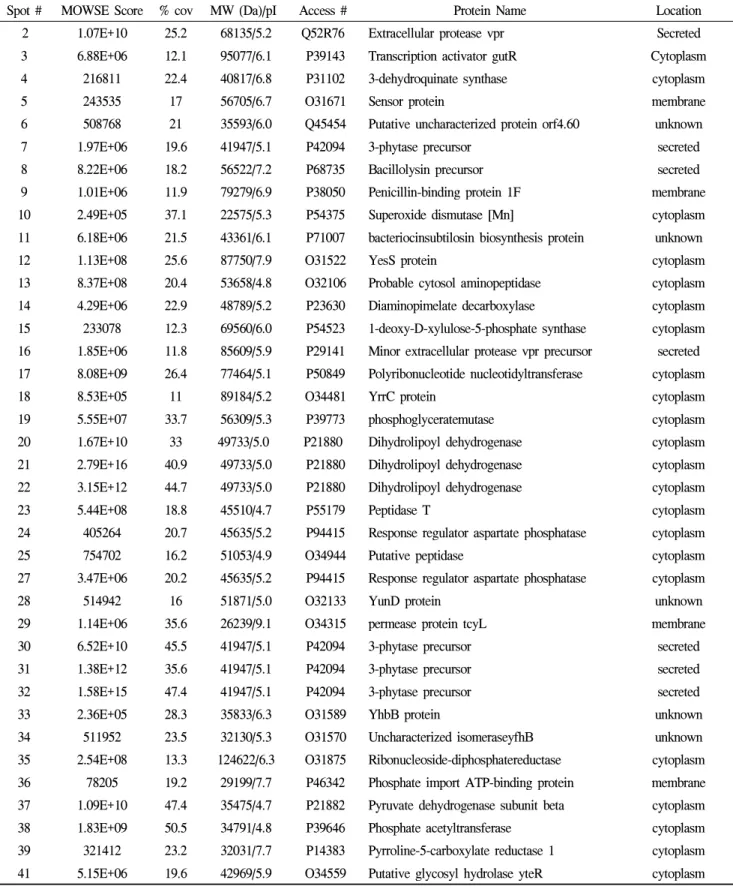
cells were grown in an LB medium and harvested during the late exponential phase. The extra-cellular proteins were precipitated, subjected to 2D gel electrophoresis at pH 4-7, and visualized with colloidal CBB staining. Based on the theoretical proteome map of allB. subtilis
proteins (Buttner et al. 2001), the proteins were separated using a single 2-DE gel with a pH range of 4–7. Thereafter, an analysis of the 2-DE images and peptide mass fingerprinting allowed the identification of 101 proteins from approximately 123 spots that were visualized on the gel ofB. subtilis
CH97 (Figure 2A) and about 100 spots visualized on the gel ofB.
subtilis
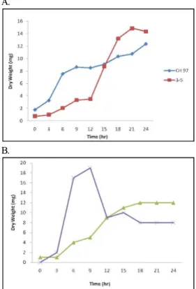
3-5 (Figure 2B).The growth of
B. subtilis
3-5 was faster than that ofB. subtilis
CH97, as theB. subtilis
3-5 strain reached the stationary phase after 21 h, whereas theB. subtilis
CH97 strain was still in the late exponential phase after 24 h (Figure 1A), which mayexplain the higher number of detected spots for the latter strain.
Notwithstanding, both strains produced extracellular proteins that were identified as cytosolic proteins.
A.
B.
Figure 1. Growth curves for
B. subtilis
(A) andB.
amyloliquefaciens
(B).B. subtilis
CH 97 (■),B. subtilis
3-5 (■),B. amyloliquefaciens
CH 51 (▴), andB.
amyloliquefaciens
86-1 (ⅹ) were grown in LB medium at 42℃.Comparison of extracellular proteomes for
B. amyloliquefaciens
CH51 andB. amyloliquefaciens
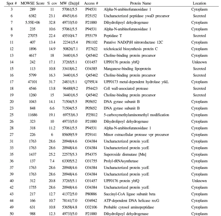
86-13)An analysis of the 2-DE images and peptide mass fingerprinting allowed the identification of 35 different proteins from the approximately 100 spots that were visualized on the gel of
B.
amyloliquefaciens
86-1 (Figure 3B) and about 200 spots visualized on the gel ofB. amyloliquefaciens
CH51 (Figure 3A). The growth ofB. amyloliquefaciens
86-1 was faster than that ofB. amyloliquefaciens
CH51, as theB. amyloliquefaciens
86-1 strain reached the stationary phase after 12 h, whereas it took 18 h for theB. amyloliquefaciens
CH51 strain (Figure 1B), which may explain the fragility of the first strain and hence the smaller number of detected spots. Overall, both strains secreted enzymes related to carbohydrates and their metabolism, transferases, sporulation-specific proteins, and transcription regulators (Figure 4B). The functions of the other identifiedproteins remain unknown.
Comparison of extracellular proteomes for
B. subtilis
andB. amyloliquefaciens
The availability of the genome sequence for
B. subtilis
in 1997 andB. amyloliquefaciens
in 2007 has enabled the prediction of all proteins containing signals for known systems of protein secretion. From the genome data, 4,107 ORFs have been identified forB. subtilis
and 5,224 ORFs forB. amyloliquefaciens
(Kunst et al. 1997; Chen et al. 2007). Based on the sesequences, 297 proteins have the potential to be translocated extracellularly from the cytoplasm ofB. subtilis
(Tjalsma et al. 2000; van Dijl et al. 2002).Figure 2. Extracellular proteins from B. subtilis CH 97 (A) and B. subtilis 3-5 (B) profiled on 2D gels. Cells were grown in LB broth at 42℃ and sampled during lateexponential growth phase.
Among the four strains used in this study,
B. subtilis
CH 97 andB. amyloliquefaciens
CH51 produced the most visible spots on the 2D gel (Figures 2 and 3). Some of the secreted proteins occurred as multiple spots, like serine protease, dihydrolipoamide, and flagellin. Altogether, 100 different proteins were identified fromB. subtilis
and 35 fromB. amyloliquefaciens.
However, not all the spots were successfully identified. In this study, the PMF identification success rate was relatively higher for the larger proteins than for the small proteins. Due to the lower number of peptides after trypsiniztion, identifying low- molecular-weight proteins is difficult using PMF. Thus, increasing the peptide numbers by efficient trypsinization and/or analyzing the proteins using LC-MS/MS is recommended.As shown in Figures 2 and 3, the protein profiles differed significantly among the four strains. In the case of
B. subtilis
, flagellin (Hag) was the main protein, whereas it was proteases in the case ofB. amyloliquefaciens
. Another major difference in the overall composition of the respective extracellular proteomes was the proteins involved in carbohydrateTable 1. Identified extracellular proteins from
B. subtilis
CH97Spot # MOWSE Score % cov MW (Da)/pI Access # Protein Name Location
24) 1.07E+10 25.2 68135/5.2 Q52R76 Extracellular protease vpr Secreted
3 6.88E+06 12.1 95077/6.1 P39143 Transcription activator gutR Cytoplasm
4 216811 22.4 40817/6.8 P31102 3-dehydroquinate synthase cytoplasm
5 243535 17 56705/6.7 O31671 Sensor protein membrane
6 508768 21 35593/6.0 Q45454 Putative uncharacterized protein orf4.60 unknown
7 1.97E+06 19.6 41947/5.1 P42094 3-phytase precursor secreted
8 8.22E+06 18.2 56522/7.2 P68735 Bacillolysin precursor secreted
9 1.01E+06 11.9 79279/6.9 P38050 Penicillin-binding protein 1F membrane
10 2.49E+05 37.1 22575/5.3 P54375 Superoxide dismutase [Mn] cytoplasm
11 6.18E+06 21.5 43361/6.1 P71007 bacteriocinsubtilosin biosynthesis protein unknown
12 1.13E+08 25.6 87750/7.9 O31522 YesS protein cytoplasm
13 8.37E+08 20.4 53658/4.8 O32106 Probable cytosol aminopeptidase cytoplasm
14 4.29E+06 22.9 48789/5.2 P23630 Diaminopimelate decarboxylase cytoplasm
15 233078 12.3 69560/6.0 P54523 1-deoxy-D-xylulose-5-phosphate synthase cytoplasm 16 1.85E+06 11.8 85609/5.9 P29141 Minor extracellular protease vpr precursor secreted 17 8.08E+09 26.4 77464/5.1 P50849 Polyribonucleotide nucleotidyltransferase cytoplasm
18 8.53E+05 11 89184/5.2 O34481 YrrC protein cytoplasm
19 5.55E+07 33.7 56309/5.3 P39773 phosphoglyceratemutase cytoplasm
20 1.67E+10 33 49733/5.0 P21880 Dihydrolipoyl dehydrogenase cytoplasm
21 2.79E+16 40.9 49733/5.0 P21880 Dihydrolipoyl dehydrogenase cytoplasm
22 3.15E+12 44.7 49733/5.0 P21880 Dihydrolipoyl dehydrogenase cytoplasm
23 5.44E+08 18.8 45510/4.7 P55179 Peptidase T cytoplasm
24 405264 20.7 45635/5.2 P94415 Response regulator aspartate phosphatase cytoplasm
25 754702 16.2 51053/4.9 O34944 Putative peptidase cytoplasm
27 3.47E+06 20.2 45635/5.2 P94415 Response regulator aspartate phosphatase cytoplasm
28 514942 16 51871/5.0 O32133 YunD protein unknown
29 1.14E+06 35.6 26239/9.1 O34315 permease protein tcyL membrane
30 6.52E+10 45.5 41947/5.1 P42094 3-phytase precursor secreted
31 1.38E+12 35.6 41947/5.1 P42094 3-phytase precursor secreted
32 1.58E+15 47.4 41947/5.1 P42094 3-phytase precursor secreted
33 2.36E+05 28.3 35833/6.3 O31589 YhbB protein unknown
34 511952 23.5 32130/5.3 O31570 Uncharacterized isomeraseyfhB unknown
35 2.54E+08 13.3 124622/6.3 O31875 Ribonucleoside-diphosphatereductase cytoplasm 36 78205 19.2 29199/7.7 P46342 Phosphate import ATP-binding protein membrane 37 1.09E+10 47.4 35475/4.7 P21882 Pyruvate dehydrogenase subunit beta cytoplasm
38 1.83E+09 50.5 34791/4.8 P39646 Phosphate acetyltransferase cytoplasm
39 321412 23.2 32031/7.7 P14383 Pyrroline-5-carboxylate reductase 1 cytoplasm 41 5.15E+06 19.6 42969/5.9 O34559 Putative glycosyl hydrolase yteR cytoplasm
Spot # MOWSE Score % cov MW (Da)/pI Access # Protein Name Location
43 1.05E+06 24.4 47737/5.4 P22326 Tyrosyl-tRNAsynthetase 1 cytoplasm
445) 135513 20.1 31811/5.2 P54504 Uncharacterized protein yqhA cytoplasm
45 95514 22.3 40136/5.6 P80862 Phosphoserine aminotransferase cytoplasm
46 261640 17 25975/5.7 Q9F4F7 4'-phosphopantetheinyl transferaseffp cytoplasm
47 97033 29.8 33851/6.6 O06973 UPF0042 protein yvcJ unknown
48 1.66E+07 15.7 79279/6.9 P38050 Penicillin-binding protein 1F membrane
49 27662 19 25844/5.7 Q06755 cytidylyltransferase cytoplasm
50 2.49E+06 40.4 22748/5.6 P54480 Putative nucleotidaseyqfW cytoplasm
55 9.74E+06 15.5 106033/5.9 P54394 helicase dinG homolog nucleus
57 8.53E+06 23.2 55473/5.3 P42176 Nitrate reductase beta chain membrane
58 166917 34.7 16355/4.8 P54332 Phage-like element PBSX protein unknown
59 4.26E+07 40.7 21824/5.5 P81100 Stress response protein SCP2 cytoplasm
61 56590 20.5 49084/5.3 P39772 Asparaginyl-tRNAsynthetase cytoplasm
62 812995 22.5 56224/5.7 P37966 Lipoprotein lplA precursor membrane
63 1.22E+06 30.2 45592/8.3 O32267 glycosyltransferasetuaH cytoplasm
65 2.52E+06 24.7 72800/5.9 P00691 Alpha-amylase precursor secreted
66 319835 21.1 91335/6.3 Q45066 DNA topoisomerase 4 subunit A cytoplasm
67 5.12E+06 22.1 119469/5.8 O08394 P-450/NADPH-P450 reductase cytoplasm
68 1.06E+07 24.1 99563/4.9 P39793 Penicillin-binding protein 1A/1B membrane
69 619649 26 101747/5.0 Q05873 Valyl-tRNAsynthetase cytoplasm
70 5.61E+06 30.4 57061/5.5 P94531 Alpha-N-arabinofuranosidase 1 cytoplasm
71 3.62E+06 26.4 73316/6.0 Q797B3 lipoteichoic acid synthase 1 membrane; secreted
72 429968 24.9 72258/4.9 P46208 Chaperone protein htpG cytoplasm
73 1.62E+06 35.8 37847/5.1 Q04797 Aspartate-semialdehyde dehydrogenase cytoplasm
74 1.13E+08 35.2 39992/5.1 P54531 Leucine dehydrogenase cytoplasm
75 4.42E+06 41.3 38411/5.3 O34499 Uncharacterized protein ykgB unknown
76 2.54E+07 29.2 56522/7.2 P68735 Bacillolysin precursor secreted
78 124809 25.6 47212/5.7 P50735 NAD-specific glutamate dehydrogenase cytoplasm
79 278216 29.3 32627/5.0 P02968 Flagellin secreted
80 5.10E+06 21.5 34615/5.4 P54327 Phage-like element PBSX protein xkdG unknown
82 50761 20.5 61838/5.3 P54551 Uncharacterized protein yqjN cytoplasm
83 1.05E+06 25 59301/4.6 P23447 Flagellar M-ring protein membrane; flagella
84 45607 23.8 45635/5.2 P94415 Response regulator aspartate phosphatase cytoplasm
87 20988 34.2 21824/5.5 P81100 Stress response protein SCP2 cytoplasm
90 102427 25.3 24791/5.8 P54601 Uncharacterized protein yhcQ spore wall
91 3.87E+07 53.5 22575/5.3 P54375 Superoxide dismutase [Mn] cytoplasm
92 3.87E+07 53.5 22575/5.3 P54375 Superoxide dismutase [Mn] cytoplasm
93 2.23E+07 45.9 25378/5.1 O34925 Purine nucleoside phosphorylase cytoplasm
95 137011 33.5 21364/5.9 P54390 UPF0302 protein ypiB unknown
Spot # MOWSE Score % cov MW (Da)/pI Access # Protein Name6) Location
96 129609 28.1 25132/7.7 P38491 Uncharacterized protein ypfA unknown
101 4.09E+07 27.1 56522/7.2 P68735 Bacillolysin precursor secreted
102 3.77E+07 41.1 56522/7.2 P68735 Bacillolysin precursor secreted
104 4.32E+08 36.2 79079/9.0 P39814 DNA topoisomerase 1 cytoplasm
105 1.84E+07 27 119469/5.8 O08394 P-450/NADPH-P450 reductase 1 cytoplasm
107 1.24E+07 51.6 33418/6.2 O34385 lipoprotein mntA precursor membrane
108 4.28E+06 30.1 99528/5.2 Q798L9 YwqA protein
110 1.44E+06 43 27464/6.7 O31614 Bis(5'-nucleosyl)-tetraphosphatase Spore
111 1.25E+08 35.7 92100/5.4 P05653 DNA gyrase subunit A
112 1.68E+06 27.3 39468/9.0 P00783 Subtilisinamylosacchariticus precursor secreted
113 7.48E+07 36.5 31497/8.7 O07921 Chitosanase precursor secreted
114 1.54E+06 26.8 77464/5.1 P50849 Polyribonucleotide nucleotidyltransferase cytoplasm
115 4.13E+06 47.5 17515/5.0 O05396 Uncharacterized protein yrhD Unknown
116 6.44E+06 35.3 24876/9.6 O05411 Uncharacterized protein yrpD Unknown
117 6.44E+06 35.3 24876/9.6 O05411 Uncharacterized protein yrpD Unknown
118 9.13E+06 30.1 45635/5.2 P94415 Response regulator aspartate phosphatase Cytoplasm
119 4.63E+07 30.9 41947/5.1 P42094 3-phytase precursor Secreted
120 4.80E+09 59.7 32417/4.9 P70999 Agmatinase Cytoplasm
123 3.92E+07 11.4 120530/5.9 P39774 Subtilinbiosynthesis protein spaB Membrane
and enolase.
B. subtilis
secreted a higher number of these carbohydrate-degrading enzymes thanB. amyloliquefaciens
. Conversely,B. amyloliquefaciens
secreted a higher number of peptidases and proteases.Figure 3. Extracellular proteins from
B. amyloliquefaciens
CH 51 (A) andB. amyloliquefaciens
86-1 (B) profiled on 2D gel. Cells were grown in LB Broth at 42℃ and sampled during late exponential growth phase.Overall, the extracellular proteomesfor
B. subtilis
andB.
amyloliquefaciens
included numerous unpredicted secreted proteins (cytoplasmic, membrane, and flagellar). Hag migrated as the largest spot on the 2D gel of extracellular proteinpreparations. The secretion of Hag is not seemingly assisted by SRP and Sec translocase in
E. coli
(Hueck, 1998; Namba et al. 1989). The appearance of cytosolic proteins in the extracellular preparations was probably due to breakage of the cells or the leakage of cytoplasmic proteins by an unknown function. The cells ofB. subtilis
3-5 andB. amyloliquefaciens
86-1 were easily lysed, as shown by their shorter growth curves compared to the other two strains (Figures 1 and 3).Figure 4. Functional classifications of identified extracellular proteins from
B. subtilis
(A) andB. amyloliquefaciens
(B).The 101 identified extracellular proteins from
B. subtilis
included 16 proteins with unknown functions (Table 1).Table 2. Identified extracellular proteins from
B. amyloliquefaciens
86-1Spot # MOWSE Score % cov MW (Da)/pI Access # Protein Name Location
3 289 11 57061/5.5 P94531 Alpha-N-arabinofuranosidase 1 Cytoplasm
6 6382 23.1 49451/6.6 P25152 Uncharacterized peptidase ywaD precursor Secreted
7 5.55E+06 32.8 49733/5.0 P21880 Dihydrolipoyl dehydrogenase Cytoplasm
8 235 10.6 57061/5.5 P94531 Alpha-N-arabinofuranosidase 1 Cytoplasm
9 27075 22.4 45510/4.7 P55179 Peptidase T Secreted
11 407 13.4 22341/5.4 P81102 Putative NAD(P)H nitroreductase 12C Cytoplasm
12 1896 14.9 50826/7.1 P27622 teichoicacid biosynthesis protein C Cytoplasm
13 4617 18 34401/6.5 Q45462 Choline-binding protein precursor Cytoplasm
14 242 17.1 37265/5.1 O31457 UPF0176 protein ybfQ Unknown
15 113 10.8 33418/6.2 O34385 Manganese-binding lipoprotein Secreted
16 5799 16.3 34401/6.5 Q45462 Choline-binding protein precursor Secreted
17 47101 31.7 24831/5.1 Q795U4 UPF0173 metal-dependent hydrolase ytkL Cytoplasm
18 4546 13.8 96488/9.2 P54423 Cell wall-associated protease Secreted
19 1260 15 34401/6.5 Q45462 Choline-binding protein precursor Secreted
20 1043 14.1 71504/5.5 P05652 DNA gyrase subunit B Cytoplasm
23 848 6.6 71504/5.5 P05652 DNA gyrase subunit B Cytoplasm
25 11686 19.1 69753/6.3 P25812 5-carboxymethylaminomethyl modification Cytoplasm
26 323 10 49733/5.0 P21880 Dihydrolipoyl dehydrogenase Cytoplasm
28 318 11.2 57061/5.5 P94531 Alpha-N-arabinofuranosidase 1 Cytoplasm
27 226 6 85609/5.9 P29141 Minor extracellular protease vpr precursor Cytoplasm
31 1763 28.6 20948/4.6 O34384 Uncharacterized protein yceE Cytoplasm
33 1763 28.6 20948/4.6 O34384 Uncharacterized protein yceE Cytoplasm
35 1437 25.2 22575/5.3 P54375 Superoxide dismutase [Mn] Cytoplasm
36 137 7.4 63305/5.2 O31755 Prolyl-tRNAsynthetase Cytoplasm
37 1763 28.6 20948/4.6 O34384 Uncharacterized protein yceE Cytoplasm
39 1763 28.6 20948/4.6 O34384 Uncharacterized protein yceE Cytoplasm
40 312 20.8 37265/5.1 O31457 UPF0176 protein ybfQ Unknown
42 1755 28.6 20948/4.6 O34384 Uncharacterized protein yceE Cytoplasm
43 217 12.7 41372/5.0 P80886 Succinyl-CoA ligase subunit beta Cytoplasm
44 166 10.7 78141/7.0 O34942 ATP-dependent DNA helicase recG Cytoplasm
49 631 10.8 53658/4.8 O32106 Probable cytosol aminopeptidase Cytoplasm
50 988 12.3 49733/5.0 P21880 Dihydrolipoyl dehydrogenase Cytoplasm
7)
Interestingly, the identified extracellular proteins from the Bacillus strains in this study included enzymes related to the metabolism of carbohydrates, lipid and amino acids enzymes involved in protein synthesis and folding and the synthesis and decay of nucleic acids, and enzymes involved in transcriptional regulations. This also supports the idea that the proteins in the
extracellular media possibly originated from the lysis of the bacterial cells.
The metabolic enzymes identified, such as hydrolase, are known to beinvolved in several pathways for carbohydrate utilization and activated during the fermentation process (Antelmann et al. 2004). Meanwhile, some of the other enzymes play a role
in the translocation machinery of the protein secretion by
B.
subtilis
. For example, one of the secreted proteins, an alpha- amylase (AmyE) precursor, is a probable presecretory protein that is recognized by a chaperone and targeted to a Sec protein translocase for transport across the membrane (Hirose et al.2000). Furthermore, the search also identified sporulation proteins, such as lipoproteins, synthesized as precursors that function as an extracellular folding catalyst in protein folding on the membrane surface (Hecker et al. 2004; Bunai et al. 2004).
The increased proteins during the growth phases at 37℃ (Figure 1A) and 42℃ (Figure 1B) showed similar changing patterns, although the growth rate at 42℃ as slightly slower than that at 37℃. The results also contradicted the previous assertion that about 75% of the secreted proteins from Bacillus have N-terminal signal peptides or specific retention signals, while the rest of the secreted proteins are transported by independent signal peptide systems, including holin-transport, flagella- transport, and cell lysis (von Heijne, 1998; Tjalsma et al. 2004).
The present results indicated that cytosolic proteins were secreted from the cells throughout the growth stages by an unknown mechanism, cell lysis, or by both mechanisms. Another possibility is that the identified unchanged or increased (Figure 2) secreted proteins are so stable that they remain in the cells (Tullius et al. 2001).8)
As secretory proteins, such as proteases and metabolic enzymes, can affect the fermentation process, secretory protein profiles can be used to select the proper microorganisms or strains for optimizingfermentation conditions.
Acknowledgment
The authors would like to thank Dr. Jeong Hwan Kim from Gyeongsang University for providing the Bacillus strains used in this study.
References
Ahn YS, Kim YS, Shin DH (2006) Isolation, identification, and fermentation characteristics of Bacillus sp. with high protease activity from traditional Cheonggukjang.
Korean J Food Sci Technol
38: 82-87.Antelmann H, Sapolsky R, Miller B, Ferrari E, Chotani G, Weyler W, Gaertner A, Hecker M (2004) Quantitative proteome profiling during the fermentation process of pleiotropic
Bacillus subtilis
mutants.Proteomics
4:2408-2424.
Bunai K, Ariga M, Inoue T, Nozaki M, Ogane S, Kakeshita H, Nemoto T, Nakanishi H, Yamane K (2004) Profiling and comprehensive expression analysis of ABC transporter
solute-binding proteins of
Bacillus subtilis
membrane based on a proteomic approach.Electrophoresis
25: 141-155.Büttner K, Bernhardt J, Scharf C, Schmid R, Mäder U, Eymann C, Antelmann H, Völker A, Völker U and Hecker M. (2001) A comprehensive two-dimensional map of cytosolic proteins of
Bacillus subtilis
.Electrophoresis
22: 2908-2935.Han GH, Cho TY, Yoo MS, Kim CS, Kim JM, Kim HA, Kim MO, Kim SC, Lee SA, Ko YS, Kim SH, Kim DB (2007) Biogenic amines formation and content in fermented soybean paste (Cheonggukjang).
Korean J Food Sci Technol
39: 541-545.Hecker M, Volker U (2004) Towards a comprehensive understanding of
Bacillus subtilis
cell physiology by physiological proteomics.Proteomics
4: 3727-3750.Hirose I, Sano K, Shioda I, Kumano M, Nakamura K, Yamane K (2000) Proteome analysis of
Bacillus subtilis
extracellular proteins: a two-dimensional protein electrophoretic study.Microbiology
146: 65-75.Hueck, CJ. (1998) Type III protein secretion systems in bacterial pathogens of animals and plants.
Microbiol Mol Biol Rev
62: 379-433.Kunst F, Ogasawara N, MoszerI, Albertini AM, Alloni G, et al. (1997) The complete genome sequence of the gram-positive bacterium
Bacillus subtilis
.Nature
90:249-256.
Pappin DJ, Hojrup P, Bleasby AJ (1993) Rapid identification of proteins by peptide-mass fingerprinting.
Curr Biol
3:327-332.
Tjalsma H, Antelmann H, Jongbloed JD, Braun PG, Darmon E, Dorenbos R, Dubois JY, Westers H, Zanen G, Quax WJ, Kuipers OP, Bron S, Hecker M, van Dijl JM (2004) Proteomics of protein secretion by Bacillus subtilis:
separating the “secrets” of the secretome.
Microbiol Mol Biol Rev
68: 207-233.Tjalsma H. (2007) Feature-based reappraisal of the
Bacillus subtilis
exoproteome.Proteomics
1: 73-81.Tullius MV, Harth G, Horwitz MA (2001) High extracellular levels of
Mycobacterium tuberculosis
glutamine synthetaseand superoxide dismutase in actively growing cultures are due to high expression and extracellular stability rather than to a protein-specific export mechanism.Infect Immun
69: 6348-6363.von Heijne G (1998) Life and death of a signal peptide.