INTRODUCTION
Gallstone ileus is an infrequent cause of intestinal obstruc- tion, responsible for 1-4% of all mechanical intestinal obstruc- tions (1). It is due to a gallstone larger than 2.5 cm in diam- eter that usually obstructs terminal jejunum. The gallstone usually enters the bowel via a cholecystoenteric fistula (2), resulting from recurrent attacks of cholecystitis. Gallstone ileus is usually a disease of the elderly and occurs more fre- quently in females (3).
The present case is interesting because, 1) the patient was a young male; 2) the ileus occurred 3 yr after cholecystecto- my and 3) the localization of the obstruction was an old side- to-side ileoileic anastomosis due to a diverticulectomy follow- ing intussusception of Meckels’ diverticulum at the age of 3.
CASE REPORT
A 44-yr-old man presented to the emergency department with lower abdominal pain of 3 days’ duration, associated with nausea, diarrhea and failure to pass flatus and feces. The last year he presented again with an episode of severe abdom- inal pain, but the abdominal examination was unremarkable.
His medical anamnesis reports Gilbert syndrome, appen- dicectomy and Meckel’s diverticulectomy due to intussus-
ception at the age of three, and laparoscopic cholecystecto- my three years before admission.
Physical examination showed mild abdominal distension, tenderness in the epigastrium, and dehydration with break- ing of the skin and glue tongue. Auscultation revealed high- pitched abdominal sounds. Laboratory studies revealed a hae- matocrit of 35.4%, hemoglobin of 11.7 g/dL and white blood cell count 8.700/μL with a predominance of polymorph nucle- ar cells (66.4%). His liver function tests were all within nor- mal limits with total bilirubin of 7.6 mg/dL.
Plain films of the abdomen showed multiple air-fluid inter- faces in the small intestine and colon full of gas. There was no evidence of pneumobilia, but there were surgical clips at the anatomic area of the right upper quadrant. Two calcified opacities were detected in the left iliac fossa at the supine radio- graph (RX), which changed location at the erect RX (Fig. 1).
At profile plain RX of the abdomen the two intraluminal abnormalities appeared in the dilated small bowel of termi- nal ileum (Fig. 1), compatible with gallstones.
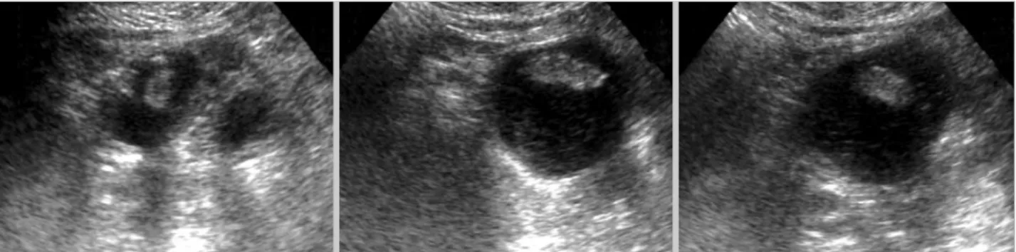
Ultrasonography (US) was performed revealing edema and thickening of the bowel wall and dilatation of the loops of small bowel, which showed active peristalsis diagnostic of mechanical small bowel obstruction. Gallstones were iden- tified within the dilated bowel as floating hyper-echoic foci producing distal acoustic shadowing reflections, which moved when the patient’s position was, changed (Fig. 2).
1216
TS Papavramidis1, S Potsi2, D Paramythiotis1, A Michalopoulos1, VN Papadopoulos1, V Douros1, A Pantoleon2, A Foutzila-Kalogera2, I Ekonomou2, and N Harlaftis1
1st Propedeutic Surgical Clinic1, Department of Radiology2, Aristotle University of Thessaloniki, A.H.E.P.A. University Hospital, Thessaloniki, Greece
Address for correspondence Theodossis Papavramidis, M.D.
1st Propedeutic Surgical Clinic, Aristotle University of Thessaloniki, A.H.E.P.A. University Hospital, 30 Korytsas Str, Panorama, Thessaloniki 55236, Greece Tel : +30-2310-344835, Fax : +30-6944536972 E-mail : [email protected] J Korean Med Sci 2009; 24: 1216-9
ISSN 1011-8934
DOI: 10.3346/jkms.2009.24.6.1216
Copyright � The Korean Academy of Medical Sciences
Gallstone Obstructive Ileus 3 Years Post-cholecystectomy to a Patient with an Old Ileoileal Anastomosis
The present case is one of gallstone obstructive ileus due to gallstones 3 yr after laparoscopic cholecystectomy. It is interesting because of the sex of the patient, the fact that ileus occurred 3 yr after cholecystectomy and that the localization of the obstruction was an old side-to-side ileoileal anastomosis due to a diverticulectomy following intussusception of Meckels’ diverticulum at the age of 3.
Key Words : Gallstones; Ileus
Received : 22 October 2007 Accepted : 22 June 2008
Gallstone Obstructive Ileus Post-cholecystectomy 1217
The computed tomography (CT) showed multiple air-fluid interfaces, distended small bowel loops and two big ectopic peripheral calcified stones with radiolucent centers in the dilat- ed small bowel of terminal ileum. One of them had a thick calcified surface and hypo-dense center which the form is like Mercedes-Benz figure. The stones had an average diameter
of 3 cm (Fig. 3).
The patient underwent exploratory laparotomy. A restrict- ed side-to-side ileoileic anastomosis was found at the termi- nal ileus together with multiple adhesions. Adhesiolysis was performed and excision of the previous anastomosis. A new laterolateral ileoileic anastomosis was performed. In the excised
Fig. 1. Two calcified opacities were detected in the left iliac fossa at the supine radiograph (RX), (A) which changed location at the erect RX. (B) At profile plain RX of the abdomen these two intraluminal abnormalities appeared in the dilated small bowel of terminal ileum, (C) compatible with gallstones.
A B C
Fig. 2. Gallstones were identified within the dilated bowel as floating hyper-echoic foci producing distal acoustic shadowing reflec- tions, which moved when the patient’s position was changed.
Fig. 3. CT scans showed multiple air-fluid interfaces the distended small bowel loops and two big ectopic peripheral calcified stones with radiolucent centers in the dilated small bowel.
1218 T.S. Papavramidis, S. Potsi, D. Paramythiotis, et al.
intestine two calculi sized approximately 3 cm were removed by enterotomy. The patient’s postoperative course was unevent- ful (Fig. 4). Pathology revealed bilirubin stones. Furthermore, in order to have chemical analysis the stones were powdered in a pestle and mortar and dissolved in different solvents de- pending upon the type of chemical constituent to be ana- lyzed. The results of the chemical analysis are presented in Table 1.
DISCUSSION
The pathogenesis of gallstone ileus necessitates the pres- ence of a gallstone in the intestinal lumen, the most common route of entry being a cholecystenteric fistula resulting from recurrent attacks of cholecystitis. A cholecystoduodenal fis- tula was demonstrated in 68% of patients with gallstone ileus by Clavien et al. (4). Stones passing spontaneously through the ampulla of Vater have also been reported to cause gall- stone ileus (5), besides the fact that more than 80% of gall- stones entering the gut are excreted uneventfully (6). The present patient had recurrent attacks of cholelithiasis and underwent laparoscopic cholecystectomy. The existence or not of a bilioenteric fistula is unknown. Concerning the pre- sent patient, we can’t identify the way through which the stones arrived to the intestine, but we are pretty sure that they enlarged in the gut, at the site of the old anastomosis.
The stones had probably remained in the anastomosis and under dyskinesis and stasis the bacterial overgrowth caused
large gallstone formation. The localization of the stones fur- ther explained the symptoms of the patient-notably inter- mittent partial obstruction- during the last two year. Proba- bly the bilirubin stones impacted and disimpacted within the old anastomosis that resulted in presentation and relief of the symptoms.
Rigler’s criteria (7): 1) air or contrast medium in the biliary tree, 2) visualization of the stone in the intestine, 3) change in position of a previously observed stone, and 4) radiologic evidence of intestinal obstruction are pathognomonic in the diagnosis of gallstone ileus. In the present case three out of these four criteria were fulfilled and, even if the images were typical, the diagnosis of gallstone ileus was many times chal- lenged due to the prior cholecystectomy. At this point we have to agree with Lassandro et al. (8) that CT shows impor- tant details such as: the evidence of endoluminal stones, their size and their number. CT may also detect ectopic stones and allow the diagnosis of gallstone ileus before severe intestinal obstruction from stone impaction occurs.
Gallstone ileus usually requires urgent surgery to relieve intestinal obstruction. Although enterolithotomy alone re- mains the popular operative method in most reports, the one- stage procedure composed of enterolithotomy, cholecystec- tomy and repair of fistula is necessary, if indicated (9). Tan et al. (10) compared the two surgical strategies of enterolitho- tomy alone and enterolithotomy with cholecystectomy for the emergent treatment of gallstone ileus, and concluded that both procedures are safe with no mortality, but the better surgical option is enterolithotomy. Recently, Chou et al. (11) proposed endoscopic approach to remove gallstones, but their size constitute a technical difficultly in order to apply. Bowel resection is only indicated when there is intestinal perforation or ischemia (12). In the present case, no dilemma involving cholecystectomy was posed since the organ was priory remov- ed. Furthermore, bowel resection was necessary due to the stenosis of the old anastomosis and the multiple adhesions.
In conclusion, gallstone obstructive ileus has to be consid- ered in a differential diagnosis of mechanical ileus, even in the absence of gallbladder. Furthermore, even when not all of the Ringler’s criteria are fulfilled then the biliary stone has to be considered as etiologic factor. Finally, when suspicion
Organic mg/g Inorganic mg/g
Total cholesterol 513.26 Total bilirubin 4.97
Bile acids 20.53
Fatty acids 13.98 Triglycerides 31.57 Phospholipids 6.54 Soluble proteins 102.87
K+ 3.17
Na+ 0.58
Ca2+ 28.20
Mg2+ 11.03
P 8.98
Oxalate 6.99
Table 1. Chemical analysis of the gallstones. Values are expre- ssed at mg/g of dry weight of stone powder
Fig. 4. Side-to-side ileoileic anastomosis. The stone’s colour was black and their size was about 3 cm in diameter.
Gallstone Obstructive Ileus Post-cholecystectomy 1219
for gallstone ileus appears then CT has to be the diagnostic modality to be employed.
REFERENCES
1. Lassandro F, Gagliardi N, Scuderi M, Pinto A, Gatta G, Mazzeo R.
Gallstone ileus analysis of radiological findings in 27 patients. Eur J Radiol 2004; 50: 23-9.
2. Masannat Y, Masannat Y, Shatnawei A. Gallstone Ileus: a review.
Mt Sinai J Med 2006; 73: 1132-4.
3. Gelbman A. Clinical quiz. Gallstone ileus with cholecystocolonic fistula. Emerg Radiol 2006; 12: 199-200.
4. Clavien PA, Richon J, Burgan S, Rohner A. Gallstone ileus. Br J Surg 1990; 77: 737-42.
5. Deitz DM, Standage BA, Pinson CW, McConnell DB, Krippaehne WW. Improving the outcome in gallstone ileus. Am J Surg 1986;
151: 572-6.
6. Piedad OH, Wels PB. Spontaneous internal biliary fistula, obstruc-
tive and nonobstructive types: twenty-year review of 55 cases. Ann Surg 1972; 175: 75-80.
7. Rigler LG, Borman CN, Noble JF. Gallstone obstruction: pathogen- esis and roentgen manifestations. JAMA 1941; 117: 1753-9.
8. Lassandro F, Romano S, Ragozzino A, Rossi G, Valente T, Ferrara I, Romano L, Grassi R. Role of helical CT in diagnosis of gallstone ileus and related conditions. AJR Am J Roentgenol 2005; 185: 1159-65.
9. Zuegel N, Hehl A, Lindemann F, Witte J. Advantages of one-stage repair in case of gallstone ileus. Hepatogastroenterology 1997; 44:
59-62.
10. Tan YM, Wong WK, Ooi LL. A comparison of two surgical strate- gies for the emergency treatment of gallstone ileus. Singapore Med J 2004; 45: 69-72.
11. Chou JW, Hsu CH, Liao KF, Lai HC, Cheng KS, Peng CY, Yang MD, Chen YF. Gallstone ileus: report of two cases and review of the literature. World J Gastroenterol 2007; 13: 1295-8.
12. Syme RG. Management of gallstone ileus. Can J Surg 1989; 32:
61-4.