INTRODUCTION
Lung cancer is one of the most lethal malignant tumors with more than 1.3 million estimated deaths annually. In the United States, approximately 175,000 new cases are diagnosed each year, 75-80% of which are non-small cell lung cancer (1).
In 2004, it has been estimated that there will be 68,200 lung cancer deaths among women and 93,100 among men; there appears to be a leveling off in the incidence of lung cancer and the death rate among males in the U.S.A. although the incidence and mortality in females continues to increase (2).
According to the Korean National Cancer Registry, lung cancer is the second most common malignant tumor follow- ing gastric cancer, and accounts for the 11.9% of total num- ber of newly reported cancers (3). Recently, lung cancer death rates have continued to increase since 1999 and lung cancer is the leading cause of all cancer deaths in Korea. Lung can- cer death rates of Korean population increased from 17.4 per 100,000 in 1993 to 26.4 per 100,000 in 2003, and accounted for 15.7% of all cancer deaths in 1993 and 20.0% in 2003 (4).
Overall, 5-yr survival rates for lung cancer are about 14%
in the United States and ranges from 5.5% to 14% in the Europe (5-7). It has been reported that the 5-yr survival rate after resection of stage IA is more than 70%, whereas that
for stage IV lung cancer is less than 3% (8, 9).
Trials to reduce mortality rates from lung cancer have been performed over many years using various methods (10-13).
However, there is no good evidence that screening for lung cancer by using chest radiography or sputum cytology can re- duce lung cancer mortality (14). Recently, lung cancer screen- ing using low-dose CT (LDCT) has been reported to be effec- tive at detecting small lung cancer (15-17), thus increasing the expectation that this method will reduce lung cancer mor- tality. However, in terms of demonstrating definite improve- ments in cancer mortality, the results of randomized controlled trials using this screening method are awaited (18).
The purpose of our study was to assess the overall detection rates of lung cancers using LDCT screening and to compare differences in the histopathologic and imaging findings of detected cancers between asymptomatic Korean high-risk and low-risk groups.
MATERIALS AND METHODS Study population
From August 1999 to September 2003, 6,406 individu- Semin Chong, Kyung Soo Lee,
Myung Jin Chung, Tae Sung Kim, Hojoong Kim*, O Jung Kwon*, Yoon-Ho Choi�, Chong H. Rhee�
Department of Radiology and Center for Imaging Science and *Division of Pulmonary and Critical Care Medicine, Department of Medicine, �Center for Health Promotion, Samsung Medical Center, Sungkyunkwan University School of Medicine, Seoul, Korea
Address for correspondence Kyung Soo Lee, M.D.
Department of Radiology, Samsung Medical Center, Sungkyunkwan University School of Medicine, 50 Irwon-dong, Gangnam-gu, Seoul 135-710, Korea Tel : +82.2-3410-2518, Fax : +82.2-3410-2559 E-mail : [email protected]
*This manuscript was presented as a scientific paper in 2004 RSNA scientific assembly (Paper number:
SSA04-03).
402
Lung Cancer Screening with Low-Dose Helical CT in Korea:
Experiences at the Samsung Medical Center
To determine overall detection rates of lung cancer by low-dose CT (LDCT) screen- ing and to compare histopathologic and imaging differences of detected cancers between high- and low-risk groups, this study included 6,406 asymptomatic Kore- an adults with ≥45 yr of age who underwent LDCT for lung cancer screening. All were classified into high- (≥20 pack-year smoking; 3,353) and low-risk (3,053; <20 pack-yr smoking and non-smokers) groups. We compared CT findings of detected cancers and detection rates between high- and low-risk. At initial CT, 35% (2,255 of 6,406) had at least one or more non-calcified nodule. Lung cancer detection rates were 0.36% (23 of 6,406). Twenty-one non-small cell lung cancers appeared as solid (n=14) or ground-glass opacity (GGO) (n=7) nodules. Cancer likelihood was higher in GGO nodules than in solid nodules (p<0.01). Fifteen of 23 cancers occurred in high-risk group and 8 in low-risk group (p=0.215). Therefore, LDCT screening help detect early stage of lung cancer in asymptomatic Korean popula- tion with detection rate of 0.36% on a population basis and may be useful for dis- covering early lung cancer in low-risk group as well as in high-risk group.
Key Words : Mass Screening; Lung Neoplasms; Tomography, Spiral Computed; Smoking
Received : 14 December 2004 Accepted : 18 January 2005
als with ≥45 yr of age (mean±SD; 55±6.7, range; 46-85) underwent LDCT for lung cancer screening. Our institution- al review board approved the lung cancer screening study, and written informed consent was obtained from all partici- pants for the CT study. All subjects were asymptomatic and none had any history of cancer within the previous 5 yr. They were 5,530 men and 876 women. We evaluated the individu- al smoking history with the answer sheets for all participants, who were classified into two groups; high-risk group who had smoked 20 pack-years or more and low-risk group who had smoked less than 20 pack-years or who never smoked.
Screening procedure
All scans were performed on multi- or single-slice CT scan- ners from lung apex to base without contrast enhancement.
The scanning parameters were 120 kVp and 48 (60 mA/0.8 sec) or 50 (50 mA/1.0 sec) mAs. CT machines with four dif- ferent detector numbers were used, namely, single-detector (HiSpeed Advantage scanner, GE Medical Systems, Milwau- kee, WI, U.S.A.; pitch 1.7:1, 5-mm thickness reconstruction with 4-mm intervals), four-detector (LightSpeed QXi, GE Medical Systems; 20-mm beam collimation, beam pitch 1.5, 5-mm thickness reconstruction with 4-mm intervals), eight- and 16-detector (LightSpeed Ultra or Ultra16, GE Medical Systems; 20-mm beam collimation, beam pitch 1.375, 5-mm thickness reconstruction with 4-mm intervals).
We evaluated all images shown on lung (window width 1,500 HU, window level -700 HU) and mediastinal (window width 342 HU, window level 56 HU) windows on PACS (Centricity 1.0, General Electric Medical Systems Integrated Imaging Solutions, Mt. Prospect, IL, U.S.A.) monitors using cine mode by one of three chest radiologists (S.C., M.J.C., and K.S.L. with two years, eight years and 15 yr of experience, respectively, in thoracic imaging). We assessed the attenua- tion, size and number of nodules, which were classified into the three types of attenuation: non-calcified solid, ground- glass opacity or calcified nodule, and were categorized into three groups of size according to their greatest dimensions:
<5 mm, 5-10 mm, and >10 mm in diameter.
For non-calcified nodules detected on baseline CT scans, we recommended the follow-up protocol: if the detected nodule was solid and 10 mm or more in diameter, immediate inter- vention (percutaneous needle biopsy, video-assisted thoraco- scopic surgery or open lung biopsy) was given to obtain a tis- sue diagnosis after obtaining thin-section CT scans through- out the nodule (for the marginal and internal nodule charac- terization of a nodule; volume scanning using multislice CT, 0.8-sec gantry rotation time, 120 kVp, 90 mA, 2.5-mm beam collimation, beam pitch 1.375, 2.5-mm thickness reconstruc- tion with 2.5-mm intervals); if the detected nodule was less than 10 mm in diameter, patients were recommended to have followed-up CT scans 6 months after the initial examination.
If the nodule did not show any growth over a follow-up period
of six months, an annual follow-up study was recommended.
For ground-glass opacity (GGO) nodules, if more than 10 mm in diameter, immediate intervention was recommended after obtaining thin-section CT scans throughout the nodule, however, if less than 10 mm in diameter, thin-section CT scans were obtained within a short-term follow-up period of 2 months, then after six-months, and annually thereafter.
Of all subjects, 3,638 subjects underwent LDCT scan only once, 1,537 subjects twice, 704 subjects three times and 527 subjects four times or more.
Cancer assessment
During screening studies, when a nodule was suggestive of lung cancer with a diameter exceeding 10 mm, a tissue diagnosis was established by obtaining tumor tissue. For 21 peripheral nodules, tissue diagnosis was made by trans-tho- racic needle biopsy in 12 patients, video-assisted thoracoscop- ic surgery in four, open lung biopsy in two, bronchoscopic biopsy in two, and sputum cytology in one. In two patients with small cell lung cancer, diagnosis was made in each by bronchoscopic biopsy and percutaneous needle biopsy. For all pathologic specimens, one lung pathologist with nine years of experience evaluated all histopathologic specimens.
For staging of non-small cell lung cancers (NSCLC), we used the revised International System for Staging Lung Can- cer adopted by the American Joint Committee on Cancer (AJCC) and the Union Internationale Contre le Cancer (UICC) system (19). For staging of small cell lung cancer (SCLC), we used the system adopted from the Veterans Affairs Research Service Lung Group (20).
We also obtained the tumor doubling time of lung cancer using the maximum diameter and volume of each tumor, which were determined from initial and follow-up CT images.
Tumor volume (V) was calculated using the following equa- tion, assuming a spherical form:
V=[4/3× ×a×b×(a+b)/2]×1/8
where a indicates the maximum tumor diameter and b indi- cates the minimum tumor diameter. The tumor doubling time (Td) was calculated using the following equation:
Td=(T-T0)×log2/logV-logV0
where T-T0indicates the length of time between two mea- surements and V0and V denote the tumor volumes at these times (21).
For all patients with lung cancers, we assessed the treat- ment given to the patients and described the follow-up out- comes after treatment when available.
Statistical analysis
Overall lung cancer detection rates were described on an individual basis and on a detected non-calcified nodule basis.
Rates were also calculated according to stratified subjects’ ages (≥45 yr, >50 yr, or finally >60 yr). Differences in the preva-
lence of all lung cancers between high- and low-risk groups were compared using the chi-square test. Differences in the prevalence of stage IA cancers between high- and low-risk groups, in the incidence of stage IA cancers between the ini- tial and follow-up screenings, and in the incidence of lung cancers between GGO and solid nodules were compared using the Fisher’s exact test. Differences of tumor doubling time in high- and low-risk groups were compared by the Mann- Whitney test. We used the SPSS software package (SPSS 11.0.0, SPSS Inc., Chicago, IL, U.S.A.) and considered to be statistically significant at a p value of <0.05.
RESULTS
Of 6,406 subjects, 3,353 subjects belonged to high-risk
group who had smoked 20 pack years or more (39±15.9 pack years). Low-risk group included 3,053 subjects; 1,581 smokers who smoked less than 20 pack-years (13±4.7 pack years) and 1,472 non-smokers. All smokers had the mean smoking history of 33.3 pack years.
Table 1 shows the characteristics of non-calcified nodules detected on LDCT scans. At CT, 35% (2,255 of 6,406) of screened subjects had at least one or more non-calcified nod- ules (n=4,037); 2,085 subjects had 3,783 solid nodules (mean, 1.8 nodules per subject) and 170 subjects 254 GGO nodules (mean, 1.5 nodules per subject).
Table 2 shows the characteristics of lung cancers detected on LDCT scans. Twenty-three primary lung cancers were detected with an overall detection rate of 0.36% (23 of 6,406) on an individual basis and 0.57% (23 of 4,037) on a non-cal- cified nodule basis. The prevalence of lung cancer according
Numbers in parenthesis are number of subjects and numbers in bracket are number of nodules per person. GGO, ground-glass opacity.
Total GGO
Solid Group
<5 mm 5-10 mm >10 mm Total <5 mm 5-10 mm >10 mm Total
High risk 1,887 (950) 191 (144) 28 (26) 2,106 (1120) 46 (21) 65 (50) 26 (23) 137 (94) 2,243 (1,214) Low risk 1,479 (816) 174 (125) 24 (24) 1,677 (965) 52 (24) 53 (40) 12 (12) 117 (76) 1,794 (1,041) Total 3,366 (1,766) 365 (269) 52 (50) 3,783 [1.81] (2,085) 98 (45) 118 (90) 38 (35) 254 [1.49] (170) 4,037 [1.79] (2,255) Table 1.Characteristics of non-calcified nodules detected by low-dose screening CT
Td*, doubling time in months; Size�, size of nodule at initial or final follow-up CT, single number denotes diameter at initial CT; LAP, lymphadenopathy;
RT, radiation therapy; CCRT, concurrent chemoradiation therapy; GGO, ground-glass opacity; BAC, bronchioloalveolar carcinoma; ca, carcinoma;
NA, not applicable.
No. Sex Histologic type TNM staging Group Treatment Outcome Size�(mm)
Initial Final Type of
Attenuation
Td*
(mo) Age
(yr)
1 M 73 Adenocarcinoma T1N0 IA GGO High Lobectomy Good 9 15 1
2 M 63 Squamous cell ca T1N0 IA Solid High Lobectomy Good 29
3 M 72 Adenocarcinoma T1N0 IA GGO Low No treatment Unknown 6 29 8
4 M 61 Squamous cell ca T2N3 IIIB Solid Low Chemotherapy, Palliative Death, 1 yr later 32 5 M 69 Adenocarcinoma T1N2 IIIA GGO High Lobectomy RT due to recurrence 1 yr later 18
6 M 46 Adenocarcinoma T1N0 IA Solid High Lobectomy Good 10 17 7
7 M 62 SCLC Extensive LAP Low Chemotherapy, Palliative Good NA
8 F 60 Adenocarcinoma T2N3M1 IV Solid Low No treatment Unknown 42
9 M 58 Adenocarcinoma T1N1 IIA Solid High Lobectomy Good 8 18 3
10 M 47 Squamous cell ca T3N2 IIIA Solid High No treatment Unknown 27
11 M 80 Adenocarcinoma T3N1 IIIA Solid High No treatment Death, 3 months later 40
12 F 56 Adenocarcinoma T1N0 IA Solid Low Lobectomy Good 18
13 M 60 Squamous cell ca T2N3M1 IV Solid High No treatment Unknown 34
14 F 60 Adenocarcinoma T1N0 IA Solid Low Lobectomy Good 6 13 3
15 M 54 Adenocarcinoma T1N0 IA Solid Low Lobectomy Pneumonectomy, due to 7 12 49
recurrence 3 yr later
16 M 53 BAC T1N0 IA GGO High Lobectomy Good 9 19 29
17 M 63 BAC T1N0 IA GGO High Lobectomy Good 8 13 27
18 M 56 Adenocarcinoma T1N0 IA GGO High Lobectomy Good 25
19 M 48 Adenocarcinoma T1N0 IA GGO High Lobectomy Good 10 12 21
20 M 61 Squamous cell ca T1N0 IA Solid Low Lobectomy Good 8 13 40
21 M 73 Pleomorphic ca T1N2 IIIA Solid High Lobectomy Good 14 16 3
22 M 56 SCLC Limited LAP High CCRT Good NA
23 M 65 Squamous cell ca T1N0 IA Solid High Lobectomy Good 5 10 3
Table 2.Characteristics of detected lung cancers by low-dose screening CT
to stratified subjects’ ages was 0.36% (23 of 6,406) in those of ≥45 yr in age, 0.47% (20 of 4,254) in those of >50 yr, and 0.91% (11 of 1,215) in those of >60 yr.
Adenocarcinoma was found in 12 patients (Fig. 1), squa- mous cell carcinoma in 6 (Fig. 2), bronchioloalveolar carci- noma in two, pleomorphic carcinoma in one and small cell
carcinoma in two.
On CT scans, 21 NSCLCs presented as a solid nodule in 14 patients and GGO in seven. The likelihood of lung can- cer was higher in GGO nodules (7 of 254 GGO nodules, 3%) than in solid nodules (14/3,783, 0.4%) (p<0.01) (Fisher’s exact test).
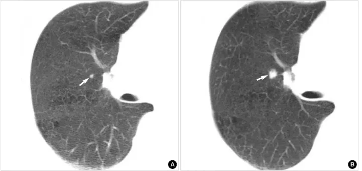
Fig. 1.A 48-yr-old man with adenocarcinoma. (A) Lung window of initial screening low-dose CT scan obtained at level of right upper lobar bronchus shows 10-mm-sized ground-glass opacity nodule (arrow) in right upper lobe. (B) Lung window of thin-section (2.5-mm thick- ness) CT scan obtained at similar level to A shows clearly ground-glass opacity nature of nodule (arrow). Right upper lobectomy disclosed adenocarcinoma.
A B
Fig. 2.A 65-yr-old man with squamous cell carcinoma. (A) Lung window of initial screening low-dose CT (5-mm collimation) scan obtained at level of bronchus intermedius shows 5-mm-sized nodule (arrow) in bottom of anterior segment of right upper lobe. (B) Repeat CT scan obtained at same level to and 6 months after A shows interval increase in nodule size (arrow). Right upper lobectomy disclosed squa- mous cell carcinoma.
A B
The pathologic stages of the 21 NSCLCs were as follows:
T1 in 16 (76%) patients, T2 in 3 (14%), and T3 in 2 (10%);
N0 in 13 (62%), N1 in 2 (10%), N2 in 3 (14%), and N3 in 3 (14%); and stage IA in 13 (62%), IIA in 1 (5%), IIIA in 4 (19%), IIIB in 1 (5%), and IV in 2 (10%).
Sixty-five percents (15 of 23) of lung cancers occurred in high-risk group and 35% (8 of 23) in low-risk group (p=
0.215) (chi-square test). Of 13 stage IA NSCLCs, 8 (57% of 14 NSCLCs in high-risk group) were observed in high-risk group and 5 (71% of 7 NSCLCs in low-risk group) in low- risk group (p=0.656) (Fisher’s exact test).
Of 23 lung cancers, 11 lung cancers were detected during initial screening and 12 during repeat study. In terms of repeat studies, 10 lung cancers were detected during the second fol- low-up study (mean 25.3 months) and 2 during the third follow-up study (mean 27.3 months). Of 9 NSCLCs detected at initial screening, three (33%) were stage IA cancers, where- as of 12 NSCLCs detected at repeat study, 10 (83%) were stage IA cancers, i.e., stage IA cancers were much more preva- lent in the repeat study than in the initial study (p=0.032) (Fisher’s exact test).
Of the 12 lung cancers for which the tumor doubling time was calculable, the average doubling time was 16±16.7 months (range; 1-49 months). Although this tumor doubling time was twice as long in low-risk (25±23.2, median; 24, range; 3-49) group as in high-risk (12±11.9 months, medi- an; 5.0, range; 1-29) group, it was not statistically significant (p=0.368) (Mann-Whitney test).
Of 23 patients with lung cancer, lobectomy was performed in 15. Two patients underwent chemotherapy. Another patient underwent concurrent chemoradiation therapy. Five patients were discharged without any therapy because they declined
treatment. Seventeen (74%) of 23 patients were still alive for 4 yr, 2 died, and 4 were lost during follow-up studies. Of the 17 living patients, 15 (65% [15/23]) were alive without tumor recurrence.
DISCUSSION
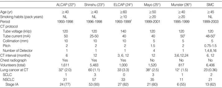
Comparisons in inclusion criteria, screening parameters, and results of LDCT screening for lung cancer are summa- rized in Table 3 (22-26).
In our study, the proportion (35%) of individuals with non- calcified nodules was higher than that (23%) reported in the Early Lung Cancer Action Project (ELCAP) study (24) and was lower than those (43% and 69%, respectively) reported in the Germany study by Diederich et al. (26) and in the Mayo study by Swensen et al. (25), respectively. The difference may be due to the 5-mm collimation used in the present study versus 10-mm collimation used in the ELCAP study. How- ever, the lower detection rates in our study compared to those of Mayo and German studies (25, 26), although they used the same 5-mm collimation, cannot be explained simply. Despite Korea has been known for endemic areas of acute or chronic granulomatous disease, the reason for this discrepancy is not clear.
The prevalence of lung cancer in the present study was 0.36% (23 of 6,406) in the study population of ≥45 yr in age, 0.47% (20 of 4,254) in that of >50 yr, and 0.91% (11 of 1,215) in that of >60 yr. These results of increasing ten- dency of cancer detection rates with older age are parallel to those of the German study (26); i.e., 1.5% (12 of 817) in the study population older than 40 yr, 2.3% (12 of 519) in that
ALCAP, anti-lung cancer association project; ELCAP, early lung cancer action project; SMC, Samsung Medical Center; NL, no limitation; SCLC, small cell lung cancer; NSCLC, non-small cell lung cancer. Numbers in parenthesis are percentages.
*, reference number; �, approximate date that is 6-18 months after baseline screening completed in 1998; �, mAs; �, number of lung cancer detected at low-dose CT; ‖, included both lung cancers considered synchronous primary tumors.
ALCAP (22*) Shinshu (23*) ELCAP (24*) Mayo (25*) Munster (26*) SMC
Age (yr) ≥40 ≥40 ≥60 ≥50 ≥40 ≥45
Smoking habits (pack years) NL NL ≥10 ≥20 ≥20 NL
Period 1993-1998 1996-1998 1993-1999� 1999-2001 1995-1999 1999-2003
CT protocol
Tube voltage (kVp) 120 120 140 120 120 120
Tube current (mA) 50 25-50 40 40 50� 48-50�
Collimation (mm) 10 10 10 5 5 5
Pitch 2 2 2 1.5 2 0.75-1.5
Number of Detector 1 1 1 4 1 1,4,8,16
CT interval (months) 6 12 3, 6, 12 12 3,6,12,24 6, 12
Chest radiograph Yes Yes Yes No No No
Volunteers (total) 1,611 5,483 1,000 1,520 817 6,406
Lung cancer at CT 32�(2.0) 60 (1.1) 33 (3.3) 38�(2.5) 12‖(1.5) 23 (0.36)
SCLC 1 3 0 3 1 2
NSCLC 31 57 33 35 11 21
Stage IA 24 (77) 53 (93) 27 (82) 21 (60) 6 (55) 13 (62)
Table 3.Comparison of inclusion criteria, screening parameters, and the results of low-dose screening CT at various institutions
older than 50 yr, and 3.9% (8 of 206) in that older than 60 yr. Although the observed prevalence was less in our study, it is clear that the prevalence of lung cancer in younger popu- lations is lower.
Nakata et al. (27) reported that persistent GGO for sever- al months is an indicator of early adenocarcinoma or its pre- cursor. The present study showed that nodules showing GGO on CT scans have a higher risk of harboring lung cancer (2.8%) than solid nodules (0.37%). Additionally, Kakinuma et al.
(28) evaluated screening CT-detected pure GGO nodules at follow-up CT and noticed that pure GGO of lung cancer nod- ule change in size and attenuation over time (GGO nodules increase in size and solid component appears within GGO nodule indicating the evolution of pure bronchioloalveolar carcinoma to adenocarcinoma with bronchioloalveolar carci- noma component). They recommended close follow-up until solid component appears for the management of such a focal GGO. We also emphasize that ‘a short-term follow up and immediate intervention with growth’ policy are needed for the management of GGO nodules detected by LDCT screening.
In the current study, 13 (62%) of 21 non-small cell lung cancers were stage IA. Stage IA cancers were much more fre- quently detected in the repeat study than in the initial study (p=0.032). The proportion of stage IA cancers in our study was higher than those in the Mayo and German Mayo studies, in which the rates of stage IA were 60% and 55%, respective- ly (25, 26), but lower than those recorded by the Anti-Lung Cancer Association Project (ALCAP), Shinshu and ELCAP studies, in which the rates of stage IA were 77%, 93%, and 82%, respectively (22-24). The high incidence of stage IA cancer in the ALCAP and Shinshu studies may be due to the fact that more cancers are detected by repeat study, and thus less advanced lung cancers are detected. In fact, in the present study, 57% of detected NSCLCs were discovered by repeat study, and 31% and 9% in the Mayo and German studies (25, 26), respectively; and 58% and 60% in the ALCAP and Shin- shu studies (22, 23). Moreover, in ALCAP, Shinshu, ELCAP and our study, most of stage IA cancers can be detected at repeat study. Therefore, we suggest that the repeat screening studies help detect early stage of lung cancer.
Most previous studies have included smokers who have consumed at least 10 packs a year or more (24-26). However, in our study and in the Shinshu study (23), subjects of 45 yr or more of age were included regardless of smoking history.
Our study population was classified into high- and low-risk groups according to smoking status and evaluated the respec- tive prevalence of lung cancer accordingly. We found that there were no significant differences in the prevalence of lung cancer, doubling time or stage IA of lung cancers between high- and low-risk groups.
Some limitations of our study should be mentioned. First, not all patients enrolled in this study were on regular and recommended followed-up study, therefore there might be some subjects who had lung cancer after the initial study but
not included in the present study. Second, a single radiolo- gist read images, and thus some nodules might have been missed.
In conclusion, LDCT screening help detect early stage of lung cancer in asymptomatic Korean population with detec- tion rate of 0.36% on a population basis and may be useful for discovering early lung cancer in low-risk group as well as in high-risk group. The possibility of lung cancer is high- er when persistent nodular areas of GGO are present at LDCT than when solid nodules are seen, therefore, a careful follow- up study should be scheduled for the detection of early lung cancer.
REFERENCES
1. Greenlee RT, Murray T, Bolden S, Wingo PA. Cancer statistics, 2000.
CA Cancer J Clin 2000; 50: 7-33.
2. Cancer statistics 2004. American Cancer Society Web site. Available at: www.cancer.org/downloads/PRO/Cancer%20Statistics%202004.
ppt. [accessed 20 July 2004].
3. Annual Report of the Korea Central Cancer Registry. National Can- cer Center Web site. Available at: www.ncc.re.kr/files/cancerStat/
2002_cancer_regi.ppt. [accessed 20 July 2004].
4. Annual Report on the Case of Death Statistics (Based on Vital Reg- istration). Korean National Statistical Office Web site. Available at:
www.nso.go.kr/newnso/upload_file/upload2/svca0300.pdf. [accessed 23 October 2004].
5. Cancer facts and figures 2003. American Cancer Society Web site.
Available at: www.cancer.org/downloads/STT/CAFF2003PWSe- cured.pdf. [accessed 20 July 2004].
6. Berrino R, Capocaccia R, Esteve J. Survival of cancer patients in Europe: the EUROCARE-2 study. Lyon, France: IARC Scientific Publications 1999; 1-572.
7. NHS Performance Indicators National Figures: February 2002. Depart- ment of Health Web site. Available at: www.performance.doh.gov.uk/
nhsperformanceindicators/hlpi2002/NationalDocument.pdf. [accessed 20 July 2004].
8. Flehinger BJ, Kimmel M, Melamed MR. The effect of surgical treat- ment on survival from early lung cancer. Implications for screening.
Chest 1992; 101: 1013-8.
9. Noguchi M, Morikawa A, Kawasaki M, Matsuno Y, Yamada T, Hiro- hashi S, Kondo H, Shimosato Y. Small adenocarcinoma of the lung.
Histologic characteristics and prognosis. Cancer 1995; 75: 2844-52.
10. Frost JK, Ball WC Jr, Levin ML, Tockman MS, Baker RR, Carter D, Eggleston JC, Erozan YS, Gupta PK, Khouri NF. Early lung can- cer detection: results of the initial (prevalence) radiologic and cyto- logic screening in the Johns Hopkins study. Am Rev Resp Dis 1984;
130: 549-54.
11. Fontana RS, Sanderson DR, Taylor WF, Woolner LB, Miller WE, Muhm JR, Uhlenhopp MA. Early lung cancer detection: results of the initial (prevalence) radiologic and cytologic screening in the Mayo Clinic Study. Am Rev Resp Dis 1984; 130: 561-5.
12. Melamed MR, Flehinger BJ, Zaman MB, Heelan RT, Perchick WA,
Martini N. Screening for early lung cancer: results of the Memorial Sloan-Kettering study in New York. Chest 1984; 86: 44-53.
13. Kubik A, Polak J. Lung cancer detection: results of a randomized prospective study in Czechoslovakia. Cancer 1986; 57: 2427-37.
14. Eddy DM. Screening for lung cancer. Ann Int Med 1989; 111: 232-7.
15. Sone S, Li F, Yang ZG, Takashima S, Maruyama Y, Hasegawa M, Wang JC, Kawakami S, Honda T. Characteristics of small lung can- cers invisible on conventional chest radiography and detected by population based screening using spiral CT. Br J Radiol 2000; 73:
137-45.
16. Kaneko M, Eguchi K, Ohmatsu H, Kakinuma R, Naruke T, Suema- su K, Moriyama N. Peripheral lung cancer: screening and detec- tion with low-dose spiral CT versus radiography. Radiology 1996;
201: 798-802.
17. Rusinek H, Naidich DP, McGuinness G, Leitman BS, McCauley DI, Krinsky GA, Clayton K, Cohen H. Pulmonary nodule detection: low- dose versus conventional CT. Radiology 1998; 209: 243-9.
18. Swensen SJ, Jett JR, Sloan JA, Midthun DE, Hartman TE, Sykes AM, Aughenbaugh GL, Zink FE, Hillman SL, Noetzel GR, Marks RS, Clayton AC, Pairolero PC. Screening for lung cancer with low- dose spiral computed tomography. Am J Respir Crit Care Med 2002;
165: 508-13.
19. Mountain CF, Dresler CM. Regional lymph node classification for lung cancer staging. Chest 1997; 111: 1718-23.
20. Green RA, Humphrey E, Close H. Alkylating agents in bronchogenic carcinoma. Am J Med 1969; 46: 516-25.
21. Ozono S, Miyao N, Igarashi T, Marumo K, Nakazawa H, Fukuda M, Tsushima T, Tokuda N, Kawamura J, Murai M. Tumor doubling time of renal cell carcinoma measured by CT: collaboration of Japanese Society of Renal Cancer. Jpn J Clin Oncol 2004; 34: 82-5.
22. Sobue T, Moriyama N, Kaneko M, Kusumoto M, Kobayashi T, Tsuchiya R, Kakinuma R, Ohmatsu H, Nagai K, Nishiyama H, Mat- sui E, Eguchi K. Screening for lung cancer with low-dose helical computed tomography: anti-lung cancer association project. J Clin Oncol 2002; 20: 911-20.
23. Sone S, Li F, Yang ZG, Honda T, Maruyama Y, Takashima S, Hase- gawa M, Kawakami S, Kubo K, Haniuda M, Yamanda T. Results of three-year mass screening programme for lung cancer using mobile low-dose spiral computed tomography scanner. Br J Cancer 2001;
84: 25-32.
24. Henschke CI, Yankelevitz DF, Libby DM, McCauley D, Pasmantier M, Altorki NK, Smith JP, Miettinen OS. Early lung cancer action project: annual screening using single-slice helical CT. Ann N Y Acad Sci 2001; 952: 124-34.
25. Swensen SJ, Jett JR, Hartman TE, Midthun DE, Sloan JA, Sykes AM, Aughenbaugh GL, Clemens MA. Lung cancer screening with CT: Mayo Clinic experience. Radiology 2003; 226: 756-61.
26. Diederich S, Wormanns D, Semik M, Diederich S, Wormanns D, Semik M, Thomas M, Lenzen H, Roos N, Heindel W. Screening for early lung cancer with low-dose spiral CT: prevalence in 817 asymptomatic smokers. Radiology 2002; 222: 773-81.
27. Nakata M, Saeki H, Takata I, Segawa Y, Mogami H, Mandai K, Eguchi K. Focal ground-glass opacity detected by low-dose helical CT. Chest 2002; 121: 1464-7.
28. Kakinuma R, Ohmatsu H, Kaneko M, Kusumoto M, Yoshida J, Nagai K, Nishiwaki Y, Kobayashi T, Tsuchiya R, Nishiyama H, Matsui E, Eguchi K, Moriyama N. Progression of focal pure ground-glass opac- ity detected by low-dose helical computed tomography screening for lung cancer. J Comput Assist Tomogr 2004; 28: 17-23.