Prevalence and Seroprevalence of Low-Risk Human Papillomavirus in Korean Women
Little is known about the prevalence and seroprevalence of low-risk human papillomavirus (HPV) and the risk factors for HPV infection in Korean women. We determined the prevalence of low-risk HPV among 902 women aged 20-59 yr and the seroprevalence of low-risk HPV subtypes 6 and 11 among 1,094 women aged 9-59 yr in the general population. Genital low-risk HPV DNA was assessed by liquid hybridization and polymerase chain reaction. Antibody titers against HPV 6 and 11 were measured by a multiplexed competitive luminex technique. The prevalence of genital low-risk HPV was 4.9%. It reached its highest peak of 10.3% at 20-29 yr of age and a second peak of 3.2% at 50-59 yr of age. The seroprevalence of HPV 6 or 11 was 9.4%. It reached its highest peak of 12.7% at 25-29 yr of age and a second peak of 12.3% at 50-59 yr of age. In multivariable analysis, the number of lifetime sexual partners and past history of sexually transmitted diseases were associated with the seroprevalence but not prevalence of HPV. It is suggested that younger women should receive prophylactic HPV vaccination before they become sexually active and exposed to HPV in their 20s. This study provides baseline data for developing HPV vaccination programs and monitoring vaccine efficacy in Korea.
Key Words: Low-Risk Human Papillomavirus; Prevalence; Seroprevalence Min-A Kim1, Jin-Kyoung Oh2,
Bo Wook Kim1, Doobyung Chay3, Dong Choon Park4, Seok Mo Kim5, Eun-Suk Kang6, Jae-Hoon Kim1, Chi-Heum Cho7, Hai-Rim Shin8, and Kyung Seo1
1Department of Obstetrics and Gynecology, Gangnam Severance Hospital, Yonsei University College of Medicine, Seoul; 2Research Institute for National Cancer Control & Evaluation, National Cancer Center, Seoul; 3Department of Obstetrics and Gynecology, Yongin Severance Hospital, Yonsei University College of Medicine, Yongin; 4Department of Obstetrics and Gynecology, St. Vincent’s Hospital, The Catholic University of Korea, Suwon; 5Department of Obstetrics and Gynecology, Chonnam National University Medical School, Gwangju; 6Department of Laboratory Medicine, Samsung Medical Center, Sungkyunkwan University School of Medicine, Seoul; 7Department of Obstetrics and Gynecology, Keimyung University School of Medicine, Daegu, Korea; 8Noncommunicable Diseases and Health Promotion, World Health Organization, Western Pacific Regional Office, Manila, the Philippines
Received: 6 September 2011 Accepted: 22 May 2012 Address for Correspondence:
Kyung Seo, MD
Department of Obstetrics and Gynecology, Gangnam Severance Hospital, Yonsei University College of Medicine, 211 Eonju-ro, Gangnam-gu, Seoul 135-720, Korea
Tel: +82.2-2019-3433, Fax: +82.2-3462-8209 E-mail: [email protected]
This work was supported by the intramural research fund from the Korea Centers for Disease Control and Prevention (2008- E00306-00) and a grant from a faculty research grant of Yonsei University College of Medicine for 2008 (6-2008-0147).
http://dx.doi.org/10.3346/jkms.2012.27.8.922 • J Korean Med Sci 2012; 27: 922-928
INTRODUCTION
Human papillomavirus (HPV) is the most common sexually transmitted infection. In one study, approximately half of the women without HPV infection became infected with HPV with- in 3 yr after initiating sexual activity (1). Currently, more than 100 different HPV subtypes have been identified, which mostly infect genital epithelial cells. HPV subtypes are divided broadly into two groups according to their epidemiological association with cervical cancer. The low-risk HPV group, including HPV
subtypes 6 and 11, is associated with benign lesions, genital warts or recurrent respiratory papillomatosis (2). The high-risk HPV group, including HPV subtypes 16 and 18, induces precancer- ous lesions such as cervical intraepithelial neoplasia (CIN), cer- vical cancer or ano-genital cancer (3, 4).
After sexual contact, genital warts may occur in the uterine cervix, vagina, vulva, anus and oral cavity. Approximately 90%
of genital warts are related to low-risk HPV types 6 and 11 (5-7).
The incidence of genital warts has gradually increased since the 1950s (6), and has become a major cause of nononcogenic HPV-
related morbidity. In addition, low grade squamous intraepi- thelial lesions are associated with not only high-risk HPV infec- tion but also low-risk HPV 6 and 11 infection.
Our current epidemiological knowledge of HPV infection is based on the genital HPV DNA test which only identifies the current HPV infection (8). In addition, the HPV DNA test has been applied limitedly because of the difficulty of collecting samples from males and the reluctance to gynecologic exami- nation among females (9-11). In contrast, serum antibody to HPV is a useful, although not perfect, complementary marker reflecting cumulative HPV exposure (8, 12, 13).
In the general population of Korea, very little data exist on the epidemiology of low-risk HPV 6 and 11, which are two of the four types targeted by the quadrivalent HPV vaccine (10, 14, 15). We estimated the prevalence and seroprevalence of low- risk HPV infection in Korean women in order to help establish vaccine policy specific to the characteristics of Korean women and assess vaccine efficacy.
MATERIALS AND METHODS Study subjects
We conducted a survey of the prevalence and seroprevalence of low-risk HPV in Korean women between July 2008 and May 2009. A total of 1,143 women, 9-59 yr of age, who visited our in- stitutions for a regular medical check-up were eligible for this study. All study samples were obtained using a complex, strati- fied, multistage probability cluster design in order to select a nationally representative sample. For females from 9 to 19 yr of age, the low-risk HPV serologic test was performed without a questionnaire study or a genital HPV DNA test in light of the relatively late age of sexual debut and conservative sexual cul- ture found in Korea. Females 20-59 yr of age were asked to fill out a self-reporting questionnaire containing questions relating to socio-demographic characteristics and lifestyle habits asso- ciated with HPV infection, such as sexual behavior, history of sexually transmitted disease (STD), usage of tobacco and usage of oral contraceptives.
Of a total of 1,143 women, the prevalence of low-risk HPV was analyzed in a total of 902 women aged 20-59 yr. This excludes 241 women; one who did not agree to the study, four who did not respond to the questionnaire, eight whose results regarding Hybrid Capture II (HC II) were missing and 228 who were 9-19 yr of age. Of the 1,130 women 9-59 yr of age, comprised of the 228 who were 9-19 yr of age along with the 902 who were 20-59 yr of age, an analysis was performed for the seroprevalence of low-risk HPV in a total of 1,094 females. This excludes 36 women 20 yr of age or older from whom serum samples could not be collected.
Specimen collection
Cervicovaginal cells were collected from 902 women 20-59 yr
of age using a Cytobrush (Cervibrush, Cell-Path, Herte, United Kingdom). These cells were collected in an Eppendrof tube that contained 20 mL of phosphate buffer solution (PBS). Blood sam- ples of 10 mL were collected from each of 1,094 women 9-59 yr of age using the vacutainer system. Blood samples were centri- fuged at 1,500 g for 10 minutes and the serum was separated from the cells. All samples were transported to a central labora- tory and stored at -70°C until processing.
Low-risk HPV DNA testing by HC II
To confirm low-risk HPV group (consisting of HPV subtypes 6, 11, 42, 43, 44) infection and viral load, cervicovaginal cells were harvested using a dacron swab, added to the collection kit (Di- gene, Gaithersburg, MD, USA) and stored at -20°C until analysis.
RNA probes of low-risk HPV were hybridized with denatured, single-stranded DNA. This reaction mixture was transferred to a tube coated with anti-DNA-RNA hybrid antibodies. The im- mobilized hybrids were then incubated with an alkaline phos- phatase-conjugated antihybrid monoclonal antibody. After rins- ing, these reactants were treated with Lumi-Phospho 530, which reacts to alkaline phosphatase, a dioxetane-based chemilumi- nescent substrate. The light generated by the reaction was mea- sured by luminometer and the intensity was recorded using units which were relative to the reaction. The relative unit for the light intensity was defined as the degree of illumination rel- ative to the positive control group. A positive cutoff value was set at 1 pg HPV DNA per milliliter in each specimen.
Low-risk HPV DNA testing by polymerase chain reaction (PCR)
For typing of low-risk HPV subtypes 6 and 11, DNA was extract- ed using the Qiagen DNA extraction kit (Qiagen, Valencia, CA, USA). β-globin PCR was performed to check the DNA quality.
GP5+/6+ primer was used on all of the β-globin PCR positive samples to confirm whether they were positive for low-risk HPV subtypes 6 and 11. Samples testing positive for low-risk HPV were examined by PCR using HPV 6 and 11 primer to determine the specific subtype.
Low-risk HPV serologic test
The antibodies to low-risk HPV subtypes 6 and 11 were mea- sured by Merck and Co. Inc., using a multiplexed competitive Luminex® assay. The antibody titration method was measured according to the HPV antibody measurement method reported by Opalka et al. in 2003 (16), and reported in milli-Merck units per mL. The monoclonal antibodies used in the HPV multi- plexed competitive Luminex assay included H6.M48 and K11.
B2 for low-risk HPV subtypes 6 and 11, respectively. Cutoff val- ues for HPV seropositivity were at least 20 and at least 16 mMU/
mL for HPV subtypes 6 and 11, respectively (17).
Statistical analysis
Odds ratios (ORs) and corresponding 95% confidence intervals (CIs) were calculated using unconditional multiple logistic re- gression to assess the associations between HPV DNA/seropos- itivity and demographic and behavioral characteristics. For all analyses, a significance level of P < 0.05 was chosen.
Ethics statement
All participants signed and submitted an informed consent for this study. The study protocol was approved by the institutional review board of Gangnam Severance Hospital (IRB No. 3-2008- 0097), Seoul.
RESULTS
Table 1 shows the overall and age-specific prevalence of low- risk HPV in the study population. Of the 902 Korean women 20- 59 yr of age that were tested for prevalence of HPV, 4.9% (44 of 902) were shown to be positive for low-risk HPV DNA. The age- standardized prevalence, calculated by applying the female population given by the National Statistics Office for Korea in 2008, was shown to be 4.9% (95% CI 2.3-7.6). The prevalence of low-risk HPV reached its highest peak of 10.3% (95% CI 6.2-14.5) among females 20-29 yr of age and decreased thereafter. It then reached a second peak of 3.2% (95% CI 0.9-5.6) among females 50-59 yr of age.
Of the 44 HC II positive samples, only 8 (18.2%) had subtypes confirmed by PCR; the subtypes of the remaining 36 samples were not confirmed. Of the 8 samples whose subtypes were con- firmed, 8 samples (18.2%) were positive for HPV 6 and 2 sam- ples (4.5%) were additionally positive for HPV 11, meaning there was simultaneous co-infection with HPV 6. The type-specific prevalence for HPV 6 and HPV 11 were 0.9% and 0.2%, respec- tively (data not shown).
Of 1,094 females 9-59 yr of age tested for seroprevalence of HPV, the seroprevalence of either low-risk HPV subtypes 6 or 11 was 9.4% (95% CI 7.7-11.3), and the age-standardized serop- revalence was shown to be 9.6% (95% CI 7.8-11.4). The age-spe- cific seroprevalence of low-risk HPV reached its highest peak of 12.7% (95% CI 7.7-19.3) among females 25-29 yr of age. It de- creased thereafter but reached a second peak of 12.3% (95% CI 8.2-17.6) among females 50 to 59 yr of age (Fig. 1). The type- specific seroprevalences of low-risk HPV are shown in Table 2.
HPV 6 seroprevalence was 8.1% (95% CI 6.6-9.9), and HPV 11 seroprevalence was 3.9% (95% CI 2.9-5.3). Peak seroprevalence occurred in the 30-39 yr-old age group for HPV 6 (11.4%, 95%
CI 7.5-16.3) and in the 25-29 yr-old age group for HPV 11 (7.0%,
Fig. 1. Seroprevalence for low-risk HPV types in 1,094 Korean women by age group.
Seroprevalence of low risk HPV (%)
Age (yr)
09-14 15-19 20-24 25-29 30-39 40-49 50-59 14.0
12.0 10.0 8.0 6.0 4.0 2.0 0.0
HPV 6 or 11 HPV 6 HPV 11 HPV 6 and 11
Table 1. Prevalence of genital low-risk HPV in 902 Korean women by age group
Age (yr) No. of
subjects
No. of subjects Low-risk HPV (+)
Prevalence of Low-risk HPV, % (95% CI)
20-29 203 21 10.3 (6.2-14.5)
30-39 235 10 4.3 (1.7-6.8)
40-49 246 6 2.4 (0.5-4.4)
50-59 218 7 3.2 (0.9-5.6)
Overall 902 44 4.9 (3.5-6.3)
Age-standardized
prevalence* 4.9 (2.3-7.6)
*Prevalence was standardized by age on the basis of the female population of Korea in 2008. HPV, human papillomavirus; CI, confidence interval.
Table 2. Seroprevalence for low-risk HPV types in 1,094 Korean women by age group
Age (yr) No. of subjects Positive rates for, % (95% CI)
HPV 6 HPV 11 HPV 6 or 11 HPV 6 and 11
9-14 150 2.0 (0.4-5.7) 1.3 (0.2-4.7) 2.0 (0.4-5.7) 1.3 (0.2-4.7)
15-19 78 2.6 (0.3-9.0) 2.6 (0.3-9.0) 3.8 (0.8-10.8) 1.3 (0.03-6.9)
20-24 55 7.3 (2.0-17.6) 5.5 (1.1-15.1) 9.1 (3.0-19.9) 3.6 (0.4-12.5)
25-29 142 10.6 (6.0-16.8) 7.0 (3.4-12.6) 12.7 (7.7-19.3) 4.9 (2.0-9.9)
30-39 220 11.4 (7.5-16.3) 3.6 (1.6-7.0) 11.4 (7.5-16.3) 3.6 (1.6-7.0)
40-49 239 7.9 (4.8-12.1) 3.3 (1.5-6.5) 9.6 (6.2-14.1) 1.7 (0.4-4.2)
50-59 210 10.0 (6.3-14.9) 4.8 (2.3-8.6) 12.3 (8.2-17.6) 2.4 (0.8-5.5)
Overall 1,094 8.1 (6.6-9.9) 3.9 (2.9-5.3) 9.4 (7.7-11.3) 2.6 (1.8-3.8)
Age-standardized prevalence* 8.3 (6.6-10.0) 4.0 (2.8-5.2) 9.6 (7.8-11.4) 2.7 (1.7-3.7)
*Prevalence was standardized by age on the basis of the female population of Korea in 2008. HPV, human papillomavirus; CI, confidence interval.
95% CI 3.4-12.6). Just like the overall seroprevalence of low-risk HPV, the type-specific seroprevalences for HPV 6 and 11 reached second peaks in the 50-59 yr-old age group. Seropositivity for both low-risk HPV 6 and 11 was rare (2.6%, 95% CI 1.8-3.8) and
reached its highest peak among females aged 25-29 yr.
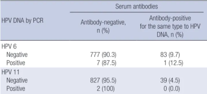
Table 3 presents the concordance between low-risk HPV DNA positivity and seropositivity. A total of 868 women aged 20-59 yr were tested with both HC II for the presence of low-risk HPV DNA and a HPV serologic test for seropositivity of low-risk HPV subtypes 6 and 11. Of the 44 HC II positive samples, only 8 had subtypes confirmed by PCR. Among females who were PCR- positive for HPV 6 and 11, the proportions who were also sero- positive to the respective type were 1/8 (12.5%) and 0/2 (0%), respectively. Among PCR-negative for HPV 6 and 11, the pro- portions that were seropositive to the respective type were 83/
860 (9.7%) and 39/866 (4.5%), respectively.
We analyzed the relationship between socio-demographic and behavioral characteristics and the seroprevalence of low- risk HPV. Seroprevalence of low-risk HPV was significantly as- sociated with the lifetime number of sexual partners and past Table 3. Concordance between genital low-risk HPV 6 and 11 infection and serum
antibodies among Korean women
HPV DNA by PCR
Serum antibodies Antibody-negative,
n (%)
Antibody-positive for the same type to HPV
DNA, n (%) HPV 6
Negative
Positive 777 (90.3)
7 (87.5) 83 (9.7)
1 (12.5) HPV 11
Negative
Positive 827 (95.5)
2 (100) 39 (4.5)
0 (0.0) HPV, human papillomavirus; CI, confidence interval.
Table 4. Odds ratios and 95% confidence intervals according to risk factors of seropositivity for low-risk HPV (HPV 6 or 11) in Korean women
Risk factors No. Low-Risk HPV
Seropositive, n (%) OR (95% CI) Adjusted OR*
(95% CI) P value
Age (yr) 9-14 15-19 20-24 25-29 30-39 40-49 50-59
150 78 55 142 220 239 210
3 (2.0) 3 (3.8) 5 (9.1) 18 (12.7) 25 (11.4) 23 (9.6) 26 (12.4)
- - Reference 1.5 (0.5-4.1) 1.3 (0.5-3.5) 1.1 (0.4-2.9) 1.4 (0.5-3.9)
- - Reference 7.0 (0.7-67.3) 4.0 (0.4-42.4) 3.8 (0.3-42.2) 5.4 (0.5-60.6)
0.664
New sexual partner in recent 6 months No
Yes
759 72
79 (10.4) 14 (19.4)
Reference 2.1 (1.1-3.9)
Reference 1.1 (0.4-3.2)
0.800
Lifetime number of sexual partners 1
2 3 or more
578 101 108
50 (8.7) 10 (9.9) 19 (17.6)
Reference 1.2 (0.6-2.4) 2.3 (1.3-4.0)
Reference 1.2 (0.5-2.6) 2.5 (1.1-5.6)
0.028
Age at sexual debut 18 or younger 19-24 25 or older
40 454 291
13 (32.5) 46 (10.1) 24 (8.3)
5.4 (2.4-12.0) 1.3 (0.7-2.1)
Reference
2.1 (0.6-7.0) 1.2 (0.6-2.2) Reference
0.310
Tobacco use No Yes
795 45
85 (10.7) 9 (20.0)
Reference 2.1 (0.97-4.5)
Reference 1.5 (0.5-4.3)
0.483
History of STDs No Yes
773 57
78 (10.1) 16 (28.1)
Reference) 3.5 (1.9-6.5)
Reference 3.6 (1.6-8.2)
0.001
Oral contraceptive use No
Yes
699 118
69 (9.9) 20 (17.0)
Reference 1.9 (1.1-3.2)
Reference 1.0 (0.5-2.2)
0.923
Marital status Married Others†
642 198
69 (10.7) 23 (11.6)
Reference 1.1 (0.7-1.7)
Reference 0.5 (0.2-1.3)
0.073
Education level Less than high school
University or higher 434
409 59 (13.6)
36 (8.8) 1.6 (1.04-2.5)
Reference 0.8 (0.4-1.5) Reference
0.570
Childbirth Never
Ever 204
639 22 (10.8)
72 (11.3) Reference
1.1 (0.6-1.7) Reference 1.0 (0.3-2.8)
0.887
*Adjusted for age, new sexual partner in recent 6 months, lifetime number of sexual partners, age at sexual debut, tobacco use, history of STDs, oral contraceptive use, marital status, education level, and childbirth. †Single, divorced, separated or widowed. HPV, human papillomavirus; OR, odds ratio; CI, confidence interval; STDs, sexually transmitted diseases.
history of STD. The seroprevalence of low-risk HPV increased from 8.7% among females with 1 lifetime sexual partner to 17.6%
among those with 3 lifetime sexual partners (P = 0.028). We also found that past history of STD was significantly associated with the seroprevalence of low-risk HPV (P = 0.001). The effects of the remaining risk factors were not statistically significant (Table 4).
DISCUSSION
This is the first study to describe the seroprevalence along with the prevalence of low-risk HPV in Korean women. The preva- lence of genital low-risk HPV infection detected by HC II was shown to be 4.9% (the age-standardized prevalence was 4.9%).
This is comparable to a low-risk HPV prevalence of 4.1% report- ed by Shin et al. in 2003 (15) using the PCR method and lower than that reported in the United States (18). Low-risk HPV prev- alence in men was 0%-43% in several studies and 2.6% in Korean university students (10). In regard to the age-specific prevalence of low-risk HPV, it reached its highest peak at 20-29 yr of age, and afterward, it showed a tendency to decrease, but reaching a second peak at 50-59 yr of age. This is similar to previously re- ported results (19). The age-specific pattern we found can be explained by multiple factors. One possible explanation is that middle-aged women have new sexual partners as they become more sexually active, and thus the possibility of new HPV infec- tion becomes high. Another is the reactivation of latent HPV in- fection due to the deterioration of immunity as a result of hor- monal deficiency during the perimenopausal period. Host fac- tor such as immune-compromised status can play a role in the HPV infection (20). A third explanation is the cohort effect: prev- alence appears to be high, but in reality patients were unaware of an infection that occurred in their 20s which was detected only from the results of tests performed later (19, 21, 22).
Concerning the distribution of low-risk HPV subtypes, among 44 HC II positive samples, HPV 6 was detected in 8 cases and HPV 11 was detected in 2 cases. 81.8% (36/44) of the HC II posi- tive samples had subtypes that could not be determined. This may be due to that the PCR primers used in our study were only for HPV subtypes 6 and 11, and thus the remaining subtypes could not be detected.
Because the clearance rate for low-risk HPV was higher than that for high-risk HPV, with a shorter duration of infection, the prevalence of low-risk HPV might reflect a pattern of new HPV infections (23). On the other hand, HPV seroprevalence is a good marker of cumulative HPV exposure, although due to a low seroconversion rate after HPV infection, HPV seropreva- lence does not indicate the current state of HPV infection (8, 12).
Worldwide, the seroprevalence of low-risk HPV varies accord- ing to HPV subtype, age, and regional differences. Nonetheless, it has been reported to be 10%-20% (24, 25). The seroprevalence
of low-risk HPV subtypes 6 and 11 was 9.4% among females aged 9-59 yr (the age-standardized prevalence was 9.6%), which was lower than those of western countries. In the present study, the seroprevalence of low-risk HPV subtypes 6 and 11 accord- ing to age showed its highest peak in the 20s and a second peak in the 50s, similar to the age-specific pattern of the prevalence of HPV infection. It was observed in previous reports that general- ly, as age increased, HPV seroprevalence also increased or main- tained a plateau level (25, 26). Additionally, it has been reported that HPV prevalence reaches a peak within 5-10 yr of sexual de- but and that HPV seroconversion occurs frequently between 6 and 18 months of DNA detection (12, 27, 28). Therefore, we can infer that first exposure to HPV among Korean women is occur- ring earlier, and that the sexual lives of middle-aged women have become more active.
The concordance between low-risk HPV DNA positivity and seropositivity was poor. Only 12.5% of HPV 6 PCR-positive wom- en were seropositive for HPV 6. In case of HPV 11, two PCR-pos- itive samples had no antibody for HPV 11. This discrepancy can be explained by the effect of HPV-specific immune-evasion mechanism, waning of detectable antibodies, a long lag time required for seroconversion (29, 30).
In regard to behavioral risk factors, there were significant dif- ferences in seroprevalence of low-risk HPV by both lifetime number of sexual partners and history of STD. It had previously been reported that HPV seroprevalence is associated with the age of sexual debut, the lifetime number of sex partners, marital status, and education level (25, 31).
Although clinical trials has not demonstrated remarkable ef- ficacy in prevention of invasive cervical cancer from HPV vacci- nation, it has shown that the vaccine is almost 100% effective in preventing infection related to the vaccine-specific HPV geno- types. According to previous studies, teenagers without sexual experience certainly benefit from HPV vaccination. Kim et al.
reported that HPV vaccine is highly immunogenic and well tol- erated in Korean girls aged 10-14 yr (32). Among sexually active women, cases where individuals were infected with all four types of HPV vaccine were rare. Therefore, even women infect- ed with one or more HPV subtypes prior to vaccination can be protected from uninfected subtypes by vaccination. Consider- ing that as women become older or more sexually active, the possibility of more sexual partners and the exposure to HPV in- creases, most women could benefit from HPV vaccination.
There are limitations in our study. First, although the low-risk HPV group is often associated with low grade squamous intraep- ithelial lesions and non-malignant genital tract lesions (genital warts), such correlation between the low-risk HPV positivity and cervical pathology was not analyzed in our study. Thereby, a further study is needed to monitor the presence and persis- tence of cervical pathology in low-risk HPV infected women.
Second, in our study, PCR assay was performed to classify low-
risk HPV subtypes 6 and 11 among various HC II positive sam- ples. It is possible that some false negative samples of HC II were missed while other low risk HPV types except HPV 6 and 11 could not have been detected. For these reasons, we may be underes- timating the low-risk HPV prevalence in our study.
In conclusion, our results confirm that the prevalence and seroprevalence of low-risk HPV tend to increase rapidly in their 20s, when women are becoming sexually active. In order to achieve optimal efficacy, it would be reasonable to vaccinate women in their teens prior to HPV exposure.
However, our study shows that seroprevalence reaches its peak earlier than it has been previously thought, implying that the pattern of sexual behavior among Korean teenagers is chang- ing rapidly. Therefore, also considering the lag period of sero- conversion, the optimal age for catch-up vaccination should be lowered. In addition, our results show that a second peak of both the prevalence and seroprevalence of low-risk HPV occurs in middle aged women. Also considering that the probability of women who are sexually active being infected with all four HPV vaccine types is low, it may be necessary to adjust the recom- mended age of catch-up vaccination and discuss the vaccina- tion strategy in middle aged woman. However, future research will be needed to define the clinical significance and natural history of seronegative infection of low-risk HPV. Our data will help to establish a policy to reduce an important fraction of dis- ease burden of low-risk HPV 6 and 11 targeted by the quadriva- lent vaccine and to monitor vaccine efficacy.
REFERENCES
1. Moscicki AB. Impact of HPV infection in adolescent populations. J Ado- lesc Health 2005; 37: S3-9.
2. Lacey CJ, Lowndes CM, Shah KV. Chapter 4: Burden and management of non-cancerous HPV-related conditions: HPV-6/11 disease. Vaccine 2006; 24 Suppl 3: S3/35-41.
3. Bosch FX, de Sanjose S. Chapter 1: Human papillomavirus and cervical cancer--burden and assessment of causality. J Natl Cancer Inst Monogr 2003: 3-13.
4. Walboomers JM, Jacobs MV, Manos MM, Bosch FX, Kummer JA, Shah KV, Snijders PJ, Peto J, Meijer CJ, Munoz N. Human papillomavirus is a necessary cause of invasive cervical cancer worldwide. J Pathol 1999;
189: 12-9.
5. Greer CE, Wheeler CM, Ladner MB, Beutner K, Coyne MY, Liang H, Langenberg A, Yen TS, Ralston R. Human papillomavirus (HPV) type distribution and serological response to HPV type 6 virus-like particles in patients with genital warts. J Clin Microbiol 1995; 33: 2058-63.
6. Koutsky LA, Galloway DA, Holmes KK. Epidemiology of genital human papillomavirus infection. Epidemiol Rev 1988; 10: 122-63.
7. Wiley D, Masongsong E. Human papillomavirus: the burden of infec- tion. Obstet Gynecol Surv 2006; 61: S3-14.
8. Ho GY, Studentsov YY, Bierman R, Burk RD. Natural history of human papillomavirus type 16 virus-like particle antibodies in young women.
Cancer Epidemiol Biomarkers Prev 2004; 13: 110-6.
9. Dunne EF, Nielson CM, Stone KM, Markowitz LE, Giuliano AR. Preva- lence of HPV infection among men: a systematic review of the literature.
J Infect Dis 2006; 194: 1044-57.
10. Shin HR, Franceschi S, Vaccarella S, Roh JW, Ju YH, Oh JK, Kong HJ, Rha SH, Jung SI, Kim JI, et al. Prevalence and determinants of genital in- fection with papillomavirus, in female and male university students in Busan, South Korea. J Infect Dis 2004; 190: 468-76.
11. Sukvirach S, Smith JS, Tunsakul S, Munoz N, Kesararat V, Opasatian O, Chichareon S, Kaenploy V, Ashley R, Meijer CJ, et al. Population-based human papillomavirus prevalence in Lampang and Songkla, Thailand.
J Infect Dis 2003; 187: 1246-56.
12. Carter JJ, Koutsky LA, Hughes JP, Lee SK, Kuypers J, Kiviat N, Galloway DA. Comparison of human papillomavirus types 16, 18, and 6 capsid antibody responses following incident infection. J Infect Dis 2000; 181:
1911-9.
13. Castle PE, Wacholder S, Lorincz AT, Scott DR, Sherman ME, Glass AG, Rush BB, Schussler JE, Schiffman M. A prospective study of high-grade cervical neoplasia risk among human papillomavirus-infected women.
J Natl Cancer Inst 2002; 94: 1406-14.
14. Clifford GM, Shin HR, Oh JK, Waterboer T, Ju YH, Vaccarella S, Quint W, Pawlita M, Franceschi S. Serologic response to oncogenic human papil- lomavirus types in male and female university students in Busan, South Korea. Cancer Epidemiol Biomarkers Prev 2007; 16: 1874-9.
15. Shin HR, Lee DH, Herrero R, Smith JS, Vaccarella S, Hong SH, Jung KY, Kim HH, Park UD, Cha HS, et al. Prevalence of human papillomavirus infection in women in Busan, South Korea. Int J Cancer 2003; 103: 413-21.
16. Opalka D, Lachman CE, MacMullen SA, Jansen KU, Smith JF, Chirmule N, Esser MT. Simultaneous quantitation of antibodies to neutralizing epitopes on virus-like particles for human papillomavirus types 6, 11, 16, and 18 by a multiplexed luminex assay. Clin Diagn Lab Immunol 2003; 10: 108-15.
17. Dias D, Van Doren J, Schlottmann S, Kelly S, Puchalski D, Ruiz W, Boerckel P, Kessler J, Antonello JM, Green T, et al. Optimization and validation of a multiplexed luminex assay to quantify antibodies to neu- tralizing epitopes on human papillomaviruses 6, 11, 16, and 18. Clin Di- agn Lab Immunol 2005; 12: 959-69.
18. Dunne EF, Unger ER, Sternberg M, McQuillan G, Swan DC, Patel SS, Markowitz LE. Prevalence of HPV infection among females in the United States. JAMA 2007; 297: 813-9.
19. Herrero R, Hildesheim A, Bratti C, Sherman ME, Hutchinson M, Mo- rales J, Balmaceda I, Greenberg MD, Alfaro M, Burk RD, Wacholder S, Plummer M, Schiffman M. Population-based study of human papillo- mavirus infection and cervical neoplasia in rural Costa Rica. J Natl Cancer Inst 2000; 92: 464-74.
20. Lee YH, Choe JY, Park SH, Park YW, Lee SS, Kang YM, Nam EJ, Park W, Kwon SR, Bae SC, et al. Prevalence of human papilloma virus infections and cervical cytological abnormalities among Korean women with sys- temic lupus erythematosus. J Korean Med Sci 2010; 25: 1431-7.
21. Hankins C, Coutlee F, Lapointe N, Simard P, Tran T, Samson J, Hum L.
Prevalence of risk factors associated with human papillomavirus infec- tion in women living with HIV. Canadian Women’s HIV Study Group.
CMAJ 1999; 160: 185-91.
22. Trottier H, Franco EL. The epidemiology of genital human papillomavi- rus infection. Vaccine 2006; 24: S1-15.
23. Goodman MT, Shvetsov YB, McDuffie K, Wilkens LR, Zhu X, Thomp-
son PJ, Ning L, Killeen J, Kamemoto L, Hernandez BY. Prevalence, ac- quisition, and clearance of cervical human papillomavirus infection among women with normal cytology: Hawaii Human Papillomavirus Cohort Study. Cancer Res 2008; 68: 8813-24.
24. Skjeldestad FE, Mehta V, Sings HL, Ovreness T, Turpin J, Su L, Boerckel P, Roberts C, Bryan J, Jansen KU, et al. Seroprevalence and genital DNA prevalence of HPV types 6, 11, 16 and 18 in a cohort of young Norwegian women: study design and cohort characteristics. Acta Obstet Gynecol Scand 2008; 87: 81-8.
25. Markowitz LE, Sternberg M, Dunne EF, McQuillan G, Unger ER. Serop- revalence of human papillomavirus types 6, 11, 16, and 18 in the United States: National Health and Nutrition Examination Survey 2003-2004. J Infect Dis 2009; 200: 1059-67.
26. Newall AT, Brotherton JM, Quinn HE, McIntyre PB, Backhouse J, Gil- bert L, Esser MT, Erick J, Bryan J, Formica N, et al. Population seroprev- alence of human papillomavirus types 6, 11, 16, and 18 in men, women, and children in Australia. Clin Infect Dis 2008; 46: 1647-55.
27. Carter JJ, Koutsky LA, Wipf GC, Christensen ND, Lee SK, Kuypers J, Kiviat N, Galloway DA. The natural history of human papillomavirus type 16 capsid antibodies among a cohort of university women. J Infect
Dis 1996; 174: 927-36.
28. Munoz N, Manalastas R Jr, Pitisuttithum P, Tresukosol D, Monsonego J, Ault K, Clavel C, Luna J, Myers E, Hood S, et al. Safety, immunogenicity, and efficacy of quadrivalent human papillomavirus (types 6, 11, 16, 18) recombinant vaccine in women aged 24-45 years: a randomised, dou- ble-blind trial. Lancet 2009; 373: 1949-57.
29. Scott M, Nakagawa M, Moscicki AB. Cell-mediated immune response to human papillomavirus infection. Clin Diagn Lab Immunol 2001; 8:
209-20.
30. Stanley M. Immune responses to human papillomavirus. Vaccine 2006;
24: S16-22.
31. Hariri S, Dunne EF, Sternberg M, Unger ER, Meadows KS, Karem KL, Markowitz LE. Seroepidemiology of human papillomavirus type 11 in the United States: results from the third National Health And Nutrition Examination Survey, 1991--1994. Sex Transm Dis 2008; 35: 298-303.
32. Kim YJ, Kim KT, Kim JH, Cha SD, Kim JW, Bae DS, Nam JH, Ahn WS, Choi HS, Ng T, et al. Vaccination with a human papillomavirus (HPV)- 16/18 AS04-adjuvanted cervical cancer vaccine in Korean girls aged 10- 14 years. J Korean Med Sci 2010; 25: 1197-204.