J Korean Soc Radiol 2016;75(6):480-486 https://doi.org/10.3348/jksr.2016.75.6.480
INTRODUCTION
Magnetic resonance arthrography (MRA) of the shoulder is widely used despite the pain and discomfort associated with its use, because it provides high sensitivity and specificity for rota- tor cuff tears (1-4). It can also provide clear discrimination be- tween rotator cuff tears and small anatomical variations (5, 6).
Intra-articular injection into the glenohumeral joint is an im- portant procedure performed prior to shoulder MRA (7). Cur- rently, the use of digital radiology allows the entire procedure of fluoroscopic arthrography (FA) to be recorded as cine images.
Arthrographic findings showing contrast leakage into the subacromial-subdeltoid bursal space can be used to investigate the pathologic condition of supraspinatus tendon (SST) tears,
Discrepancy between Fluoroscopic Arthrography and Magnetic Resonance Arthrography in Patients with Arthroscopically
Confirmed Supraspinatus Tendon Tears: The Additional Benefit of Cine Fluoroscopic Arthrography Images
관절경으로 확인된 극상건 파열 환자들에게 있어서 투시 관절 조영술과 자기공명 관절 조영술의 불일치: 영화 투시 관절 조영술의 추가적 이점
Seok Hahn, MD
1,2, Young Han Lee, MD
1, Jin-Suck Suh, MD
1*
1Department of Radiology, Research Institute of Radiological Science, Medical Convergence Research Institute, and Severance Biomedical Science Institute, Yonsei University College of Medicine, Seoul, Korea
2Department of Radiology, Inje University College of Medicine, Haeundae Paik Hospital, Busan, Korea
Purpose: To determine the additional diagnostic benefits of fluoroscopic arthrogra- phy (FA) in patients with full-thickness supraspinatus tendon (SST) tears by compar- ing FA images with magnetic resonance arthrography (MRA) images.
Materials and Methods: This study included FA and MRA images of 53 patients who were confirmed to have full-thickness SST tears by arthroscopy. In the FA anal- ysis, the presence of contrast leakage into the subacromial-subdeltoid bursa was re- corded. In the MRA analysis, contrast leakage, retraction of a torn tendon, width and length of the tear, and supraspinatus atrophy were evaluated. Patients were di- vided into the concordant group or the discordant group based on the presence of contrast leakage to compare the characteristics of SST tears. We used Fisher’s exact test and two-sample t-test for the comparison.
Results: Of the 53 patients, 34 were included in the concordant group and 19 were in- cluded in the discordant group. In the concordant group, the grades of retraction were higher than those in the discordant group; the width and length of the tears were larg- er. Muscle atrophy was more severe in the concordant group.
Conclusion: A full-thickness SST tear did not always exhibit contrast leakage on FA, particularly small SST tears or tears with low-grade retraction. FA can provide diag- nostic information regarding the severity of full-thickness SST tears by itself.
Index terms Shoulder Arthrography
Magnetic Resonance Imaging Fluoroscopy
Received December 15, 2015 Revised May 3, 2016 Accepted June 6, 2016
*Corresponding author: Jin-Suck Suh, MD Department of Radiology, Research Institute of Radiological Science, Medical Convergence Research Institute, and Severance Biomedical Science Institute, Yonsei University College of Medicine, 50-1 Yonsei-ro, Seodaemun-gu, Seoul 03722, Korea.
Tel. 82-2-2228-7420 Fax. 82-2-393-3035 E-mail: [email protected]
This is an Open Access article distributed under the terms of the Creative Commons Attribution Non-Commercial License (http://creativecommons.org/licenses/by-nc/3.0) which permits unrestricted non-commercial use, distri- bution, and reproduction in any medium, provided the original work is properly cited.
such as a full-thickness tear. However, in some cases, unexpect- ed discrepancies occur between arthrography and MRA, show- ing no contrast leakage on arthrography but a full-thickness SST tear on MRA.
To the best of our knowledge, no study has examined the dif- ferences between arthrography and MRA for shoulder patholo- gy. The purpose of this study is to determine if there are addi- tional diagnostic benefits of FA in patients with arthroscopically confirmed full-thickness SST tears by comparing FA images with MRA images.
MATERIALS AND METHODS
Patient Selection
Our retrospective study was approved by the hospital’s Insti- tutional Review Board. From September 2012 to March 2013, we identified 150 patients who underwent arthroscopy for shoul- der pain and had an SST tear reported in their arthroscopic op- eration notes. Ninety-seven patients were excluded because of the following criteria: 1) no available preoperative MRA and FA data, including conventional magnetic resonance image (MRI) without arthrography (n = 22), lack of FA (n = 14), and magnet- ic resonance (MR) imaging performed in other hospitals (n = 57), and 2) partial-thickness SST tear (n = 4). After exclusion, 53 patients (age range, 35–76 years; mean age, 64.3 years) with an arthroscopically confirmed full-thickness SST tear were en- rolled in this study. Of them, seven patients had an articular-sid- ed partial thickness tear of the infraspinatus tendon (IST) and two patients had a full thickness tear of the IST, In their opera- tion records, there was no fibrous adhesion at the tear site or in the adjacent area. The mean interval between MR examination and arthroscopic surgery was 32.7 days (range, 27–36 days). Of the patients enrolled, 21 were men (39.6%) and 32 were women (60.4%), and 39 patients (73.6%) affected on the right side and 14 patients (26.4%) affected on the left side were evaluated.
Fluoroscopic Arthrography and Magnetic Resonance Arthrography
All patients provided written informed consent for the pro- cedure. A 23-gauge needle was inserted into the glenohumeral joint via an anterior approach under pulsed fluoroscopic guid- ance (Zexira DREX-ZX80, Toshiba Medical Systems Corpora-
tion, Otawara, Japan). The injection was performed with the pa- tient supine in a straight anteroposterior position. The injection consisted of approximately 20 cc of the following mixture: 2 cc of ioxitalamic acid (Telebrix 30 Meglumine, Guerbet, Aulnay- sous-Bois, France), 0.08 cc gadopentetate dimeglumine (Magn- evist, Bayer Schering Pharma AG, Berlin, Germany), and 18 cc normal saline. Standard sterile management was applied in all procedures. Approximately 16–18 cc of contrast mixture was in- jected into the glenohumeral joint and cine images were ob- tained during the injection period by FA.
After arthrography, patients were escorted to the MRI room, and a shoulder MRA was performed within 20 minutes. One of the three 3T MRI systems with a dedicated shoulder coil were used (Achieva or Achieva TX, Philips Healthcare, Best, the Neth- erlands, n = 25; Discovery MR 750, GE Healthcare, Milwaukee, WI, USA, n = 8; and Trio, Siemens Healthcare, Erlangen, Ger- many, n = 20). Conventional 2D images, including fat saturation T1-weighted axial images (repetition time/echo time, 690/20 ms in Philips, 570/7 ms in Siemens, and 580/10 ms in GE; slice thickness/interslice gap, 3/0.3 mm; field of view, 140 × 140 mm), oblique coronal and oblique sagittal images (repetition time/echo time, 640/10 ms in Philips, 530/7 ms in Siemens, and 580/10 ms in GE; slice thickness/interslice gap, 3/0.3 mm; field of view, 140
× 140 mm), T2-weighted oblique coronal images (repetition time/echo time, 3300/70 ms in Philips, 3500/80 ms in Siemens, and 3300/70 ms in GE; slice thickness/interslice gap, 3/0.3 mm;
field of view, 140 × 140 mm), and T1-weighted oblique sagittal images (repetition time/echo time, 690/8 ms in Philips, 620/8 ms in Siemens, and 530/8 ms in GE; slice thickness/interslice gap, 3/1 mm; field of view, 140 × 140 mm), were obtained.
Imaging Interpretation
Two musculoskeletal radiologists (one radiologist with more than 8 years of experience in musculoskeletal imaging and one radiologist with musculoskeletal radiology fellowship) indepen- dently reviewed both FA images and MRA images in a random order within a two-week time span. They kept a two-week in- terval between evaluation of each set of FA and MRA images.
They were blinded to radiologic reports, arthroscopic findings, and any clinical information. In both FA and MRA analyses, presence of contrast leakage into the subacromial-subdeltoid bursa was checked. We measured the width of tears on fat satu-
ration T1-weighted oblique coronal images, and the length of tears on fat saturation T1-weighted oblique sagittal images. We categorized SST retraction into 3 grades using a published meth- od for determining the Patte score on T2-weighted oblique cor- onal images: little, humeral head level, and glenoid level (8). We then assessed supraspinatus muscle atrophy using the tangent sign and occupation ratio. We considered that the tangent sign was absent when the superior margin of the supraspinatus mus- cle was superior to the line tangential to the coracoid and scapu- lar spine (9, 10). Occupation ratio was measured according to the method described by Thomazeau et al. (11) and Khoury et al.
(12), which was the ratio between the cross section of the supra- spinatus muscle belly and that of its fossa on the T1-weighted oblique sagittal image crossing through the medial border of the coracoid process of the scapula. Lines were drawn as close as possible to the supraspinatus outer margin, inner margins of the coracoid process and scapular spine, and superior limits of the supraspinatus fossa.
We accepted consensus for the presence and grading and considered the average of the two readers’ values for measure- ments. Finally, we divided the patients into the following two groups: Group 1, concordant group (leakage into the subacro- mial-subdeltoid bursa on both MRA and FA) and Group 2, dis- cordant group (leakage on MRA, but no leakage on FA). All measurements were performed using a picture archiving and communication system (PACS, Centricity Radiology RA 1000;
General Electric Healthcare, Chicago, IL, USA).
Statistical Analyses
Fisher’s exact test was used to compare categorical variables, and two-sample t-test was used to compare continuous variables between the groups. SPSS software version 20.0 (IBM Corp., Armonk, NY, USA) was used for statistical analyses. Findings were considered statistically significant when the p-value was less than 0.05.
RESULTS
Of the 53 patients, 34 (64.2%) were included in the concor- dant group (Fig. 1), and the remaining 19 (35.8%) were includ- ed in the discordant group (Fig. 2, Table 1). None of the patients showed leakage on FA only. The mean values of the width and length of SST were significantly larger in Group 1 than in Group 2 (p = 0.01 and 0.02, respectively). With respect to the SST retrac- tion grade, there was a significant difference between the two groups (p = 0.03). Both the tangent sign and occupation ratio showed significant differences between the two groups (p < 0.01, both).
All patients with tendon retraction at the glenoid level (8/53, 15.1%) belonged to Group 1. There were a total of 16 patients with confirmed massive SST tears. Of them, 15 patients who had mas- sive SST tears (15/53, 28.3%) belonged to Group 1. One patient
A B C
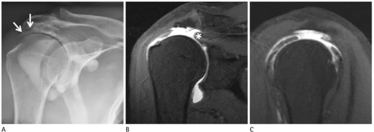
Fig. 1. A 75-year-old male patient in Group 1 (concordant group).
A. Contrast leakage into the subacromial-subdeltoid bursa is shown on a FA spot image (arrows).
B, C. Oblique coronal and sagittal T1-weighted fat saturation MRA show a full-thickness SST tear (width: 23.6 mm, length: 37.6 mm) with retrac- tion of the tendon (asterisk) at the level of the glenohumeral joint.
FA = fluoroscopic arthrography, MRA = magnetic resonance arthrography, SST = supraspinatus tendon
with a massive SST tear who belonged to Group 2 also had a full-thickness subscapularis tendon tear (Fig. 3).
DISCUSSION
To evaluate SST tears using shoulder MRA, direct arthrogra- phy is a necessary and important procedure for glenohumeral in- jection. It can be uncomfortable for patients because of pain, time consumption, and radiation if the procedure is performed under
fluoroscopic guidance. In general, this intra-articular injection could be regarded as a brief procedural step prior to MRA. How- ever, more information can be obtained from the digitally stored cine FA images. In practice, we usually find concordant results between FA and MRA, but discordant results were observed in some patients. We reviewed the literature and identified the dif- ferences between FA and MRA, but we did not find any reports specifically related to this subject. In this study, we tried to deter- mine if there are any differences between FA and MRA findings
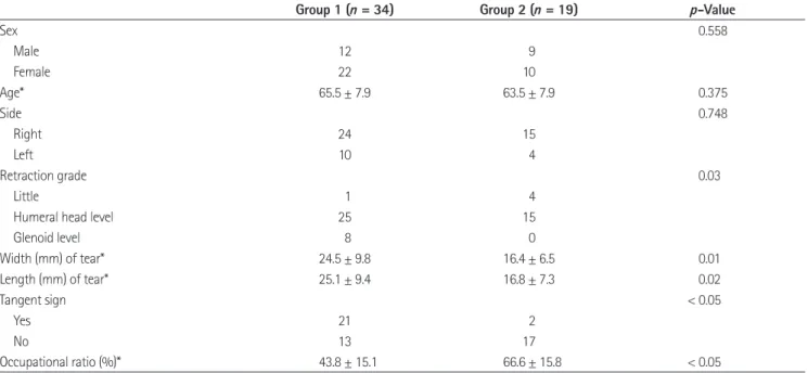
Table 1. Comparison between Group 1 and Group 2
Group 1 (n = 34) Group 2 (n = 19) p-Value
Sex 0.558
Male 12 9
Female 22 10
Age* 65.5 ± 7.9 63.5 ± 7.9 0.375
Side 0.748
Right 24 15
Left 10 4
Retraction grade 0.03
Little 1 4
Humeral head level 25 15
Glenoid level 8 0
Width (mm) of tear* 24.5 ± 9.8 16.4 ± 6.5 0.01
Length (mm) of tear* 25.1 ± 9.4 16.8 ± 7.3 0.02
Tangent sign < 0.05
Yes 21 2
No 13 17
Occupational ratio (%)* 43.8 ± 15.1 66.6 ± 15.8 < 0.05
*Mean ± standard deviation.
A B C
Fig. 2. A 48-year-old female patient in Group 2 (discordant group).
A. There is no contrast leakage into the subacromial-subdeltoid bursa on a FA spot image.
B, C. Oblique coronal and sagittal T1-weighted fat saturation MRA show a full-thickness SST tear (width: 13.4 mm, length: 10.1 mm) with little retraction of the tendon (asterisk).
FA = fluoroscopic arthrography, MRA = magnetic resonance arthrography, SST = supraspinatus tendon
and to observe if there are additional benefits of FA images.
We compared the FA and MRA findings between the con- cordant and discordant groups with respect to confirmed full- thickness SST tears. As mentioned in the results, the concordant group showed not only wider and longer SST tears but also high- er grades of SST retraction than the discordant group. Because the dimensions of SST tears and retraction of SST reflect the ov- erall defect size of SST tears, our results indicate that if SST tears are large, the possibility of leakage on FA images is high. The concordant group also had more patients with a positive tangent sign and showed a lower occupation ratio than the discordant group. A positive tangent sign and a low occupation ratio dem- onstrate atrophy of the supraspinatus, which can be correlated with the chronicity of the tear. We think that patients in the con- cordant group had more advanced supraspinatus atrophy and it would take longer for detection after the event of a SST tear.
We can explain the difference between the concordant and discordant groups by flap tears or a one-way check valve mecha- nism. If the SST tear is small, the edge of the torn tendon or fi- brosis that develops after the tear can act as a check valve that blocks the flow of injected contrast from the glenohumeral joint cavity into the subacromial-subdeltoid bursa during FA. On the other hand, during preparation for MRA, the physical movement required (e.g., walking, raising the affected arm, or lying down)
can cause a change in the pressure gradient between the joint and the bursa. The pressure change allows a greater flow of the con- trast into the bursal space; therefore, leakage appears on MRA images. Conversely, if the defect is large, the valve-like action is not sufficient to interrupt the flow. This mechanism can explain the difference between the concordant and discordant groups.
The results of our study can be helpful in situations when only shoulder FA s available. Corticosteroid or nonsteroidal anti-in- flammatory drug injection into the glenohumeral joint under flu- oroscopic guidance is one of these clinically useful instances. Flu- oroscopic guidance is generally used for glenohumeral injection and it has several advantages such as a wider view of bony struc- tures or confirmation of successful injection when a mixture with contrast media is used.
There are many conditions that can cause shoulder pain such as osteoarthritis, adhesive capsulitis, rotator cuff disease, and la- bral pathology (13). Intra-articular corticosteroid injection has shown a short- or medium-term therapeutic effect because cor- ticosteroids are powerful anti-inflammatory drugs (14). If con- trast leakage is found in the subacromial-subdeltoid bursa on FA images during injection, we can infer that the patient has a full-thickness rotator cuff tear and it is relatively large. Moreover, we can inform the patient regarding his or her current shoulder status and recommend the appropriate treatment rather than
A B
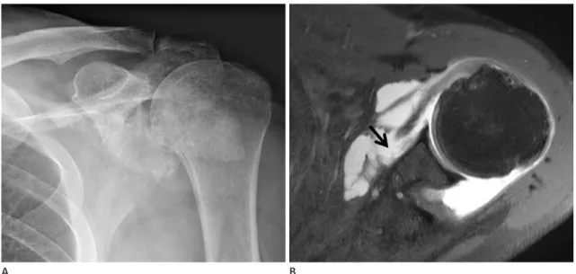
Fig. 3. A 62-year-old female patient in Group 2 (discordant group).
A. There is no contrast leakage into the subacromial-subdeltoid bursa on a FA spot image.
B. An axial T1-weighted fat saturation MRA shows a subscapularis tear (arrow). A moderate amount of contrast leaked primarily into the extra- articular space via the tear site.
FA = fluoroscopic arthrography, MRA = magnetic resonance arthrography
corticosteroid injection.
However, there are several limitations to this study. First, it was a retrospective study, but all patients had full-thickness tears confirmed by arthroscopic surgery. Second, we focused on only full-thickness SST tears because it is difficult to detect partial- thickness tears with FA images. Third, the injection into the gle- nohumeral joint consisted of only approximately 16–18 cc of con- trast mixture, and the unique case (a case of a massive SST tear in the discordant group) in which leakage was not visible on FA images occurred due to this reason (Fig. 3). The injected contrast mixture leaked into another site of severe injury in this patient;
hence, leakage into the subacromial-subdeltoid bursa did not oc- cur. If more amount of the contrast mixture was injected into the joint, the leakage would have been visible on FA images. Fourth, two patients had a full thickness tear of the IST in our study.
The situation that contrast leakage occurs into the subacromial- subdeltoid bursa via the IST tear site cannot be excluded. Final- ly, a small number of variables were used to compare the con- cordant and discordant groups.
In conclusion, not all full-thickness SST tears showed con- trast leakage on FA images, especially small SST tears or tears with low-grade retraction. Because the concordance between FA and MRA is more frequent in patients with larger SST tears and greater worsening of supraspinatus atrophy, FA can provide di- agnostic information to determine the severity of full-thickness SST tears by itself and it has the potential to be used as another imaging modality to evaluate these tears.
Acknowledgments
This work was supported by a National Research Foundation (NRF) grant funded by the Korea government, Ministry of Sci- ence, ICT & Future Planning (MSIP, 2015R1A2A1A05001887).
REFERENCES
1. Spick C, Szolar DH, Reittner P, Preidler KW, Tillich M. MR arthrography of the shoulder: do we need local anesthe- sia? Eur J Radiol 2014;83:980-983
2. de Jesus JO, Parker L, Frangos AJ, Nazarian LN. Accuracy of MRI, MR arthrography, and ultrasound in the diagnosis of rotator cuff tears: a meta-analysis. AJR Am J Roentgenol 2009;192:1701-1707
3. Elentuck D, Palmer WE. Direct magnetic resonance ar- thrography. Eur Radiol 2004;14:1956-1967
4. Jacobson JA, Lin J, Jamadar DA, Hayes CW. Aids to suc- cessful shoulder arthrography performed with a fluoro- scopically guided anterior approach. Radiographics 2003;
23:373-378; discussion 379.
5. Hodler J, Kursunoglu-Brahme S, Snyder SJ, Cervilla V, Kar- zel RP, Schweitzer ME, et al. Rotator cuff disease: assess- ment with MR arthrography versus standard MR imaging in 36 patients with arthroscopic confirmation. Radiology 1992;182:431-436
6. Flannigan B, Kursunoglu-Brahme S, Snyder S, Karzel R, Del Pizzo W, Resnick D. MR arthrography of the shoulder:
comparison with conventional MR imaging. AJR Am J Roentgenol 1990;155:829-832
7. Jbara M, Chen Q, Marten P, Morcos M, Beltran J. Shoulder MR arthrography: how, why, when. Radiol Clin North Am 2005;43:683-692, viii
8. Patte D. Classification of rotator cuff lesions. Clin Orthop Relat Res 1990;(254):81-86
9. Mellado JM, Calmet J, Olona M, Esteve C, Camins A, Pérez Del Palomar L, et al. Surgically repaired massive rotator cuff tears: MRI of tendon integrity, muscle fatty degener- ation, and muscle atrophy correlated with intraoperative and clinical findings. AJR Am J Roentgenol 2005;184:
1456-1463
10. Warner JJ, Higgins L, Parsons IM 4th, Dowdy P. Diagnosis and treatment of anterosuperior rotator cuff tears. J Shoul- der Elbow Surg 2001;10:37-46
11. Thomazeau H, Rolland Y, Lucas C, Duval JM, Langlais F. At- rophy of the supraspinatus belly. Assessment by MRI in 55 patients with rotator cuff pathology. Acta Orthop Scand 1996;67:264-268
12. Khoury V, Cardinal E, Brassard P. Atrophy and fatty infil- tration of the supraspinatus muscle: sonography versus MRI. AJR Am J Roentgenol 2008;190:1105-1111
13. Hegedus EJ, Zavala J, Kissenberth M, Cook C, Cassas K, Hawkins R, et al. Positive outcomes with intra-articular glenohumeral injections are independent of accuracy. J Shoulder Elbow Surg 2010;19:795-801
14. Malfair D. Therapeutic and diagnostic joint injections. Ra- diol Clin North Am 2008;46:439-453, v
관절경으로 확인된 극상건 파열 환자들에게 있어서 투시 관절 조영술과 자기공명 관절 조영술의 불일치:
영화 투시 관절 조영술의 추가적 이점
한 석
1,2· 이영한
1· 서진석
1*
목적: 관절경으로 확인된 극상건 완전 파열 환자들에 대해서 투시 관절 조영술 영상과 자기공명 관절 조영술 영상을 비교 함으로써 투시 관절 조영술의 추가적인 진단적 이점을 알고자 하는 것이다.
대상과 방법: 이 연구에는 관절경을 통해 확인된 53명의 극상건 완전 파열 환자들이 포함되었다. 투시 관절 조영술 분석 에서는 견봉하-삼각근하 점액낭으로의 누출을 기록하였다. 자기공명 관절 조영술의 분석에서는 견봉하-삼각근하 점액낭 으로의 조영제 누출, 파열건의 수축, 파열의 너비과 길이, 극상근의 위축 등을 평가하였다. 극상건 파열의 특징을 비교하기 위해 환자들을 조영제 누출 유무에 따라 일치군과 불일치군으로 나누었다. 비교를 위해 피셔의 정확검정과 두 표본 t-검 정을 사용하였다.
결과: 총 53명의 환자 중 34명이 일치군이었고, 19명이 불일치군이었다. 일치군이 파열건 수축의 정도가 불일치군보다 높 았으며, 파열의 너비와 길이도 일치군이 불일치군보다 컸다. 근육 위축은 일치군에서 더 심했다.
결론: 극상건 완전 파열은 투시 관절 조영술에서 조영제 누출이 항상 보이지 않았는데, 특히 극상건 파열이 작거나 파열건 수축이 심하지 않는 경우 보이지 않았다. 투시 관절 조영술은 이 검사만으로 극상건 완전 파열의 심한 정도에 대해 진단적 정보를 제공할 수 있다.
1연세대학교 의과대학 영상의학교실, 2인제대학교 의과대학 해운대백병원 영상의학과