Received August 6, 2019, Revised October 10, 2019, Accepted for publication October 14, 2019
*These authors have equally contributed to the article.
Corresponding author: Jie Liu, Department of Dermatology, Peking Union Medical College Hospital, Chinese Academy of Medical Sciences and Peking Union Medical College, No. 1 Shuaifuyuan Wangfujing Dongcheng District, Bejing 100730, China. Tel: 86-010-69151536, Fax: 86-010-6915 1502, E-mail: liujie04672@pumch.cn
ORCID: https://orcid.org/0000-0001-8219-2429
Yuehua Liu, Department of Dermatology, Peking Union Medical College Hospital, Chinese Academy of Medical Sciences and Peking Union Medical College, No. 1 Shuaifuyuan Wangfujing Dongcheng District, Bejing 100730, China. Tel: 86-010-69151536, Fax: 86-010-69151502, E-mail:
yuehualiu@263.net
ORCID: https://orcid.org/0000-0002-2660-0675
This is an Open Access article distributed under the terms of the Creative Commons Attribution Non-Commercial License (http://creativecommons.
org/licenses/by-nc/4.0) which permits unrestricted non-commercial use, distribution, and reproduction in any medium, provided the original work is properly cited.
Copyright © The Korean Dermatological Association and The Korean Society for Investigative Dermatology
Ann Dermatol Vol. 32, No. 1, 2020 https://doi.org/10.5021/ad.2020.32.1.14
ORIGINAL ARTICLE
Mycosis Fungoides and Variants of Mycosis Fungoides:
A Retrospective Study of 93 Patients in a Chinese Population at a Single Center
Yixin Luo*, Zhaorui Liu*, Jie Liu, Yuehua Liu, Wei Zhang1, Yan Zhang1
Departments of Dermatology and 1Hematology, Peking Union Medical College Hospital, Chinese Academy of Medical Sciences and Peking Union Medical College, Beijing, China
Background: Mycosis fungoides (MF) is the most common types of cutaneous T cell lymphoma. It typically presents with erythematous patches and plaques in the early stage and tumors and extracutaneous involvement with possibly fatal outcomes in the late stage. To facilitate early and accurate di- agnosis of MF, it is essential to be knowledgeable of classic and variants of this disease. However, there is limited pub- lished data in the Chinese population. Objective: To charac- terize our patient group and to provide additional insight into these malignancies. Methods: Patients diagnosed with my- cosis fungoides and its variants from October 2012 to January 2018 were retrospectively analyzed. Disease-specif- ic survival (DSS) rate and curve according to early and ad- vanced stages were also calculated. Results: The mean age at diagnosis of ninety-three MF patients was 38.9±1.73 years
(range: 6∼77). Forty-five males (48.4%) and 48 females (51.6 %) were included in this study. The DSS rate of ear- ly-stage MF was 98.6%, while that of advanced stage MF was 88.9%. There was a significant difference in DSS rate be- tween early stage and advanced stage MF (p=0.042, log- rank test). The median age of 10 patients with hypo- pigmented MF (hMF) was 10.5 years (range: 6∼28). The age of hMF was younger than that of classical MF patients (p
<0.05). Conclusion: Early-stage MF has a better prognosis than advanced stage and hMF affects younger people than classic MF among Chinese. This study provides an insight in- to mycosis fungoides and its variants in a Chinese population. (Ann Dermatol 32(1) 14∼20, 2020)
-Keywords-
Cutaneous, Lymphoma, Mycosis fungoides, Retrospective studies, T-Cell
INTRODUCTION
Mycosis fungoides (MF) is the most common type of cuta- neous T cell lymphoma, accounting for about 50% of all primary cutaneous lymphoma and 60% of cutaneous T cell lymphoma1. Classic mycosis fungoides is defined as an indolent non-Hodgkin lymphoma that characterized with patches and plaques preferentially located on the covered sites such as buttocks clinically in early stage and tumors and extracutaneous involvement with possibly fa- tal outcomes in the late stage. Numerous variants of MF have been reported in recent decades2-4. In 2018 update of the World Health Organization-The European Organi- sation for Research and Treatment of Cancer (WHO-EORTC) classification5 for primary cutaneous T cell lymphomas,
folliculotropic MF (FMF), pagetoid reticulosis and gran- ulomatous slack skin (GSS) are described as variants of MF due to their distinctive clinical and histopathologic features.
Meanwhile, there are clinical variants of MF such as hypo- pigmented MF (hMF), ichthyosiform MF, and poikiloderm- atous MF, which have unique clinical but similar histo- pathologic features to classic MF.
The epidemiological features of mycosis fungoides and its variants may vary by geographical area. To date, few stud- ies on MF from Asian6-8 did not focus on MF variants. Be- sides, few studies reported characteristics of mycosis fun- goides and its variants in a Chinese population. Therefore, the present study aimed to evaluate the features of my- cosis fungoides and its variants in China.
MATERIALS AND METHODS
Patient selection and clinical parameters
In this retrospective analysis, we searched our institutional clinical registry and pathology database between October 2012 and January 2018 for patients diagnosed with MF.
Clinical course and diagnostic results were reviewed. Pa- tients who were confirmed clinically and pathologically at our institution were included. MF was diagnosed accord- ing to the 2018 updated version of the WHO-EORTC clas- sification for primary cutaneous T cell lymphomas sys- tem5. Patients who had diagnosed biopsy with hypopig- mented skin lesions, ichthyosiform skin lesions or poikilo- dermatous skin lesion were defined as hMF, ichthyosiform MF, and poikilodermatous MF respectively. Patients were exclusion based on the criteria: clinical records were in- complete; the patients followed at our hospital for less than one year unless they died within that period. The fol- lowing data were retrieved from the database: age at diag- nosis, sex, duration between symptom onset and diag- nosis, duration of follow-up (from the first diagnosis to the last follow-up record), tumor-node-metastasis-blood (TNMB) classification stage at the time of diagnosis, treat- ment modality, and follow-up data. This study was ap- proved by the research ethics committee of the hospital (IRB no. S-K839).
Statistical analysis
All statistical calculations were performed using IBM SPSS ver. 23.0 (IBM Corp., Armonk, NY, USA). The date of the first diagnostic biopsy was considered as the date of diag- nosis. Disease-specific survival (DSS) rate was calculated from the date of diagnosis to death from MF or last fol- low-up. For surviving patients, the study endpoint was 31 December, 2018. Kaplan–Meier plots were used for sur- vival analysis. Comparison between curves was made by
log-rank testing. Moreover, Mann–Whitney tests were used as appropriate. p values below 0.05 were considered significant.
RESULTS
Clinical characteristics of mycosis fungoides
A total of 93 patients diagnosed with mycosis fungoides from October 2012 and January 2018. All patients were Chinese. The age at the diagnosis was 38.9±1.7 years (range: 6∼77). Forty-five males (48.4%) and 48 females (51.6%) were included in this study, with male to female ratio 1:1.1. The range of duration of symptoms was six months to 360 months (median, 72 months). Moreover, the range of duration of follow-up was 8∼120 months (median, 44.5 months). All patients were evaluated clinical stage by using the TNMB staging system. Of the 93 patients, the ear- ly stage (stage IA, IB, and IIA) included 75 cases (80.6%), and the advanced stage (stage IIB, III, and IV) included 18 cases (19.4%) (Table 1).
Patients were treated with the stage-directed treatment modalities. In general, patients with early-stage MF were mainly treated with skin-directed therapy, including top- ical corticosteroid, phototherapy. Psoralen with ultraviolet A (UVA) and UVA combination with ultraviolet B (UVB) are the common choice in phototherapy. Patients with ad- vanced stage MF were treated with systemic modalities combined with skin-directed therapy. The systemic modal- ities included interferon α, methotrexate, systemic steroid, acitretin, and radiotherapy.
Among the 93 patients, 76 patients (81.7%) were classi- fied as classic MF, 1 patient (1.1%) was FMF, 2 patients (2.2%) were GSS, 10 patients (10.8%) were hMF, 3 patients (3.2%) were ichthyosiform MF, and 1 patient (1.1%) was poikilo- dermatous MF.
Survival outcomes
The follow-up period of all patients ranged from 8∼120 months (median, 44.5 months). During the period of fol- low-up, three patients died from mycosis fungoides. Kaplan–
Meier estimated of survival early stage and advanced stage of mycosis fungoides were calculated (Fig. 1). Of 93 my- cosis fungoides patients, the DSS rate of all MF patients was 96.7%. In the early stage MF, the DSS rate was 98.6%, while in advanced stage MF, the DSS rate was 88.9%
(p=0.042, log-rank test).
Epidemiology data of patients based on variants of mycosis fungoides
According to the several MF variants, the characteristics of patients were stratified. Among the classic MF (76 patients,
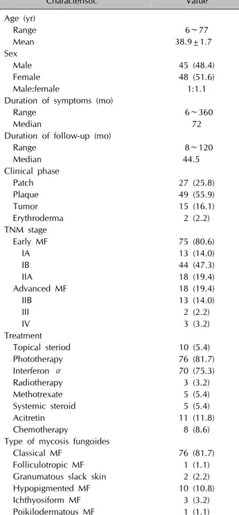
Table 1. Clinical characteristics of 93 patients with mycosis fungoides
Characteristic Value
Age (yr)
Range 6∼77
Mean 38.9±1.7
Sex
Male 45 (48.4)
Female 48 (51.6)
Male:female 1:1.1
Duration of symptoms (mo)
Range 6∼360
Median 72
Duration of follow-up (mo)
Range 8∼120
Median 44.5
Clinical phase
Patch 27 (25.8)
Plaque 49 (55.9)
Tumor 15 (16.1)
Erythroderma 2 (2.2)
TNM stage
Early MF 75 (80.6)
IA 13 (14.0)
IB 44 (47.3)
IIA 18 (19.4)
Advanced MF 18 (19.4)
IIB 13 (14.0)
III 2 (2.2)
IV 3 (3.2)
Treatment
Topical steriod 10 (5.4)
Phototherapy 76 (81.7)
Interferon α 70 (75.3)
Radiotherapy 3 (3.2)
Methotrexate 5 (5.4)
Systemic steroid 5 (5.4)
Acitretin 11 (11.8)
Chemotherapy 8 (8.6)
Type of mycosis fungoides
Classical MF 76 (81.7)
Folliculotropic MF 1 (1.1)
Granumatous slack skin 2 (2.2)
Hypopigmented MF 10 (10.8)
Ichthyosiform MF 3 (3.2)
Poikilodermatous MF 1 (1.1)
Values are presented as number only, mean±standard deviation, or number (%).
TNM: tumor, node, metastasis, MF: mycosis fungoides.
Fig. 1. Disease-specific survival curve according to early-stage mycosis fungoides (MF) and advanced-stage MF.
81.7%), there were 33 males and 43 females. The mean age of classic MF was 42.6±1.7 years. For hMF, there were 6 males and 4 females. The median age was 10.5 years (range: 6∼28). In addition, there was a significant difference between the age of hMF and that of classic MF
(p<0.05, Mann–Whitney test). The hMF patients were younger and had a significant difference than that of clas- sical MF patients (Table 2).
Clinical data of 10 patients with hypopigmented mycosis fungoides
From a population of 93 patients diagnosed with MF, ten patients were included in the sample as hMF patients: one adult man, one adult woman, five boys, and three girls.
Two patients only had hypopigmented lesions. Eight pa- tients had both hypopigmented lesions and erythematous lesions.
The clinical description is shown in Table 3. For the clin- ical stage, all patients were either stage IA or IB. Four pa- tients had stage IA, and six patients had stage IB.
The primary treatment modalities employed were photo- therapy, including narrowband UVB, UVB and UVB com- bination with UVA. Three patients underwent photother- apy with interferon α. All of these ten patients went into remission. Seven patients responded completely, and three patients responded partially. Moreover, no patient was with recurrences documented.
DISCUSSION
Mycosis fungoides usually has the disease progression as a slow evolution from patches, plaques to tumors and ex- tracutaneous involvement with possibly fatal ends, which is by the long duration of symptoms and cutaneous lesions at presentation1. Besides classic MF, there are still several variants of MF. The wide range of clinical and histopatho- logic presentations of MF, especially early-stage MF, re- quires a broad differential diagnosis. It helps to establish a correct diagnosis of MF, particularly in the early stages of
Table 2. Epidemiology data of patients based on variants of mycosis fungoides Characteristic Classic
MF
Folliculotropic MF
Granumatous slack skin
Hypopigmented MF
Ichthyosiform MF
Poikilodermatous MF Sex
Male 33 2 6 2 1
Female 43 1 - 4 1 -
Male:Female 1:1.3 All female All male 1.5:1 2:1 All male
Age (yr)
Mean 42.6 - 29.0 13.4 38.3 -
Median 41.5 48.0 29.0 10.5 42.0 28.0
Range 10~77 - 21~37 6~28 29~44 -
MF: mycosis fungoides, -: not available.
Table 3. Clinical data of 10 patients with hypopigmented mycosis fungoides No. Sex
Age at diagnosis
(yr)
Lesion type Location Clinical stage
Duration of symptoms
(mo)
Treatment Response
1 M 11 Hypopigmented patches Trunk IA 13 nbUVB, topical
corticosteroids
Complete response 2 M 13 Hypopigmented patches
and erythematous lesions
Multisites IB 48 Interferon α, UVA+UVB, topical corticosteroids
Complete response
3 M 28 Hypopigmented patches and erythematous lesions
Multisites IB 7 UVA+UVB Complete
response 4 M 13 Hypopigmented patches
and erythematous lesions
Back, low extremities
IA 60 UVB Complete
response 5 M 10 Hypopigmented patches
and erythematous lesions
Trunk IA 13 UVB Complete
response
6 M 8 Hypopigmented patches
and erythematous lesions
Multisites IB 36 nbUVB Complete
response
7 F 8 Hypopigmented patches
and erythematous lesions
Trunk IA 197 UVB Partial
response
8 F 9 Hypopigmented patches
and erythematous lesions
Multisites IB 60 Interferon α, nbUVB, topical corticosteroids
Partial response
9 F 6 Hypopigmented patches
and erythematous lesions
Multisites IB 18 nbUVB Partial
response
10 F 28 Hypopigmented patches Multisites IB 60 Interferon α,
nbUVB
Complete response M: male, F: female, nbUVB: narrow band ultraviolet B, UVA: ultraviolet A, UVB: ultraviolet B.
the disease, that being aware of the distinctive character- istics of this disease. Because of the rarity of the disease, there are few studies about subtype distribution of mycosis fungoides and its variants9. Although studies focused on specific subtype were published, relevant data in Chinese population are seldom reported.
In our study, the mean age at diagnosis of all MF patients and classic MF patients were 39.0 years and 42.6 years, respectively, which were both younger compared with other studies from western countries and Asian countries7,10,11, while in accordance with the research from Singapore9.
The possible reasons might be that patients could get ac- cess to dermatologists directly due to the imperfect referral system in China and that data, from a single center, might be biased. Also, there was not a male preponderance in the sex distribution of MF patients, in accordance with our previous study8 but in contrast to other studies11,12.
The stage is related to prognostic significance in mycosis fungoides13. According to this study, the majority of our patients (75 patients, 80.6%) were in the early stage, and 18 patients (19.4%) were in an advanced stage. The prog- nosis of advanced stage MF was significantly worse than
early-stage MF. The survival analysis of our study also con- firmed the favorable prognosis of MF with a 96.7% DSS rate. Besides stage, older age was considered as a risk fac- tor for MF, but it is controversial. Based on the study from Lebowitz et al.14, older age at diagnosis of MF does not predict worse disease-specific outcomes. We speculated the main reason is the difficulty of careful monitoring in daily life.
The therapy of mycosis fungoides should follow a step- wise, stage-adapted approach. The consensus recommen- dation from the European Organisation for Research and Treatment of Cancer-Cutaneous Lymphoma Task Force (EORTC-CLTF) is accepted and used widely.
In this study, although there were some patients in T1a stage, none of them chose a “watch and see” policy that recommended by consensus recommendation from EORTC- CLTF. The early stage MF patients were mainly treated with phototherapy, according to the standardized guide- lines released by the United States Cutaneous Lymphoma Consortium15. Some of the patients in this study also used phototherapy and interferon α combined therapy. Safety and efficacy have been confirmed16,17.
It has been reported that in large series, FMF accounts for approximately 10% of all patients with MF13,18. Among 93 patients in our study, one was diagnosed in FMF without any other medical history, which is less than 10%. In two studies from Korea, the percentage of FMF in all MF pa- tients accounted for around 2% and 4% respectively19,20. In a study in Argentina21, the portion was about 6%.
Those data from Asian countries might suggest that the rel- ative frequency of FMF has geographical and racial differences. Our FMF patient was in an early stage with multiple lesions on her face and got complete remission after topical steroids treatment. It is suggested that FMF pa- tients are received treatment based on stage, similar to the stepwise approach in classic MF22.
GSS is extremely rare. The two GSS patients presented with typical manifestation: pendulous folds of scaly atro- phic skin, huge ulcer, and secondary bacterial infection.
They were treated with chemotherapy and interferon α plus systemic steroids, respectively. Both of them got a partial response and were suggested to go under the long- term follow-up to exclude the increased risk of other ma- lignant lymphomas23.
hMF, ichthyosiform MF, and poikilodermatous MF are not listed in the latest WHO classification of cutaneous lym- phoma. However, they do have distinctive clinical mani- festation to lead to narrow the differential diagnosis of MF.
Ichthyosiform MF and poikilodermatous MF have the same typical pathological features as classic MF and similar de- veloping process.
Besides clinical aspects, hMF is also distinct from the clas- sic MF in epidemiological and prognostic aspects. It most commonly affects patients in childhood and adolescence.
According to the recent study from Brazil24, the average age of 20 hMF patients was 43.85 years. Rodney analyzed 20 hMF patients with an average age of 32.2 years3. Tan et al.9 included 47 hMF patients in Singapore with a me- dian age at diagnosis of 17. The present study demon- strated an average age of 13.4 years with age ranged from 6 to 28, in line with the previous findings. All patients in our study were Chinese. It is reported that white patients usually have hypopigmented lesions with an erythematous lesion. This “mixed” pattern is said to prevail in the Cauca- sian population25. However, eight patients among 10 hMF patients presented with hypopigmented lesions and eryth- ematous lesions in our study. Two patients were only with hypopigmented lesions, showing that the “mixed” pattern is also common in Chinese hMF patients. Our data show- ed all the hMF patients, in stage IA or IB, treated by photo- therapy or other skin-directed treatment and got remission.
Until the end time of follow-up, no relapse was docu- mented. As shown in the literature, the prognosis of hMF is good, and patients respond very well to skin-direct treat- ment26.
The limitations of this study include a single-center based experience and a small sample number. Despite the flaws, the study is meaningful as it gives more insight into my- cosis fungoides and its variants among Chinese patients:
early-stage MF has a better prognosis than advanced stage and hMF affects younger patients than that of classic pa- tients among Chinese patients. This study provides a new insight of mycosis fungoides and its variants in Chinese population. To be knowledgeable of variants of MF could help to give an accurate and prompt diagnosis and initiate appropriate treatment.
ACKNOWLEDGMENT
This work was supported by grants from Beijing Natural Science Foundation (Grant No.7182127), the National Key Research and Development Program of China (Grant No. 2016YFC0901500) and Milstein Medical Asian Ame- rican Partnership Foundation.
CONFLICTS OF INTEREST
The authors have nothing to disclose.
ORCID
Yixin Luo, https://orcid.org/0000-0002-5592-162X
Zhaorui Liu, https://orcid.org/0000-0002-9811-9063 Jie Liu, https://orcid.org/0000-0001-8219-2429 Yuehua Liu, https://orcid.org/0000-0002-2660-0675 Wei Zhang, https://orcid.org/0000-0002-7740-3778 Yan Zhang, https://orcid.org/0000-0003-0244-218X
REFERENCES
1. Willemze R, Jaffe ES, Burg G, Cerroni L, Berti E, Swerdlow SH, et al. WHO-EORTC classification for cutaneous lym- phomas. Blood 2005;105:3768-3785.
2. Souissi A, Ben Lagha I, Jendoubi F, Drissi H, Chelly I, Mokni M. Spiky follicular mycosis fungoides: a trichoscopic feature.
J Eur Acad Dermatol Venereol 2019;33:e252-e253.
3. Rodney IJ, Kindred C, Angra K, Qutub ON, Villanueva AR, Halder RM. Hypopigmented mycosis fungoides: a retrospec- tive clinicohistopathologic study. J Eur Acad Dermatol Ve- nereol 2017;31:808-814.
4. Pankratov O, Gradova S, Tarasevich S, Pankratov V. Poikilo- dermatous mycosis fungoides: clinical and histopathologi- cal analysis of a case and literature review. Acta Dermato- venerol Alp Pannonica Adriat 2015;24:37-41.
5. Willemze R, Cerroni L, Kempf W, Berti E, Facchetti F, Swerdlow SH, et al. The 2018 update of the WHO-EORTC classification for primary cutaneous lymphomas. Blood 2019;
133:1703-1714.
6. Ku LS, Lo KK. Mycosis fungoides--a retrospective study of 40 cases in Hong Kong. Int J Dermatol 2005;44:215-220.
7. Ishiji T, Takagi Y, Niimura M. Cutaneous lymphomas in Tokyo:
analysis of 62 cases in a dermatology clinic. Int J Dermatol 2001;40:37-40.
8. Liu J, Yu X, Liu Y, Jin H, Ma D, Qu T, et al. Relative fre- quency and survival of primary cutaneous lymphomas: a retro- spective analysis of 98 patients. Chin Med J (Engl) 2014;
127:645-650.
9. Tan EST, Tang MBY, Tan SH. Retrospective 5‐year review of 131 patients with mycosis fungoides and Sézary syndrome seen at the National Skin Centre, Singapore. Australas J Dermatol 2006;47:248-252.
10. Su C, Nguyen KA, Bai HX, Cao Y, Tao Y, Xiao R, et al.
Racial disparity in mycosis fungoides: an analysis of 4495 cases from the US National Cancer Database. J Am Acad Dermatol 2017;77:497-502.e2.
11. Eder J, Kern A, Moser J, Kitzwögerer M, Sedivy R, Trautinger F. Frequency of primary cutaneous lymphoma variants in Austria: retrospective data from a dermatology referral centre between 2006 and 2013. J Eur Acad Dermatol Venereol 2015;29:1517-1523.
12. Bradford PT, Devesa SS, Anderson WF, Toro JR. Cutaneous lymphoma incidence patterns in the United States: a popu- lation-based study of 3884 cases. Blood 2009;113:5064-5073.
13. Agar NS, Wedgeworth E, Crichton S, Mitchell TJ, Cox M, Ferreira S, et al. Survival outcomes and prognostic factors in mycosis fungoides/Sézary syndrome: validation of the revised International Society for Cutaneous Lymphomas/European Organisation for Research and Treatment of Cancer staging
proposal. J Clin Oncol 2010;28:4730-4739.
14. Lebowitz E, Geller S, Flores E, Pulitzer M, Horwitz S, Mosko- witz A, et al. Survival, disease progression and prognostic factors in elderly patients with mycosis fungoides and Sézary syndrome: a retrospective analysis of 174 patients. J Eur Acad Dermatol Venereol 2019;33:108-114.
15. Olsen EA, Hodak E, Anderson T, Carter JB, Henderson M, Cooper K, et al. Guidelines for phototherapy of mycosis fungoides and Sézary syndrome: a consensus statement of the United States Cutaneous Lymphoma Consortium. J Am Acad Dermatol 2016;74:27-58.
16. Stadler R, Otto HG, Luger T, Henz BM, Kuhl P, Zwingers T, et al. Prosepctive randomized multicenter clinical traial on the use of interferon-2α plus acitretin Photo(chemo)therapie April 2007 24 versus interferon - 2α plus PUVA in patients with cutaneous T-cell lymphoma stages I and II. Blood 1998;
92:3578-3581.
17. Olisova OY, Megna M, Grekova EV, Zaslavsky DV, Goren- kova LG, Sidikov AA, et al. PUVA and interferon α2b combined therapy for patients with mycosis fungoides at different stages of the disease: a seven-year retrospective study in Russia. J Eur Acad Dermatol Venereol 2019;33:
e72-e74.
18. van Doorn R, Van Haselen CW, van Voorst Vader PC, Geerts ML, Heule F, de Rie M, et al. Mycosis fungoides: disease evolution and prognosis of 309 Dutch patients. Arch Der- matol 2000;136:504-510.
19. Lee HS, Suh KS, Lee DY, Cho KH, Oh SH, Kim SC, et al.
Cutaneous lymphoma in Korea: a nationwide retrospective study. Acta Derm Venereol 2016;96:535-539.
20. Park JH, Shin HT, Lee DY, Lee JH, Yang JM, Jang KT, et al.
World Health Organization-European Organization for Re- search and Treatment of Cancer classification of cutaneous lymphoma in Korea: a retrospective study at a single tertiary institution. J Am Acad Dermatol 2012;67:1200-1209.
21. Abeldaño A, Enz P, Maskin M, Cervini AB, Torres N, Acosta AC, et al. Primary cutaneous lymphoma in Argentina: a re- port of a nationwide study of 416 patients. Int J Dermatol 2019;58:449-455.
22. van Santen S, van Doorn R, Neelis KJ, Daniëls LA, Horváth B, Bruijn MS, et al. Recommendations for treatment in fol- liculotropic mycosis fungoides: report of the Dutch Cutaneous Lymphoma Group. Br J Dermatol 2017;177:223-228.
23. Kempf W, Ostheeren-Michaelis S, Paulli M, Lucioni M, Wechsler J, Audring H, et al.; Cutaneous Lymphoma Histo- pathology Task Force Group of the European Organization for Research and Treatment of Cancer. Granulomatous my- cosis fungoides and granulomatous slack skin: a multicenter study of the Cutaneous Lymphoma Histopathology Task Force Group of the European Organization For Research and Treat- ment of Cancer (EORTC). Arch Dermatol 2008;144:1609- 1617.
24. Amorim GM, Niemeyer-Corbellini JP, Quintella DC, Cuzzi T, Ramos-E-Silva M. Hypopigmented mycosis fungoides: a 20-case retrospective series. Int J Dermatol 2018;57:306- 312.
25. Ardigó M, Borroni G, Muscardin L, Kerl H, Cerroni L. Hypo-
pigmented mycosis fungoides in Caucasian patients: a cli- nicopathologic study of 7 cases. J Am Acad Dermatol 2003;
49:264-270.
26. Boulos S, Vaid R, Aladily TN, Ivan DS, Talpur R, Duvic M.
Clinical presentation, immunopathology, and treatment of juvenile-onset mycosis fungoides: a case series of 34 pa- tients. J Am Acad Dermatol 2014;71:1117-1126.