Brief Report
454 Ann Dermatol
Received December 14, 2016, Revised May 20, 2017, Accepted for publication June 19, 2017
Corresponding author: Ossama Abbas, Department of Dermatology, American University of Beirut Medical Center, P.O.Box 11-0236, Riad El Solh St Beirut, Lebanon. Tel: 961-1-350000 (ext. 7915), Fax: 961-1- 745320, E-mail: [email protected]
ORCID: https://orcid.org/0000-0001-6970-8056
This is an Open Access article distributed under the terms of the Creative Commons Attribution Non-Commercial License (http://creativecommons.
org/licenses/by-nc/4.0) which permits unrestricted non-commercial use, distribution, and reproduction in any medium, provided the original work is properly cited.
Copyright © The Korean Dermatological Association and The Korean Society for Investigative Dermatology
pISSN 1013-9087ㆍeISSN 2005-3894
Ann Dermatol Vol. 31, No. 4, 2019 https://doi.org/10.5021/ad.2019.31.4.454
BRIEF REPORT
Possible Role for Plasmacytoid Dendritic Cells in Pemphigus
Nehmat Ramadan, Mazen Kurban, Ossama Abbas
Department of Dermatology, American University of Beirut Medical Center, Beirut, Lebanon
Dear Editor:
Plasmacytoid dendritic cells (pDCs) are a specialized DC population1. They display plasma cell morphology and ex- press CD4, CD123, HLA-DR, BDCA-2, and toll-like re- ceptors (TLR)7 and TLR9 within endosomal compartments.
pDCs are usually not present in normal skin, but infiltrate the skin in several cutaneous pathologies including in- flammatory/autoimmune, infectious, and neoplastic enti- ties1. Upon TLR stimulation, pDCs have the ability to se- cret type I interferons (IFNs) up to 1,000 times more than other cell types as well as proinflammatory cytokines in- cluding interleukin (IL)-6 and tumor necrosis factor (TNF)-α1. These lead mainly to an antiviral state and contribute to the regulation of the function of other immune cells such as myeloid DC, T-, B- and natural killer (NK) cells. Thus, pDCs provide protective immunity at the skin level by regulated sensing of microbial or self-nucleic acids upon skin damage. However, when excessive sensing of self-an- tigens occurs, pDCs may participate in an exaggerated self-directed immune responses contributing to the patho- genesis of different inflammatory/autoimmune cutaneous pathologies. Several studies have identified a significant role of pDCs in several inflammatory/autoimmune muco- cutaneous disorders including lupus erythematosus (LE),
psoriasis, and lichen planus (LP)1. However, the role of pDCs in the autoimmune blistering disorders has not been well-explored. Hence, we intend in this study to inves- tigate pDC role in the pemphigus group of the auto- immune blistering skin diseases. This may allow us to un- cover part of the underlying pathogenesis of these disorders.
Our institutional review board approved the study (American University of Beirut IRB protocol DER.OA.24). Forty-six pemphigus cases (including 36 pemphigus vulgaris (PV) and 10 pemphigus foliaceus types) and 32 pemphigoid cases (29 bullous pemphigoid and 3 pemphigoid ges- tationis) as comparison group were retrieved from our database. Only straightforward cases that fit the clinico- pathological and immunoflorescence features of the re- spective autoimmune blistering diseases were included.
Immunohistochemical analysis was performed on sections obtained from formalin-fixed, paraffin-embedded tissue us- ing antibodies to BDCA-2 (mouse immuno globulin G1, clone 124B3.13; Dendritics, Lyon, France) and myxovirus resistance protein A (MxA, M143; University of Freiburg, Freiburg, Germany). Anti-BDCA2 antibody is a specific pDC marker1, while anti-MxA antibody assesses type I IFN production by pDCs, since MxA is well established surro- gate marker for local type I IFN production1. A semi- quantitative scoring system was used to assess pDC re- cruitment and MxA expression (Table 1).
Results showed the pDCs to be present in all of the pem- phigus cases (n=46) and 72% (n=23) of pemphigoid cases.
However, pDCs were significantly more abundant in pem- phigus cases (Fig. 1B, E) than in pemphigoid cases (Fig.
1H) with a significantly higher pDC score (p<0.05). MxA expression was mostly patchy in both pemphigus (n=44, 96%) (Fig. 1C, F) and pemphigoid (n=23, 72%) cases (Fig.
1I).
Our hypothesis in this study concerning pDCs role in the pemphigus group is based on several observations. First,
Brief Report
Vol. 31, No. 4, 2019 455 Fig. 1. (A∼C) Pemphigus vulgaris. (A) Representative case showing suprabasal acantholysis with underlying dermal inflammatory infiltrate (H&E, ×40). (B, C) BDCA-2 immunostaining highlighted plasmacytoid dendritic cells (pDCs) in a superficial perivascular and interstitial distribution with pDCs making up more than 10% of the inflammatory infiltrate/pDC score of 2 (B: ×100, C: ×200). (D∼F) Pemphigus foliaceus. (D) Representative case showing subcorneal acantholysis with underlying dermal inflammatory infiltrate (H&E,
×40). (E, F) BDCA-2 immunostaining highlighted pDCs in a superficial perivascular and interstitial distribution with pDCs making up more than 10% of the inflammatory infiltrate/pDC score of 2 (E: ×100, F: ×200). (G∼I): Bullous pemphigoid. (G) Representative case with subepidermal blistering and underlying inflammatory infiltrate (H&E, ×100). (H, I) BDCA-2 immunostaining highlighted scattered pDCs in a perivascular and interstitial distribution with pDCs making up less than 10% of the inflammatory infiltrate/pDC score of 1 (H: ×100, I: ×100).
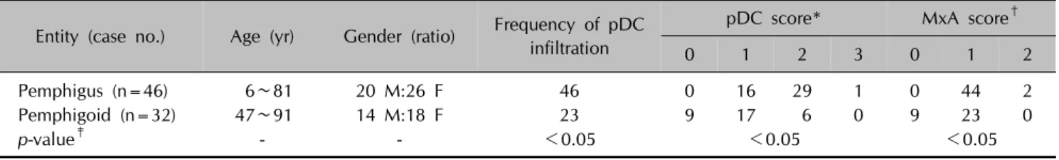
Table 1. pDCs presence and MxA expression in pemphigus versus pemphigoid group (%) Entity (case no.) Age (yr) Gender (ratio) Frequency of pDC
infiltration
pDC score* MxA score†
0 1 2 3 0 1 2
Pemphigus (n=46) 6∼81 20 M:26 F 46 0 16 29 1 0 44 2
Pemphigoid (n=32) 47∼91 14 M:18 F 23 9 17 6 0 9 23 0
p-value‡ - - <0.05 <0.05 <0.05
pDCs: plasmacytoid dendritic cells, MxA: myxovirus resistance A, M: male, F: female. *BDCA2+ pDC content was scored as percentage of total mononuclear infiltrate: 0 (no positive cells), 1 (1%∼10% positive cells), 2 (10%∼50% positive cells), 3 (>50% positive cells). †MxA staining was scored as: 0=negative, 1=patchy/weak, and 2=diffuse. ‡Statistical analysis was performed by using the Mann-Whitney test to analyze statistical differences in pDC and MxA scores between the 2 groups. A two-tailed p-value of <0.05 was considered statistically significant. Normal skin tissue served as negative control and cutaneous lupus erythematosus served as positive control.
Brief Report
456 Ann Dermatol
several reports have described induction of autoimmune blistering disorders following administration of IFN-α, the endogenous local counterpart of which is mainly pro- duced by pDCs2. Second, high-titer IFN-α antibodies have been detected in patients with autoimmune blistering dis- orders3. Third, imiquimod, an immunomodulator known to be a potent pDCs activator, and TNF inhibitors, known to be secondary inducers of IFN-α, have been reported to induce autoimmune blistering disorders4,5. Fourth, some autoimmune blistering disorders have been associated with viral infections. Since pDCs’ mainly function in an- ti-viral resistance, their involvement in such autoimmune blistering disorders would not be surprising6. Finally, auto- immune blistering disorders have been associated with several inflammatory disorders such as LE, LP and psor- iasis, in which evidence suggests significant pDC role in their underlying pathogenesis7.
Our study results support our hypothesis, especially in re- lation to a possible role for pDCs in pemphigus pathogenesis.
While the contribution of the adaptive immune system has been well studied using animal models, the earlier mecha- nisms that contribute to the initial production of autoanti- bodies and loss of tolerance have not been well inves- tigated7,8. One study demonstrated that, in the presence of Dsg3, NK cells interact with CD4+ T cells in the perile- sional skin and peripheral blood of PV patients leading to the production of several cytokines, with especially high IL-6 levels. IL-6 is a pleiotropic cytokine important in the pathophysiology of inflammation and autoimmune dis- orders such as its role in the production of anti-DNA and chromatin autoantibodies in LE8. However, the contribution of pDCs, which can also secret IL-6, was not explored in this study8. In another study investigating the local in- flammatory infiltrate in Darier’s disease and using 14 PV cases as a comparison group, the authors reported the presence of CD123+ pDCs in low percentages (<5%) in PV cases9.
The results of our study and the known multifaceted im- munological functions of the pDC indicate a possible role of this cell in the autoimmune blistering disorders, espe- cially pemphigus group1. Especially with NK cells which have been implicated in the early pathogenic steps of PV pathogenesis, pDCs have been shown to have bidirec- tional interaction10. There is evidence that NK cells, upon cell-to-cell contact, promote pDC maturation and strongly enhance pDC production of IFN-α, TNF-α, and IL-6. On the other hand, pDCs can efficiently promote NK cell activation. In addition, several studies have shown that pDCs are critical for antibody responses through their role in promoting plasma cell differentiation from naive and memory B cells1. Actually, pDCs have been shown to in-
duce plasma cell differentiation through the sequential ac- tion of type I IFNs and IL-610. This thus makes their possi- ble role in the induction of autoantibodies in the auto- immune blistering disorders unsurprising.
The authors recognize that a relatively small number of cases have been studied and that the study is performed at only one point during the course of these autoimmune blistering disorders. Hence, these observations are consid- ered preliminary.
In summary, we have shown that pDCs are recruited into the skin lesions of the autoimmune blistering disorders, with significantly higher content in the pemphigus group.
Their consistent presence speaks in favor of an important role of these cells in their pathogenesis, possibly in the ini- tial mechanisms leading to autoantibody production.
ACKNOWLEDGMENT
This research has been supported by a grant from the Medical Practice Plan (MPP) at the American University of Beirut Medical Center.
CONFLICTS OF INTEREST
The authors have nothing to disclose.
ORCID
Nehmat Ramadan, https://orcid.org/0000-0003-1082-7899 Mazen Kurban, https://orcid.org/0000-0003-1011-0687 Ossama Abbas, https://orcid.org/0000-0001-6970-8056
REFERENCES
1. Saadeh D, Kurban M, Abbas O. Update on the role of plasmacytoid dendritic cells in inflammatory/autoimmune skin diseases. Exp Dermatol 2016;25:415-421.
2. Niizeki H, Inamoto N, Nakamura K, Tsuchimoto K, Hashimoto T, Nishikawa T. A case of pemphigus foliaceus after in- terferon alpha-2a therapy. Dermatology 1994;189 Suppl 1:129-130.
3. Prümmer O, Zillikens D, Porzsolt F. High-titer interferon- alpha antibodies in a patient with pemphigus foliaceus. Exp Dermatol 1996;5:213-217.
4. Lo Schiavo A, Sangiuliano S, Puca RV, Brunetti G, Ruocco E, Cozzi R. Contact pemphigus: a side-effect of imiquimod therapy. Int J Dermatol 2008;47:765-767.
5. Boussemart L, Jacobelli S, Batteux F, Goulvestre C, Grange P, Carlotti A, et al. Autoimmune bullous skin diseases occurring under anti-tumor necrosis factor therapy: two case reports. Dermatology 2010;221:201-205.
Brief Report
Vol. 31, No. 4, 2019 457
Received April 23, 2018, Revised June 19, 2018, Accepted for publication July 17, 2018
Corresponding author: Hyun-Sun Yoon, Department of Dermatology, SMG-SNU Boramae Medical Center, 20 Boramae-ro 5-gil, Dongjak-gu, Seoul 07061, Korea. Tel: 82-2-870-2382, Fax: 82-2-831-0714, E-mail: [email protected]
ORCID: https://orcid.org/0000-0003-1401-2670
This is an Open Access article distributed under the terms of the Creative Commons Attribution Non-Commercial License (http://creativecommons.org/
licenses/by-nc/4.0) which permits unrestricted non-commercial use, distribution, and reproduction in any medium, provided the original work is properly cited.
Copyright © The Korean Dermatological Association and The Korean Society for Investigative Dermatology 6. Ruocco E, Ruocco V, Lo Schiavo A, Brunetti G, Wolf R.
Viruses and pemphigus: an intriguing never-ending story.
Dermatology 2014;229:310-315.
7. Vassileva S, Drenovska K, Manuelyan K. Autoimmune blistering dermatoses as systemic diseases. Clin Dermatol 2014;32:364-375.
8. Stern JN, Keskin DB, Barteneva N, Zuniga J, Yunis EJ, Ahmed AR. Possible role of natural killer cells in pemphigus vulgaris-preliminary observations. Clin Exp Immunol 2008;
152:472-481.
9. Miracco C, Pietronudo F, Mourmouras V, Pellegrino M, Onorati M, Mastrogiulio MG, et al. Possible implication of local immune response in Darier's disease: an immuno- histochemical characterization of lesional inflammatory infiltrate. Mediators Inflamm 2010;2010:350304.
10. Wehner R, Dietze K, Bachmann M, Schmitz M. The bidirectional crosstalk between human dendritic cells and natural killer cells. J Innate Immun 2011;3:258-263.
https://doi.org/10.5021/ad.2019.31.4.457
Factors Determining Treatment Response to Cryotherapy for Foot Warts
Do-Yeop Kim, Hyun-sun Park, Soyun Cho, Hyun-Sun Yoon
Department of Dermatology, SMG-SNU Boramae Medical Center, Seoul, Korea
Dear Editor:
Different clinical and biologic factors, such as disease du- ration, infection site, and lesion size, are associated with the treatment response to cryotherapy of cutaneous warts1-3. However, published data on the predictive factors of cry- otherapy in the treatment of cutaneous warts showed in- consistent results3,4. In addition, the majority of previous studies have not controlled for confounding variables1-4, or have included warts located in different anatomical sites1,2. Thus, we aimed to investigate the factors affecting the treatment response to cryotherapy in foot warts using multivariable analysis.
We reviewed the medical records of patients having foot warts and who started cryotherapy at the SMG-SNU Boramae Medical Center from February 2016 through January 2018. All patients were followed until we con- firmed that their warts completely disappeared, until they
were lost to follow-up, or until February 2, 2018 (date of scheduled data extraction), whichever arrived earlier. Age, sex, disease duration, infection site (toe, sole, and peri- ungual), number of lesions, the maximum diameter of le- sions, and recurrent status (primary infection vs. re-in- fection) were obtained from the medical records of the ini- tial visits. Treatment intervals, the number of cryotherapy sessions, and treatment outcomes (cleared vs. persistent) were obtained. A patient with clearance was considered a patient who no longer had visible warts and had sustained normal skin color and skin lines for at least 4 weeks after the last cryotherapy. A responder was defined as a patient having complete clearance of warts within after 6 cry- otherapy sessions5. The study protocol was approved by the Institutional Review Board of the SMG-SNU Boramae Medical Center (approval number: 30-2017-30) and the re- quirement for informed consent was waived.