pISSN 2288-0585⋅eISSN 2288-6850
Postsurgical Wound Infection Caused by Mycobacterium conceptionense Identified by Sequencing of 16S rRNA, hsp65,
and rpoB Genes in an Immunocompetent Patient
Ja Young Lee
1*, Si Hyun Kim
1,2*, Jeong Hwan Shin
1,2, Hyun-Kyung Lee
3, Young Min Lee
3, Sae Am Song
1, Il Kwon Bae
4, Chang-Ki Kim
5, Kyung Ran Jun
1, Hye Ran Kim
1, Jeong Nyeo Lee
1, Chulhun L. Chang
61
Department of Laboratory Medicine,
2Paik Institute for Clinical Research, and
3
Department of Internal Medicine, Inje University College of Medicine,
4
Department of Dental Hygiene College of Medical and Life Science, Shilla University, Busan,
5
Department of Laboratory Medicine, Korean Institute for Tuberculosis, Osong,
6
Department of Laboratory Medicine, Pusan National University School of Medicine, Yangsan, Korea
Rapidly growing mycobacteria are ubiquitous in the environment and are increasingly being recognized as opportunistic pathogens. Recently, a new species, Mycobacteium conceptionense, has been validated from the Mycobacterium fortuitum third biovariant complex by molecular analysis. However, there are few reports, and postsurgical wound infection by this species is rare. We report a case of postsurgical
wound infection caused by M. conceptionense in an immunocompetent patient that was identified by a se- quencing analysis of 16S rRNA, hps65, and rpoB genes. (Ann Clin Microbiol 2014;17:23-27)
Key Words: Gene sequencing, Mycobacterium con- ceptionense, Wound infection
23
Received 23 October, 2013, Revised 5 February, 2014, Accepted 11 February, 2014
Correspondence: Chulhun Ludgerus Chang, Department of Laboratory Medicine, Pusan National University School of Medicine, 49 Busandaehak-ro, Mulgeum-eup, Yangsan 626-770, Korea. (Tel) 82-55-360-1870, (Fax) 82-55-360-1880, (E-mail) [email protected]
*These authors contributed equally to this work.
ⓒ The Korean Society of Clinical Microbiology.
This is an Open Access article distributed under the terms of the Creative Commons Attribution Non-Commercial License (http://creativecommons.org/licenses/by-nc/3.0) which permits unrestricted non-commercial use, distribution, and reproduction in any medium, provided the original work is properly cited.
INTRODUCTION
Rapidly growing mycobacteria (RGM) are defined as non- tuberculous mycobacteria (NTM) that grow within 7 days on sol- id media. They are ubiquitous in the environment and often can be isolated from tap water. Increasingly, they are being recog- nized as opportunistic pathogens [1]. The major important RGM are Mycobacterium abscessus, Mycobacterium chelonae, and Mycobacterium fortuitum complex. The M. fortuitum complex is composed of M. fortuitum, Mycobacterium peregrinum, and M.
fortuitum third biovariant complex. Recently, a few new species of M. fortuitum group have been found exploiting advancements of molecular analysis [2-4]. Mycobacterium conceptionense was validated but there are few reports, and wound infection is rare [5-7]. We report a case of postsurgical wound infection in an im- munocompetent patient that was caused by M. conceptionense.
CASE REPORT
A 66-year-old man presented with pus and a skin defect on his lower back for one month at previous operative site. He was operated on by foraminotomy with a device for intervertebral assisted motion (DIAMTM) insertion, which was a H shaped in- terspinous spacer consisting of silicone core, poly ester mesh, fixation cables and titanium crimps, as a result of spinal stenosis eight months earlier. The lesion had started as erythematous nodule with swelling one month before and then burst. Physical examination revealed that soft tissue was exposed with yellow pus over the wound. He did not complain of tenderness or local heat. No specific medical or family histories were reported, and his general condition was good. Laboratory tests including com- plete blood count, liver function tests, and C-reactive protein showed no significant abnormalities. A Blood Quantiferon-TB
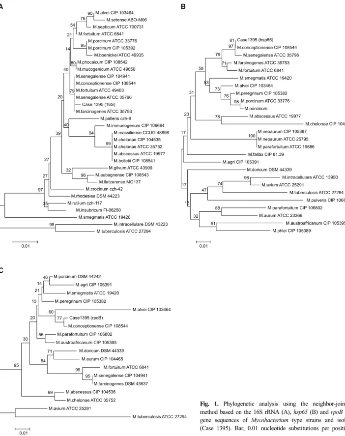
Fig. 1. Phylogenetic analysis using the neighbor-joining method based on the 16S rRNA (A), hsp65 (B) and rpoB (C) gene sequences of Mycobacterium type strains and isolate (Case 1395). Bar, 0.01 nucleotide substitutions per position.
test was negative. No active pulmonary disease was detected by chest radiography. Skin biopsy of the lesion showed a denuded epidermis with granulation tissue and granuloma with central neutrophilic aggregation, appearing as a fungal or mycobacterial
infection. The patient initially received empirical anti-myco- bacterial treatment with isoniazid, rifampicin, pyrazinamide, and ethambutol (HREZ) and wound dressing daily.
Non-pigmented strain was cultured on egg-based solid media
in 5 days at 36°C from tissue on skin-defect. Acid fast stain us- ing Ziehl-Neelsen method showed positive results. However, the isolate from tissue culture was not identified by PCR-restriction fragment length polymorphism (RFLP) analysis based on the rpoB gene. Fungal culture was negative. After two months, the patient complained of continuous drainage of yellow pus from the wound, and the anti-mycobacterial medication was stopped.
The same NTM was cultured on solid medium for mycobacteria from pus. Gam stain and acid-fast bacillus stain directly from clinical specimen were negative and this isolate was also un- identified by RFLP method. Antimycobacterial susceptibility tests were performed according to the Clinical and Laboratory Standards Institute (CLSI) M24-A, retrospectively. The results were interpreted according to the criteria for rapidly growing mycobacteria. The strain was susceptible to amikacin, cipro- floxacin, clarithromycin, doxycycline, and imipenem, however, it was intermediate to cefoxitin, and tobramycin.
We performed 16S rRNA gene sequencing to identify NTM as previously described [8], but the isolates was not identified because the sequence was indistinguishable, showing 99.38%
homology with M. fortuitum (GenBank Accession No. AY457067), 99.58% with Mycobacterium farcinogenes (GenBank AF055333), 99.79% with Mycobacterium senegalense (GenBank AF480596) and M. conceptionense (GenBank EU191913). The isolate was further investigated by sequencing using hsp65 and rpoB [9].
The hsp65 sequences of the strain showed 99% similarity with M. senegalense (GenBank AY684045) as well as 99.5% sim- ilarity with M. conceptionense (GenBank EU191920). Sequencing analysis based on rpoB showed 99.4% homology with M. con- ceptionense (GenBank AY859695), and the second closest match was Mycobacterium porcinum (GenBank AY544955), with 98.3% homology. This isolate was finally identified as M.
conceptionense by 16S rRNA, hsp65, and rpoB genes sequenc- ing and phylogenetic analysis using MEGA version 4 (Fig. 1).
After recovering of NTM from solid agar media, the anti-my- cobacterial treatment changed to minocyclin, ofloxacin, and clarithromycin and wound dressing continued daily. After 2 weeks, oral medication was stopped because the patient devel- oped dizziness. The patients refused to take anti-mycobacterial medication anymore. In spite of dressing of the wound daily, the amount of discharge around the wound increased, and local pain developed. Eventually, he was re-operated on and a new DIAM installed, and the patient is progressing well.
DISCUSSION
M. conceptionese is a non-pigmented RGM belonging to the M. fortuitum group that was recently distinguished from M.
porcinum. New species of the M. fortuitum group have steadily increased following the advancement of molecular methods [2-4]. Generally, the 16S rRNA gene has been used for the identification of unusual mycobacterial isolates. However, se- quence analysis of partial 16S rRNA gene cannot discriminate between closely related RGM species. Thus, additional genes such as sodA, hsp65, and rpoB were proposed for sequence analysis for the molecular identification of mycobacteria be- cause these genes showed greater interspecies and intraspecies sequence divergence than 16S rRNA gene [10]. In this case, we identified the isolate performing sequence analysis of rpoB and hsp65 genes as well as the 16S rRNA gene. The isolate shared more than 99.5% sequence similarity of the 16S rRNA gene with M. senegalense, M. farcinogenes, and M. conceptionense, whereaas for hsp65 analysis, the isolate showed 99.5% sequence similarity of M. conceptionense and 99.0% of M. senegalense.
Also, rpoB gene sequence of the isolate shared 99.4% similarity with M. conceptionense and 98.3% with M. porcinum. The re- sults supported the view that rpoB sequencing is the primary tool for the molecular identification of RGM, and alternative DNA targets such as hsp65 provide better resolution at the spe- cies level [10,11]. However, there are no validated interpretative criteria for the identification of RGM by sequence analysis ex- cept 16S rRNA gene [12].
The M. fortuitum groups are considered common etiologic pathogens for postsurgical wound infection [13]. Various medi- cal or surgical procedures related to wound infection by RGM include cardiac bypass surgery, augmentation mammoplasty, and subcutaneous needle injections or liposuction for cosmetic pur- poses [14]. Typically, the wound infection related to RGM oc- curred as a late postoperative complication [15]. Clinical mani- festations usually start with watery discharge, wound break- down, and low fever after 1 to 12 months. Thus, non-healing of a surgical site or dehiscence of a previously healed incision should be an important trigger to suspect mycobacterial in- fection [14,16,17]. Most reported patients were generally healthy.
Risk factors for the surgical site infection were trauma and con- tamination of surgical supplies and antiseptic solution [14]. In the initial report of skin and soft tissue infection related to M.
conceptionense, this microorganism was isolated from post- traumatic osteitis that occurred after exposure to a river [5].
Water has been mentioned as an important source because of the ubiquitous environmental distribution of RGM. However, the sources of infection in the subsequent cases were not re- vealed [6,7]. Our patient denied any contact with contaminated water, trauma, or acupuncture before the onset of symptoms. It could be assumed that the DIAM was infected, although the source in our case remains unknown. The patient underwent a surgical operation at other hospital, so we could not investigate the possibility of nosocomial infection caused by a surgical equipment contamination.
The optimal antibiotic therapy for M. conceptionese infection has not been established. In previous reports, most patients were treated with a combination of surgery and antimicrobial agents according to the antimicrobial susceptibility of the isolates [5-7].
Especially in plastic surgical sites infected by RGM such as M.
fortuitum, it is important to use adjuvant antimicrobial treatment in addition to a prompt surgical approach because patients treat- ed with surgery alone had relapses within 4 to 6 weeks [13].
The duration of antimicrobial therapy in wound infections was diverse, ranging from 3 to 18 months [7]. In our case, initial an- timicrobial therapy including minocyclin, ofloxacin, and clari- thromycin lasted only for 2 weeks because of side effects. Then he denied appropriate anti-mycobacterial treatment and incision and drainage at skin defected region. The only treatment was daily site dressing. It leaded to be the progression of the in- fection by M. conceptionense. The amount of discharge around the wound started to increase, and expanding pain developed around the wound. Eventually, he was treated by reoperation and the infection related symptoms were relieved.
We describe a case of surgical site infection in an immuno- competent patient caused by M. conceptionese. We identified the isolate performing sequence analysis of the rpoB and hsp65 genes in addition to 16S rRNA gene. In view of the limited reports of M. conceptionese infection, further investigation is needed to establish the predisposing factors, treatment, and prognosis.
ACKNOWLEDGMENTS
This work was supported by the Basic Science Research Program through the National Research Foundation of Korea (NRF), funded by the Ministry of Education, Science and Technology (2010-0011703).
REFERENCES
1. Colombo RE and Olivier KN. Diagnosis and treatment of infections
caused by rapidly growing mycobacteria. Semin Respir Crit Care Med 2008;29:577-88.
2. Adékambi T, Berger P, Raoult D, Drancourt M. rpoB gene sequence-based characterization of emerging non-tuberculous mycobacteria with descriptions of Mycobacterium bolletii sp. nov., Mycobacterium phocaicum sp. nov. and Mycobacterium aubagnense sp. nov. Int J Syst Evol Microbiol 2006;56:133-43.
3. Adékambi T and Drancourt M. Dissection of phylogenetic relationships among 19 rapidly growing Mycobacterium species by 16S rRNA, hsp65, sodA, recA and rpoB gene sequencing. Int J Syst Evol Microbiol 2004;54:2095-105.
4. Schinsky MF, Morey RE, Steigerwalt AG, Douglas MP, Wilson RW, Floyd MM, et al. Taxonomic variation in the Mycobacterium fortuitum third biovariant complex: description of Mycobacterium boenickei sp. nov., Mycobacterium houstonense sp. nov., Mycobac- terium neworleansense sp. nov. and Mycobacterium brisbanense sp.
nov. and recognition of Mycobacterium porcinum from human clinical isolates. Int J Syst Evol Microbiol 2004;54:1653-67.
5. Adékambi T, Stein A, Carvajal J, Raoult D, Drancourt M. Descri- ption of Mycobacterium conceptionense sp. nov., a Mycobacterium fortuitum group organism isolated from a posttraumatic osteitis inflammation. J Clin Microbiol 2006;44:1268-73.
6. Liao CH, Lai CC, Huang YT, Chou CH, Hsu HL, Hsueh PR.
Subcutaneous abscess caused by Mycobacterium conceptionense in an immunocompetent patient. J Infect 2009;58:308-9.
7. Thibeaut S, Levy PY, Pelletier ML, Drancourt M. Mycobacterium conceptionense infection after breast implant surgery, France.
Emerg Infect Dis 2010;16:1180-1.
8. Kim JH, Lee JY, Kim HR, Heo KW, Park SK, Lee JN, et al.
Acute lymphadenitis with cellulitis caused by Staphylococcus lugdunensis. Korean J Lab Med 2008;28:196-200.
9. Shin JH, Lee EJ, Lee HR, Ryu SM, Kim HR, Chang CL, et al.
Prevalence of non-tuberculous mycobacteria in a hospital environ- ment. J Hosp Infect 2007;65:143-8.
10. Ringuet H, Akoua-Koffi C, Honore S, Varnerot A, Vincent V, Berche P, et al. hsp65 sequencing for identification of rapidly growing mycobacteria. J Clin Microbiol 1999;37:852-7.
11. Adékambi T, Drancourt M, Raoult D. The rpoB gene as a tool for clinical microbiologists. Trends Microbiol 2009;17:37-45.
12. Clinical and Laboratory Standards Institute. Interpretive criteria for identification of bacteria and fungi by DNA target sequencing;
approved guideline. Document MM18-A. Wayne, PA; Clinical and Laboratory Standards Institute, 2010.
13. Brown-Elliott BA and Wallace RJ Jr. Clinical and taxonomic status of pathogenic nonpigmented or late-pigmenting rapidly growing mycobacteria. Clin Microbiol Rev 2002;15:716-46.
14. Murillo J, Torres J, Bofill L, Ríos-Fabra A, Irausquin E, Istúriz R, et al. Skin and wound infection by rapidly growing mycobacteria:
an unexpected complication of liposuction and liposculpture. The Venezuelan Collaborative Infectious and Tropical Diseases Study Group. Arch Dermatol 2000;136:1347-52.
15. Rodrigues C, Mehta A, Jha U, Bharucha M, Dastur FD, Udwadia TE. Nosocomial Mycobacterium chelonac infection in laparoscopic surgery. Infect Control Hosp Epidemiol 2001;22:474-5.
16. Thami GP, Kaur S, Chander J, Attri AK. Post surgical atypical mycobacterial infection due to Mycobacterium fortuitum. J Infect 2002;45:210-11.
17. Kalita JB, Rahman H, Baruah KC. Delayed post-operative wound infections due to non-tuberculous mycobacterium. Indian J Med Res 2005;122:535-9.
=국문초록=
16S rRNA, hsp65 , 및 rpoB 염기순서분석으로 동정한 Mycobacterium conceptionense 에 의한 면역능이
정상인 환자에서 발생한 수술후 창상감염
인제대학교 의과대학 1진단검사의학교실, 2백인제기념임상의학연구소, 3내과, 4신라대학교 의생명과학대학 치위생학과,
5대한결핵협회 결핵연구원 진단검사의학부, 6부산대학교 의학전문대학원 진단검사의학교실
이자영1, 김시현1,2, 신정환1,2, 이현경3, 이영민3, 송새암1, 배일권4, 김창기5, 전경란1, 김혜란1, 이정녀1, 장철훈6
신속발육항산균은 자연환경에서 흔히 검출되는 균으로, 기회감염병원체로 인체감염의 주요 원인균으로서 인식되고 있 다. M. conceptionense는 분자역학적 분석을 통해 M. fortuitum의 세번째 생체변이종에서 분리되어 새로운 종으로 명명되 었다. 그러나 이 균에 의한 감염 보고는 많지 않고 특히 수술후 창상 감염에 대한 보고는 거의 없다. 이에 저자들은 16S rRNA, hsp65 및 rpoB 유전자 염기순서분석을 이용하여 동정한 M. conceptionense에 의한 수술 후 창상감염을 보고하고자 한다. [Ann Clin Microbiol 2014;17:23-27]
교신저자 : 장철훈, 626-770, 경남 양산시 물금읍 부산대학로 49 부산대학교 의학전문대학원 진단검사의학교실 Tel: 055-360-1870, Fax: 055-360-1880
E-mail: [email protected]