60 http://www.ecevr.org/
CLINICAL
EXPERIMENTAL VACCINE
RESEARCH
Introduction
Canine rabies continues to be a major threat in many countries especially in Asia and in Africa [1]. The disease is endemic in all provinces of Morocco except the southern desert region, with the domestic dog being the main reservoir and vector [2-5] of the virus. Since 1986, about 22 human deaths have been reported yearly [6] and since 2000, an average of 376 animal cases have been recorded annually, mainly in dogs and in livestock, especially cattle [7]. The major element of rabies control strategies is regu- lar application of injectable vaccine to reach and maintain sufficient vaccination cov- erage in the field enough to stop rabies virus transmission. Moroccan authorities have set up several rabies eradication plans since 1986, but to date rabies remains a serious
© Korean Vaccine Society.
This is an Open Access article distributed under the terms of the Creative Commons Attribution Non-Com- mercial License (http://creativecommons.org/licenses/
by-nc/3.0) which permits unrestricted non-commercial use, distribution, and reproduction in any medium, pro- vided the original work is properly cited.
K O R E A N V A C C I N E S O C I E T Y
K O R E A N K O R E A N A C C I N E O C I E T Y V
S
Clin Exp Vaccine Res 2016;5:60-69 http://dx.doi.org/10.7774/cevr.2016.5.1.60 pISSN 2287-3651 • eISSN 2287-366X
Sami Darkaoui1, Ouafaa Fassi Fihri2, Jean Luc Schereffer3,
Nadia Aboulfidaa1,
Marine Wasniewski3, Karima Zouine1, Mohammed Bouslikhane2, Khadija Id Sidi Yahia1, Florence Cliquet3
1Office National de Sécurité Sanitaire des Produits Alimentaires, DPIV Rue Ikhlass CYM (BP4509 Akkari), Rabat; 2Department of Pathology and Veterinary Public Health, Agronomic and Veterinary Institute Hassan II, IAV Hassan II (BP Rabat Instituts), Rabat, Morocco; 3Nancy OIE/WHO/EU Laboratory for Rabies and Wildlife, French Agency for Food, Environmental and Occupational Health and Safety, Technopôle Agricole et Vétérinaire de Pixérécourt, Malzéville, France
Received: October 6, 2015 Revised: October 30, 2015 Accepted: November 18, 2015
Corresponding author: Sami Darkaoui, DVM DPIV Rue Ikhlass CYM (BP4509 Akkari), Rabat, Morocco
Tel: +212-537690477, Fax: +212-537690133 E-mail: darkaouisami@gmail.com
No potential conflict of interest relevant to this article was reported.
The authors sincerely thank the following people for their contribution to this study: from ONSSA: El Oufir Mohammed, Cherouite Abdellah; from ANSES: Jacques Barrat, Sébastien Kempff, Anouck Labadie, Audrey Labbe and Laetitia Tribout; from IAV Hassan II: Fatima- Ezzahra Lahkak, Yassine Youssef; from Biopharma: Bachir Harif;
from Duve Institute: Université catholique de Louvain (UCL), Belgium: Ahmed Essaghir.
The study was funded by the technical cooperation program in the field of agriculture between the govern- ments of Morocco and France.
Purpose: To fight animal rabies, Moroccan veterinary authorities organize annual dog mass vaccination campaigns using Rabivac vaccine, an inactivated adjuvanted cell culture veteri- nary rabies vaccine. Two experiments were undertaken to assess the efficacy and immuno- genicity of Rabivac.
Materials and Methods: The first experiment involved 13 caged dogs (8 vaccinated and 5 neg- ative controls). Dogs were bled at day 0 (D0) and at days D7, D14, D21, D28, D35, D49, D56, D64, D70, D77, D84, D91, D98, D105, D112, and D119 post-vaccination. At D121, a virulent challenge was performed. After 70 days monitoring period, seven out of eight vaccinated dogs survived the challenge (one dog succumbed to a mesenteric torsion accident) and four out of five con- trols succumbed. All vaccinated dogs seroconverted and the control dogs remained negative.
The second experiment consisted in a field study involving 919 owned dogs randomly selected in eight Moroccan districts located in different parts of the country. The dogs were identified and vaccinated by the parenteral route and bled on the vaccination day (D0) and on D30.
Results: Ninety-two percent of dogs developed a positive rabies virus neutralizing antibody response to vaccination and 24% were positive at D0, suggesting that dogs were previously vaccinated. The increase in rabies antibody titers was highly significant in all districts. No sig- nificant difference seemed occurring between the geographical status (rural, semiurban, or urban) of the districts on the results obtained.
Conclusion: Rabivac is efficacious both in experimental and field conditions. This supports its use in dog mass vaccination campaigns.
Keywords: Rabies vaccines, Dogs, Neutralizing antibodies, Mass vaccination, Morocco
Immunogenicity and efficacy
of Rabivac vaccine for animal
rabies control in Morocco
health problem in Morocco [8]. Mass dog parenteral vaccina- tion is an integral component of the rabies control measures [9], using an inactivated adjuvanted cell culture veterinary ra- bies vaccine produced locally since 1986 [2].
The mass annual vaccination campaigns are conducted in suburban and rural areas and organized locally by each dis- trict, with a vaccinator team visiting each house (door to door model) or present at several central points [10]. The dog vac- cination campaigns are free of charge for dog owners and cover all the country. In urban settlements, parenteral vacci- nation is ensured by private veterinarians only, based on the ownership responsibility.
In view of the current epidemiological situation and of the fact that prophylactic efforts did not lead to the expected re- sults, it appeared necessary to assess the efficacy of the vac- cine in laboratory controlled conditions and also in the field.
The World Health Organization (WHO) [11] recommends as- sessing mass dog vaccination campaigns efficacy by using well-designed serological monitoring, aiming to evaluate the vaccine potency in field conditions and also the vaccination coverage of dog population in vaccinated areas. The humoral response to rabies parenteral vaccination shows a classic profile with a latent phase, an exponential phase after first vaccination and a plateau and then a decrease in the anti- body titers [12]. In primary vaccinated dogs, the seroconver- sion occurs generally between 4 and 6 weeks [13] and it has been shown that seroconversion is an indicator of protection against rabies [12]. In the present study, we evaluated the ef- ficacy of the locally produced vaccine to protect field dogs in experimental conditions against a field dog rabies virus chal- lenge. The immunogenicity of the vaccine was also investi- gated to evaluate vaccine effectiveness in field conditions. A blood test was performed thirty days after rabies vaccination of field dogs in eight Moroccan districts and the immunologi- cal response was measured with a WHO/World Organization of Animal Health (OIE) reference antibody virus neutraliza- tion test [14] to check seroconversion rates.
Materials and Methods
Ethics statement
All animal experiments were carried out after approval of the Moroccan national veterinary and animal welfare authority (i.e., ONSSA: 040315-15 and 110118-02) and executed by competent trained veterinarians supervised by ONSSA. All efforts were made to minimize animal suffering and strict eu-
thanasia criteria were utilized. In all of the studied sites and regions, informed consent was obtained prior to each blood sampling from the owners, who were fully informed of the purpose of the study. Vaccination and blood sampling were only carried out with the owner or with a responsible adult representing the owner.
Vaccine
The Rabivac vaccine (Biopharma Laboratory, Rabat, Moroc- co) is a monovalent inactivated rabies vaccine for cats and dogs. The vaccine is produced with Pasteur virus strain on BHK21 cell culture and inactivated by beta-propiolactone then adjuvanted with aluminium hydroxide. The potency of the vaccine was checked by using the European Pharmaco- peia test [15] and the antigenic activity found was at least two International Unit (IU) per dose. The vaccine was condi- tioned in glass vials of 5 mL (5 doses) and stored between 2°C and 8°C until use. The manufacturer recommended a primo- vaccination of two shots of 1 mL each within 30 days on healthy animals and a yearly booster shot.
Dogs
First experiment
Thirteen stray dogs (7 males and 6 females), aged between 3 and 6 months, and issued from the local common breed were collected in Rabat city with the collaboration of Rabat munic- ipality veterinary service. They were housed collectively until the challenge where they were placed individually. A quaran- tine and acclimatization period of 9 months was observed prior to the start of the experiment. Every dog was implanted subcutaneously with a micro-chip on the left shoulder for in- dividual identification. Dogs were fed an industrial dog food ration once a day and provided with water ad libitum. Dogs did not receive any treatment before vaccination.
All dogs were assigned to two groups randomly: group V dogs (5 males and 3 females) were vaccinated whereas group C dogs (2 males and 3 females) were the non-vaccinated con- trols. Two serum samples were taken from all dogs at recep- tion in the experimental station and before vaccination and tested for rabies antibodies. Dogs were bled at the jugular vein. Sera were stored at -20°C until analysis.
Second experiment
A total of 919 owned dogs randomly chosen older than 3 months of age living in eight Moroccan districts (Fig. 1) Aga- dir-Chtouka (CHT), Beni-Mellal (BEN), Casablanca (CAS),
Khemisset (KHEM), Oujda (OUJ), Settat (SET), Sidi Kacem (SIDIK), Skhirat-Temara (SKT) were identified with a collar in collaboration with local veterinary services and local popula- tion. They were not pretreated for internal parasites.
Vaccine administration and blood sampling First experiment
Dogs of group V were vaccinated subcutaneously with 1 mL of Rabivac at day 0 (D0) and at day 30 (D30). Dogs from group C were not vaccinated and were kept as controls.
Dogs of both groups were bled at D0 and at days D7, D14, D21, D28, D35, D49, D56, D64, D70, D77, D84, D91, D98, D105, D112, and D119 post-vaccination.
All sera were stored at -20°C until the analysis of rabies an- tibodies.
Second experiment
Owned dogs received 1 dose (1 mL) of Rabivac at D0 by the subcutaneous route. Dogs were bled at D0 and D30.
All sera were stored at -20°C until the analysis of rabies an- tibodies.
Challenge phase and clinical observation
A challenge was undertaken on first experiment dogs 4 months (D121) after the primo-vaccination as requested by the international organizations to evaluate the efficacy of the vaccine in the target species [15,16]. The challenge virus was a canine rabies strain homogenate named ‘Ariana’ obtained in Tunisia from salivary glands of a naturally infected dog [17].
The strain was stored in liquid nitrogen until use. The day of challenge (D121), one ampoule of virus was thawed under cold running water. A dose of 1 mL of 105.6 MIC LD50 (MIC Fig. 1. Location and district names in Morocco where owned dog were vaccinated and sampled (adapted from: http://www.d-maps.com).
LD50, mouse intracerebral lethal dose 50%) was injected into the left temporal muscle of each dog. The titer of the virus was verified by back-titration in mice on the day of challenge in seven groups of five Swiss mice each intracerebrally injected with 0.03 mL of different dilutions of the virus to be tested.
Dogs were clinically monitored by a veterinarian twice a day for 70 days after the challenge. Necropsy was undertaken for dead dogs and brain samples were stored at -80°C. At the end of the observation period, all surviving dogs were hu- manely euthanatized by using pentobarbital overdosis (Dole- thal, Vetoquinol Inc., Lure, France). All brain samples were screened for the presence of rabies virus antigen by the im- munofluorescence antibody test (FAT) [18] and confirmed by the mouse inoculation test (MIT) [18] in case of FAT negative result. Double smears taken from Ammon’s horn, cerebel- lum, cerebral cortex, and medulla oblongata were stained with a fluorescent conjugate specific for rabies virus nucleo- capsid (BioRad Inc., Marnes-la-Coquette, France).
Rabies virus neutralizing antibodies
The titration of rabies virus neutralizing antibodies was car- ried out by using the fluorescent antibody virus neutralization test as previously described [14]. The WHO Second Interna- tional Standard for Rabies Immunoglobulin (NIBSC, Potters Bar, UK) was used as a positive control and the 96-well micro- plates were stained by using a fluorescein isothiocyanate anti- rabies monoclonal globulin conjugate (Fujirebio Diagnostic, Malven, PA, USA). The antibody titers of serum samples were expressed in International Unit per milliliter (IU/mL) by com- paring results obtained with those of the positive standard in- cluded in each test. A threshold of positivity of 0.24 IU/mL has been used as done in previous studies [14,19-22].
Statistical analyses
Excel 2007 Software was used to carry out calculations and graphics. R software (version 3.1.1) was used to conduct the non parametric Wilcoxon test (U test). The Bernouilli test (α=0.05) and the χ2 test were used to compare proportions obtained between different regions. All statistical analyses were undertaken with a 95% confidence limit.
Results
Kinetics of rabies virus neutralizing antibody responses of dogs maintained under experimental conditions
Table 1 and Fig. 2 show the kinetics of rabies virus neutralizing
antibodies of individual dogs in groups V and C. Seven out of eight vaccinated dogs seroconverted as soon as 7 days post- vaccination and all dogs (8/8) developed a positive serological response 14 days after vaccination with titers higher than the threshold of 0.24 IU/mL (geometric mean, 3.34 UI/mL). Vac- cinated dogs showed the maximum antibody levels 3 weeks (D21) after primo-vaccination with mean titer up to 5.81 IU/
mL. Three weeks after the booster shot (D49) the maximum level of mean antibodies 13.31 IU/mL was achieved. From two months after the primo-vaccination, the humoral re- sponse became significantly weaker and decreased regularly to reach an average rabies antibody titer of 0.57 IU/mL 4 months after vaccination.
Resistance to rabies challenge of dogs maintained in experi- mental conditions: detection of rabies virus
The back titration of the inoculated challenge virus suspen- sion on mice led to a titer calculation of 105,61 DL50 ICS/mL.
After virus challenge of all dogs 121 days after the primo-vacci- nation, four out of five dogs from group C showed a variety of rabies symptoms (anorexia, curiosity, prostration, paresis, pa- ralysis, trembling, vomit, and dyspnea). The dogs succumbed between 17 and 27 days after rabies challenge (Table 1). One surviving challenged animal (male dog No. 9) remained healthy and did not present rabies clinical symptoms.
All but one dog of group V (male dog No. 4) survived the challenge and remained healthy throughout the observation period (Table 1). The vaccinated dog that died succumbed 58 days after challenge. This dog did not exhibit any clinical sign suggestive of rabies. This dog seroconverted rapidly after vac- cination and all titers measured during the kinetic study re- vealed the presence of rabies antibodies despite a negative ti- ter (0.04 IU/mL) 28 days after primo-vaccination. In view of results found during all the kinetic study for this dog and of our experience with rabies antibody testing, we assume the fact that this result constituted an aberrant one, probably due to a mistake during the test. Unfortunately, volumes were in- sufficient to repeat the serological test.
Rabies was confirmed by FAT in all four dogs from the con- trol group that had succumbed to the challenge. The FAT was negative in the different examined brain smears of the con- trol dog (No. 9) that survived the challenge. The mice inocu- lation test failed also to detect rabies virus in brain sample and the necropsy did not reveal any particular finding.
Dogs that survived the virus challenge were euthanized and brain samples were analyzed by FAT and MIT. No virus
Table 1. Rabies virus neutralizing antibodies (IU/mL) in dog sera and response to challenge Group Animal No.Rabies virus neutralizing antibodies (IU/mL) Response to challenge
FAT result
Day 1 Day 7 Day 14 Day 21 Day 28 Day 35 Day 49 Day 56 Day 64 Day 70 Day 77 Day 84 Day 91 Day 98 Day 105 Day 112 Day 119 Day 121
Group V (vaccinated)10.040.663.463.467.921.511.150.290.290.170.220.130.040.070.070.060.04SN 20.040.170.502.623.462.6210.452.622.621.510.660.380.220.290.220.380.22SN 30.041.9910.4510.453.467.9254.8210.4510.4510.454.563.461.991.991.151.151.99SN 4≤1.154.5610.456.010.044.5631.5510.453.463.461.990.500.870.870.380.380.87D (58)N 50.043.462.6213.7713.7713.7731.5510.454.562.621.510.220.290.380.380.870.29SN 60.061.991.511.991.991.993.463.462.621.510.870.661.150.500.660.381.15SN 70.043.466.0118.1510.4523.9341.5918.152.623.461.991.991.151.151.152.621.15SN 80.040.663.464.567.926.0110.4510.4510.457.920.661.151.990.660.870.381.99SN GMT0.041.413.345.813.105.2313.315.243.122.371.110.640.570.520.450.490.57 S.D.0.011.493.595.514.367.1618.305.423.543.291.291.070.710.570.390.770.71
Group C (control not vaccinated)
90.040.040.040.070.290.060.060.040.040.040.040.060.060.040.060.040.22SN 100.040.040.040.040.040.060.040.170.040.040.040.040.040.040.040.040.04D (20)P 11≤1.150.130.040.170.060.040.060.040.040.040.040.060.040.040.040.060.04D (17)P 120.060.040.040.070.040.040.100.040.060.040.040.040.040.040.100.040.04D (27)P 130.040.040.040.040.040.040.040.040.040.040.040.040.040.040.040.040.04D (20)P GMT0.040.050.040.070.060.050.060.050.040.040.040.050.040.040.050.040.06 S.D.0.010.040.000.050.10.010.020.050.010.000.000.010.010.000.020.010.07 Sera were titrated by the fluorescent antibody virus neutralization test and titers are expressed in International Unit per milliliter (IU/mL). GMT, geometrical mean titer; S.D., standard deviation; S, survived; D, died (day of death after challenge); N, negative; P, positive.
was detected in the brain of surviving seven vaccinated dogs.
The brain tissue sample from the vaccinated dog (No. 4) which died was negative by FAT for rabies antigen in the dif- ferent examined brain smears. The MIT confirmed this result as it failed to detect rabies virus in the brain samples. The nec- ropsy of the animal revealed a mesenteric torsion accident.
Serological response of owned dogs
The assay conducted on owned dogs involved a total of 919 dogs vaccinated by the parenteral route in eight different Mo- roccan districts. The Table 2 and Fig. 3 record the distribution
1.5 1.0 0.5 0 -0.5 -1.0 -1.5 AVN rabies antibodies titer (log IU/mL)Mean F -2.0
BEN KHEM SIDIK CHT SET SKT CAS OUJ Overall mean Districtnames where owned dogs wrere vaccinated and sampled
D0 D30
Positivity threshold log 0.24 IU/mL
Fig. 3. Mean rabies neutralizing antibody titers in log (IU/mL) before and 30 days after vaccination in owned dogs. FAVN, fluorescent anti- body virus neutralization; BEN, Beni-Mellal; KHEM, Khemisset; SIDIK, Sidi Kacem; CHT, Agadir-Chtouka; SET, Settat; SKT, Skhirat-Temara;
CAS, Casablanca; OUJ, Oujda.
Positivity threshold (log 0.24 (log IU/mL)) Group C: controls
1.5 1.0 0.5 0 -0.5 -1.0 -1.5 AVN rabies antibodies titer (log IU/mL)Mean F -2.0
1 7 14 21 28 35 49 56 64 70 77 84 91 98 105 112 119 Days after vaccination
Challenge Vaccination
boost Vaccination
Fig. 2. Mean rabies neutralizing antibody titers in log (IU/mL) after vaccination of first experiment dogs. In contrast, all unvaccinated dogs (group C) did not seroconvert as titers were below the 0.24 IU/
mL threshold during all the experiment (geometric mean titer ranging from 0.04 to 0.07 IU/mL). FAVN, fluorescent antibody virus neutraliza- tion.
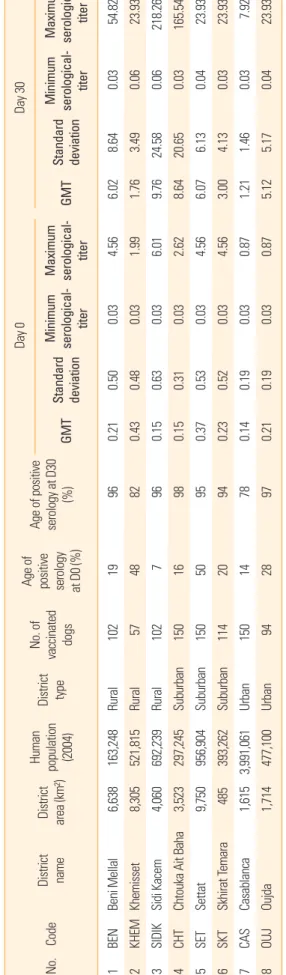
Table 2. District and owned dog characteristics: serological data (IU/mL) obtained on dogs vaccinated by the parenteral route No.Code District name District area (km²) Human population (2004)
District type
No. of vaccinated dogs
Age of positive serology at D0 (%) Age of positive serology at D30
(%)
Day 0Day 30 GMT
Standard deviation
Minimum serological- titer
Maximum serological- GMT titer
Standard deviation
Minimum serological- titer
Maximum serological- titer 0.2154.820.038.646.024.560.500.03966,638119Beni MellalBEN163,248Rural102 0.4323.930.063.491.761.990.030.488,305824857Rural521,815KhemissetKHEM2 0.15218.260.0624.589.766.010.030.634,060967102Rural692,2393Sidi KacemSIDIK 165.540.0320.658.642.620.030.310.159816150Suburban 297,2453,523Chtouka Ait BahaCHT4 23.930.040.376.136.074.560.030.53Settat95150Suburban 956,9049,750SET550 0.2323.930.034.133.004.560.030.5294SKT114Suburban 393,262485emaraSkhirat T206 0.870.030.141.211.460.037.920.1915078Urban3,991,0611,615CasablancaCAS714 0.2123.930.045.175.120.870.030.19289794Urban477,1001,714 OujdaOUJ8 Sera were titrated by the fluorescent antibody virus neutralization test and titers are expressed in International Unit per milliliter (IU/mL). D, day; GMT, geometrical mean titer.
of vaccinated dogs within the districts and the percentages of dogs that developed positive rabies antibody response after vaccination.
At the start of the assay (D0), positive percentages of sero- logical responses ranged from 7% in Sidi Kacem to 50% in Settat. At D30, the positive serology percentages ranged from 78% in Settat to 98% in Chtouka (Table 2). On an overall basis, 24% of dogs were positive at D0 and 92% of them were posi- tive 30 days post-vaccination. Highest seropositive percent- ages (α=0.05) were found in the suburban regions at D0 (29%) and also at D30 (96%) as compared with urban areas (D0, 17%; D30, 83%) and with rural areas (at D0 [20%] only; at D30 [93%], the difference was significant with the value of the ur- ban areas). The increase in rabies antibody titers at D30 (Fig.
3) was highly significant in all districts (p<0.001).
It should be noted that missing serological data were regis- tered in overall population for 11.6% dogs at D0 and 16.6%
dogs at D30, likely because of insufficient serum sample, he- molysis of the sample or loosed sample.
Discussion
In Morocco, many efforts are deployed in infected areas to fight against the disease based essentially on mass canine vaccination campaigns and dog population control [3]. The parenteral dog vaccination using injectable vaccines is the gold standard tool recommended by OIE [15] and WHO for effective rabies control in dogs [10,11]. A vaccination cover- age of 70% of the dog population is considered to be a critical threshold to achieve this goal [11,23]. The rabies vaccine to be used should be approved by the national regulatory au- thorities according to relevant high quality international standards [11,15,16].
Rabivac is a locally produced inactivated rabies vaccine in use in mass dog vaccination campaigns in Morocco. In this study, its efficacy was assessed in experimental conditions comparable to those of the European Pharmacopeia mono- graph [16], which requires a virulent challenge of the target species following vaccination. In addition, a field experiment organized in eight Moroccan districts was undertaken to check the immunogenicity of the vaccine in owned dogs.
All dogs maintained under experimental conditions were collected in the field at early age (between 3 and 6 months old), housed in an experimental kennel and kept for a long quarantine period (9 months) prior to be vaccinated since their health status as regards rabies was unknown. All dogs
had negative rabies serology (<0.24 IU/mL) at D0 except dogs Nos. 4 and 11 that had a titer ≤1.15 IU/mL. The serum sam- ples were pre-diluted prior to serological testing because of insufficient volume, hence the exact value of the titer was im- possible to calculate and the immunological status (positive or negative) was therefore unknown. All vaccinated dogs se- roconverted with titers above 0.24 IU/mL as early as 14 days post-vaccination (geometric mean, 3.34 IU/mL). This is simi- lar to the findings of other authors [24,25] who tested another commercial vaccine produced with the same virus strain and who found a mean antibody value of 2.53 IU/mL [26] and 5.0 IU/mL [24] 2 weeks post-vaccination in laboratory dogs. The same finding was demonstrated 1 month after vaccination, just before the boost, with a mean antibody levels of 3.10 UI/
mL in our study compared with 2.03 UI/mL in the study of Minke et al. [26] and 2.8 IU/mL in the study of Kallel et al. [24]
on laboratory dogs. Two weeks after the boost vaccination (D49), all dogs had high serology titers with a maximum of 41.59 IU/mL indicating an adequate immunization. The mean serological titer of the vaccinated dogs 4 months after vacci- nation (D119) was 0.57 IU/mL, with a high number of dogs still positive (5/8) for rabies antibody detection, contrary to previous studies demonstrating a decrease in time in rabies antibody levels in laboratory and owned field dogs primary vaccinated with one dose of commercial vaccines [24-29].
The longer persistence of detectable rabies antibodies in this study is due to the boost one month after vaccination. After the rabies virus inoculation, one dog (No. 4) died without any symptoms or necropsy findings related to rabies. The kinetics of the rabies antibody response of this dog was similar to that of other dogs. Rabies diagnosis using FAT and MIT provided negative results. The death was not due to rabies but to a tor- sion mesenteric accident observed during the necropsy of the animal. All other vaccinated dogs survived, demonstrat- ing the ability of the vaccine to protect dogs against a challenge virus of dog origin, as it would happen in field conditions.
All dogs of group C had negative serology during all the ex- periment except dog No. 11 who had a titer ≤1.15 IU/mL at D0. The serum was pre-diluted prior to serological testing as its volume was insufficient, as for the serum D0 of dog No. 4 (see above). Therefore, the status (positive or negative) could not be determined. All other antibody titers of this dog during the experiment were clearly below 0.24 IU/mL. This dog died of rabies 17 days after the challenge, i.e., in the same time range than other control dogs, suggesting this dog was pre- sumably not vaccinated. A percentage of 80% (4/5) of control
dogs died from rabies between 17 and 27 days after challenge, which corresponded to the classic rabies incubation period generally reported in experimentally infected dogs [17,30,31].
The observed symptoms were in concordance with rabies clinical signs described by the OIE [15]. The 80% mortality rate observed in the control group is in conformity with the requirements of WHO, OIE, and European Pharmacopeia [11,15,16] for validating the assay. The results of other studies using the same rabies vaccine strain [25] showed a compara- ble level of protection after a rabies virus challenge of vacci- nated dogs. Our results demonstrate the ability of the tested vaccine to induce a high specific humoral response and full protection of field dogs against a dog rabies virus challenge.
Of 919 serologically field owned dogs sampled in urban, suburban, and rural areas, an overall of 24% harbored a posi- tive serological response before vaccination, likely as a result of residual antibody level of a previous vaccination. Unfortu- nately, the dates of the last vaccinations before D0 were un- known. At D0, depending on districts, seropositive percent- ages varied with the highest levels obtained at Settat, Khem- isset, and Oujda (50%, 48%, and 28%, respectively) and the lowest levels found at Sidi Kacem, Casablanca, and Choutka (7%, 14%, and 16%, respectively). The vaccination coverage in rural (20%) and suburban areas (17%) were statistically similar while it was statistically higher in suburban areas (29%); however, these results suggest globally that no signifi- cant difference seems occurring between the geographical status of the regions on the seropositive results obtained. The Sidi Kacem province seemed to be less covered by mass par- enteral vaccination campaigns at the date of the experiment.
It should be noted that the cumulative numbers of human and animal rabies cases in this province are among the high- est within the country [3,7]. The seroprevalence percentages determined before vaccination provide a picture at a certain time of the level of detectable residual antibodies in dogs sampled in the different areas. The time elapsed between the last vaccination of each dog and the blood sampling at D0 was unknown. The curve of rabies antibodies after primary vaccination of laboratory or field dogs has been largely de- scribed (for review, see [12]) and consists in a rapid produc- tion of specific antibodies after the first vaccination followed by a regular decrease then a new rise after a booster to obtain a higher level then a new decrease but with a higher level than that previously achieved. The peak of rabies antibodies is generally observed between 3 and 6 weeks post-vaccina- tion [12,13,26]. A detailed review analysis of the significance
of seroconversion results coupled to those of the probability of surviving a challenge has clearly demonstrated that ani- mals seroconverting at a certain time after vaccination have a very high probability of surviving in case of rabies infection, even if they have no more detectable antibodies at the time or before the challenge [12]. Therefore, the interpretation of seroprevalence results in a field dog population sampled at different times after vaccination has a number of limitations.
The percentage of 24% of seropositive dogs at D0 obtained in our study is an indicator suggesting that dogs have been pre- viously vaccinated, and probably higher vaccination cover- age of dog populations existed as these dogs had seroconver- ted before the D0 sampling. This value corresponds to those which have been published in similar countries [5,32,33].
At D30 post-vaccination, an overall percentage of 92% of dogs were tested positive for rabies antibodies demonstrating the vaccine was highly effective in field conditions. The vacci- nation coverage was estimated at the best period, i.e., 30 days after the vaccination of dogs older than 3 months of age, and results showed in all regions significant increases in rabies antibody levels before and after vaccination. In Casablanca, where 22% of dogs were seronegative at D30, of which 9% had rabies antibodies levels less than 0.1 IU/mL, demonstrating a total absence of antibody response, and the weakest levels of rabies antibodies among different regions tested were ob- served (geometrical mean titer, 1.21 IU/mL). The reasons for that cannot be attributed to the vaccine since a better efficacy was achieved in other regions but could be linked to human deficiencies during the vaccination and sampling steps at the date of the experiment. The negative results could also be due to unhealthy living conditions as dogs were not dewormed and maybe did not receive sufficient food. It is known that ali- mentation and parasitism may influence the production of antibodies [12]. Another explanation could be that the im- mune response of certain dogs did not already reach the 0.24 IU/mL threshold 30 days after vaccination.
The serological monitoring of dogs following the parenter- al mass vaccination campaigns is no longer recommended by WHO on a routine basis [11] because of the kinetics of ra- bies antibodies showing a rapid decrease particularly in pri- mary vaccinated dogs, which represent >30% of the popula- tion because of the high turnover of the dog population [5,33]. Field well–designed seroprevalence studies are now recommended, as done here, for assessing the efficacy of a novel vaccine or if some failures are suspected during the campaigns, such as vaccine potency or cold chain mainte-
nance [11], at the best period after vaccination to check sero- conversion dates.
In view of these results obtained on field dogs and on the literature available on this topic, it is hypothesized that a booster with Rabivac undertaken 1 month after the primary vaccination is probably not justified. A single vaccine injec- tion carried out each year on all dogs, including puppies, as currently done in other places with available comparable commercial vaccines, should be sufficient to ensure adequate immunization of dogs. Studies aiming at estimating the dog population should be organized also in Morocco to update the knowledge of the dynamics of the population, estimated in the 1990s at 1.6 to 2 million [5].
These results demonstrated that Rabivac induced a com- plete protection in experimental dogs after a virulent dog ra- bies virus challenge and a satisfactory humoral response in field dogs living in different conditions. This vaccine is there- fore adequate for mass dog vaccination campaigns against rabies in Morocco.
ORCID
Sami Darkaoui http://orcid.org/0000-0002-9347-9625 Ouafaa Fassi Fihri http://orcid.org/0000-0001-8056-7979 Jean Luc Schereffer http://orcid.org/0000-0003-0888-7286 Nadia Aboulfidaa http://orcid.org/0000-0002-5161-4585 Marine Wasniewski http://orcid.org/0000-0001-5054-3634 Karima Zouine http://orcid.org/0000-0001-7871-1134 Mohammed Bouslikhane http://orcid.org/0000-0002-9682-8931 Khadija Id Sidi Yahia http://orcid.org/0000-0001-6434-5153 Florence Cliquet http://orcid.org/0000-0003-2237-1243
References
1. Knobel DL, Cleaveland S, Coleman PG, et al. Re-evaluat- ing the burden of rabies in Africa and Asia. Bull World Health Organ 2005;83:360-8.
2. Matter H, Blancou J, Benelmouffok A, Hammami S, Fassi- Fehri N. Rabies in North Africa and Malta. In: King AA, Fooks AR, Aubert M, Wandeler AI, editors. Historical per- spective of rabies in Europe and the Mediterranean Basin.
Paris: World Organisation for Animal Health; 2004. p.185- 99.
3. Fassi-Fihri O. Historique et situation épidémiologique de la rage au Maroc. Renforcement de la surveillance et des systèmes d’alerte pour la fièvre catarrhale ovine, la fièvre
du Nil occidental et la rage au Maroc, en Algérie et en Tu- nisie. Projet FAO GCP/RAB/002/FRA. Rome: Food and Agriculture Organization of the United Nations; 2008.
4. Dodet B; Africa Rabies Bureau (AfroREB). The fight against rabies in Africa: from recognition to action. Vaccine 2009;
27:5027-32.
5. El-Harrak M. Epidemiological factors and control of rabies in North Africa. In: Compendium of the OIE Global Con- ference on Rabies Control; 2011 Sep 7-9; Incheon, Korea.
Paris: World Organisation for Animal Health; 2012. p.45-9.
6. World Health Organization. Morocco [Internet]. Geneva:
World Health Organization; 2014 [cited 2015 Oct 1]. Avail- able from: http://www.who.int/rabies/epidemiology/Ra- bies_CP_Morocco_09_2014.pdf?ua=1.
7. Barkia A. Evaluation du système de surveillance de la rage humaine au Maroc [Internet]. Senegal : Journee Mondiale de la Rage; 2013 [cited 2015 Oct 1]. Available from: http://searg.
info/fichiers/afroreb/2013/presentations/afroreb2013_08.
pdf.
8. Darkaoui S, Boue F, Demerson JM, Fassi Fihri O, Yahia KI, Cliquet F. First trials of oral vaccination with rabies SAG2 dog baits in Morocco. Clin Exp Vaccine Res 2014;3:220-6.
9. Meslin FX. Strategy, approaches, guidance and resources for human and dog rabies control. In: FAO/GARC Work- shop; 2012 Nov 6-8; Rome, Italy. Rome: Food and Agricul- ture Organization of the United Nations; 2013. p.3-5.
10. World Health Organization. Oral vaccination of dogs against rabies: guidance for research on oral rabies vac- cines and field application of oral vaccination of dogs against rabies. Geneva: World Health Organization; 2007.
11. World Health Organization. WHO expert consultation on rabies: second report. Geneva: World Health Organization;
2013.
12. Aubert MF. Practical significance of rabies antibodies in cats and dogs. Rev Sci Tech 1992;11:735-60.
13. Barth R, Gruschkau H, Jaeger O. Chick-embryo-cell inac- tivated rabies vaccine for veterinary use: laboratory and field experience. In: Kuwert E, Mérieux C, Koprowski H, Bogel K, editors. Rabies in the tropics. Berlin: Springer Verlag; 1985. p.241-8.
14. Cliquet F, Aubert M, Sagne L. Development of a fluores- cent antibody virus neutralisation test (FAVN test) for the quantitation of rabies-neutralising antibody. J Immunol Methods 1998;212:79-87.
15. World Organisation for Animal Health. Rabies [Internet].
Paris: World Organisation for Animal Health; 2013 [cited
2015 Oct 1]. Available from: http://www.oie.int/fileadmin/
Home/fr/Health_standards/tahm/2.01.13_RABIES.pdf.
16. European Depatment for the Quality of Medicines, Coun- cil of Europe. Rabies vaccine (inactivated) for veterinary use. Mongraph 0451. European Pharmacopoeia. 7th ed.
Strasbourg: Council of Europe; 2013. p.1008-11.
17. Perrin P, Jacob Y, Aguilar-Setien A, et al. Immunization of dogs with a DNA vaccine induces protection against ra- bies virus. Vaccine 1999;18:479-86.
18. Meslin FX, Kaplan MM, Koprowski H. Laboratory tech- niques in rabies. 4th ed. Geneva: World Health Organiza- tion; 1996.
19. Cliquet F, Sagne L, Schereffer JL, Aubert MF. ELISA test for rabies antibody titration in orally vaccinated foxes sam- pled in the fields. Vaccine 2000;18:3272-9.
20. Millan J, Chirife AD, Kalema-Zikusoka G, et al. Serosurvey of dogs for human, livestock, and wildlife pathogens, Uganda. Emerg Infect Dis 2013;19:680-2.
21. Wasniewski M, Guiot AL, Schereffer JL, Tribout L, Mahar K, Cliquet F. Evaluation of an ELISA to detect rabies anti- bodies in orally vaccinated foxes and raccoon dogs sam- pled in the field. J Virol Methods 2013;187:264-70.
22. Hammami S, Schumacher C, Cliquet F, Tlatli A, Aubert A, Aubert M. Vaccination of Tunisian dogs with the lyophi- lised SAG2 oral rabies vaccine incorporated into the DBL2 dog bait. Vet Res 1999;30:607-13.
23. Coleman PG, Dye C. Immunization coverage required to prevent outbreaks of dog rabies. Vaccine 1996;14:185-6.
24. Kallel H, Diouani MF, Loukil H, et al. Immunogenicity and efficacy of an in-house developed cell-culture derived veterinarian rabies vaccine. Vaccine 2006;24:4856-62.
25. Mansfield KL, Burr PD, Snodgrass DR, Sayers R, Fooks
AR. Factors affecting the serological response of dogs and cats to rabies vaccination. Vet Rec 2004;154:423-6.
26. Minke JM, Bouvet J, Cliquet F, et al. Comparison of anti- body responses after vaccination with two inactivated ra- bies vaccines. Vet Microbiol 2009;133:283-6.
27. Cliquet F, Muller T, Mutinelli F, et al. Standardisation and establishment of a rabies ELISA test in European labora- tories for assessing the efficacy of oral fox vaccination campaigns. Vaccine 2003;21:2986-93.
28. Cliquet F, Verdier Y, Sagne L, et al. Neutralising antibody ti- tration in 25,000 sera of dogs and cats vaccinated against ra- bies in France, in the framework of the new regulations that offer an alternative to quarantine. Rev Sci Tech 2003;22:857- 66.
29. Tepsumethanon W, Polsuwan C, Lumlertdaecha B, et al.
Immune response to rabies vaccine in Thai dogs: a pre- liminary report. Vaccine 1991;9:627-30.
30. Perrin P, Jacob Y, Desmezieres E, Tordo N. DNA-based im- munisation against rabies and rabies-related viruses: to- wards multivalent vaccines. Dev Biol (Basel) 2000;104:151- 7.
31. Cliquet F, Barrat J, Guiot AL, et al. Efficacy and bait accep- tance of vaccinia vectored rabies glycoprotein vaccine in cap- tive foxes (Vulpes vulpes), raccoon dogs (Nyctereutes procy- onoides) and dogs (Canis familiaris). Vaccine 2008;26:4627- 38.
32. Davlin SL, Vonville HM. Canine rabies vaccination and domestic dog population characteristics in the develop- ing world: a systematic review. Vaccine 2012;30:3492-502.
33. Lembo T, Hampson K, Kaare MT, et al. The feasibility of canine rabies elimination in Africa: dispelling doubts with data. PLoS Negl Trop Dis 2010;4:e626.