Introduction
Osteoarthritis is thought by some to be unavoidable;
almost everyone older than 50 years of age is affected to some extent. Although the temporomandibular joint (TMJ) is less affected by osteoarthritis than are heavy weight- bearing joints, TMJ osteoarthritis is found in 40% of older adults at microscopic and 14% at radiographic levels.1 However, although osteoarthritis is definitely an aging phenomenon, recent research has identified osteoarthritis in a majority of young patients referred to a TMJ clinic for joint pain and dysfunction.1 An autopsy study2indi-
cated that the signs of degenerative changes on the TMJ articular surfaces were found in 28% of the younger group (16 to 39 years of age) and in 50% of the older group (55 to 78 years of age). Zhao et al3showed that radiographic TMJ osteoarthritic changes were common in adolescent and young adult patients younger than 30 years of age, especially in women, and the occurrence increased sharp- ly between the ages of 11 to 19 years.
Diagnosis of osteoarthritis most commonly depends on the findings from clinical and radiographic examinations.
The autopsy specimen study of Westesson et al4 found computed tomography (CT) to have a sensitivity of 0.75 and a specificity of 1.00 in diagnoses of bone changes with a positive predictive value of 1.0 and negative predictive value of 0.78. CT was superior to MRI in detection of osseous abnormalities. Guidelines from the RDC/TMD validation project5recommend CT for use in both clinical and research settings. The relatively new technique of using CBCT has
Condylar bony changes in patients with temporomandibular disorders: a CBCT study
Kyung-Soo Nah
Department of Oral and Maxillofacial Radiology, School of Dentistry, Pusan National University, Yangsan, Korea ABSTRACT
Purpose: Diagnosis of osteoarthritis most commonly depends on clinical and radiographic findings. The present study attempted to observe the bony changes in temporomandibular joint (TMJ) patients from all age groups.
Materials and Methods: The first-visit clinical records and cone beam computed tomography (CBCT) data of 440 TMJs from 220 consecutive TMJ patients were reviewed retrospectively.
Results: The most frequent condylar bony change observed was sclerosis (133 joints, 30.2%) followed by surface erosion (129 joints, 29.3%), flattening of the articular surface (112 joints, 25.5%), and deviation in form (58 joints, 13.2%), which included 33 TMJs in a cane-shape, 16 with a lateral or medial pole depression, 6 with posterior condy- lar surface flattening, and 3 with a bifid-shaped condyle. Fifty-three joints (12.0%) showed hypoplastic condyles but only 1 joint showed hyperplasia. Osteophyte was found in 35 joints (8.0%) and subcortical cyst in 24 joints (5.5%), 5 of which had surface erosion as well. One hundred nineteen joints (27.0%) had only one kind of condylar bony change, 66 joints (15.0%) had two, 52 joints (11.8%) had three, 12 joints (5.0%) had four, and 6 joints (1.4%) had five kinds of condylar bony changes at the same time. Eighty-five (65.9%) of 129 joints with surface erosion had pain recorded at the chief complaint.
Conclusion: With more widespread use of CBCT, more specific or detailed guidelines for osteoarthritis are needed.
(Imaging Sci Dent 2012; 42 : 249-53)
KEY WORDS: Cone-Beam Computed Tomography; Temporomandibular Joint; Osteoarthritis; Pain
*This study was supported by a Pusan National University Hospital Grant.
Received June 30, 2012; Revised July 21, 2012; Accepted September 3, 2012 Correspondence to : Prof. Kyung-Soo Nah
Department of Oral and Maxillofacial Radiology, School of Dentistry, Pusan National University, Busandaehark-ro 49, Mulgeum-eup, Yangsan-si, Gyeongsangnam-do 628- 870, Korea
Tel) 82-55-360-5260, Fax) 82-55-360-5029, E-mail) ksnah@pusan.ac.kr
Copyright ⓒ 2012 by Korean Academy of Oral and Maxillofacial Radiology
This is an Open Access article distributed under the terms of the Creative Commons Attribution Non-Commercial License (http://creativecommons.org/licenses/by-nc/3.0) which permits unrestricted non-commercial use, distribution, and reproduction in any medium, provided the original work is properly cited.
Imaging Science in Dentistry∙pISSN 2233-7822 eISSN 2233-7830
now become available. The advantage of this technique is the lower radiation dose to the patient compared with con- ventional CT. Hintze et al6compared cone beam comput- ed tomography (CBCT) with conventional tomography in detection of morphological TMJ changes, and in general, no significant differences in diagnostic accuracy were found between the two techniques. Since no evidence that con- ventional or CBCT is inferior to CT has been presented, the choice of imaging technique must depend on the avail- able equipment.7
The osseous component analysis criteria for the condy- lar head of the validation project was gross hypoplasia or hyperplasia, flattening of the articular surface, subcortical sclerosis or cysts, surface erosion, osteophytes, generaliz- ed sclerosis, loose joint bodies, and deviation in form. In the present study, an attempt was made to check the distri- bution of these bony changes observed in TMJ patients from all age groups.
Materials and Methods
The first-visit clinical records and CBCT radiographic data of 440 TMJs from 220 consecutive TMJ patients (male 64, female 156 from 11 to 78 years, mean 29±16 years, Table 1) referred from a temporomandibular disorder cli- nic in the author’s department were reviewed retrospec- tively. The condylar head was checked for hypoplasia or hyperplasia, flattening of the articular surface, subcortical sclerosis or cysts, surface erosion, osteophytes, generalized sclerosis, loose joint bodies, and deviation in form, and also condylar position was recorded following the guide- lines from the RDC/TMD Validation Project.5The chief complaints, including pain and/or noise, were reviewed to find any relationship with the radiographic changes. The CBCT images were taken by a Pax-Zenith 3D (Vatech Co., Hwaseong, Korea). The routine radiographic TMJ exami- nation included 10-12 axial and coronal skull views show- ing the condylar heads, scout views of the midsagittal, and coronal views of each condyle, and 9 corrected sagit-
tal and 4 coronal images for both sides. All images were directly interfaced with a PACS system (M-view, Infinitt Healthcare, Seoul, Korea) on a high resolution monochro- matic monitor (MFGD 5421, Barco, Kortrijk, Belguim) of
Fig. 1.Generalized sclerosis of the right condyle (35/F).
Fig. 2.Surface erosion of the right condyle (15/F).
Table 1.Age and gender distribution of TMJ patients
10-19 20-29 30-39 40-49 50-59 60-69 70-79 Total
Male 28 20 4 4 4 2 2 64
(%) (12.7) (9.1) (1.8) (1.8) (1.8) (0.9) (0.9) (29.1)
Female 62 40 17 10 13 11 3 156
(%) (28.2) (18.2) (7.7) (4.5) (5.9) (5.0) (1.4) (70.9)
Total 90 60 21 14 17 13 5 220
(%) (40.9) (27.3) (9.5) (6.4) (7.7) (5.9) (2.3) (100)
2,048×2,560 image matrices and 145.9 ft-lambert lumine- scence.
Results
The most frequent change observed was the posterior position of the condyle in the sagittal plane (299 joints, 68.0%), followed by sclerosis (133 joints, 30.2%), includ- ing 15 joints showing totally generalized sclerosis (Fig. 1).
Surface erosion was seen in 129 joints (29.3%), 5 of which had a subcortical cyst as well (Fig. 2). Flattening of the arti- cular surface was observed in 112 joints (25.5%). Devia- tion in form were observed in 58 joints (13.2%), which in- cluded 33 cane-shapes (Fig. 3), 16 medial or lateral pole depressions (Figs. 4 and 5), 6 cases of posterior condylar
surface flattening (Fig. 6), and 3 bifid-shaped condyles.
Fifty-three joints (12.0%) showed hypoplastic condyles, but only 1 joint showed hyperplasia. Osteophytes were found in 35 joints (8.0%) and subcortical cysts in 24 joints (5.5%), 5 of which had surface erosion as well.
One hundred nineteen joints (27.0%) had only one con- dylar bony change, 66 joints (15.0%) had two, 52 joints (11.8%) had three, 12 joints (5.0%) had four, and 6 joints (1.4%) had 5 kinds of condylar bony changes at the same time (Table 2).
As for the clinical relationship with the radiographic changes of TMJ, 85 (65.9%) of the 129 joints with surface erosion had pain recorded as the chief complaint.
Fig. 3.Cane-shaped remodeling of both condyles (16/F).
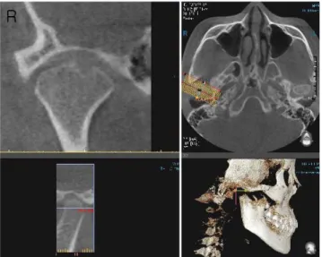
Fig. 4.Lateral pole depression of the right condyle (20/M).
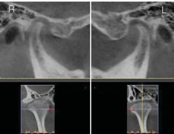
Fig. 5.Medial pole flattening of the left condyle (15/M).
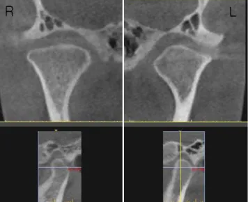
Fig. 6.Posterior surface flattening of the right condyle (61/M).
Discussion
Osteoarthritis is considered a synonyms of osteoarthro- sis, which is a non-inflammatory focal degenerative disord- er of the synovial joints, primarily affecting the articular cartilage and sub-condylar bone and initiated by the deter- ioration of the articular soft-tissue cover and exposure of bone.8This is a common degenerative and destructive altera- tion of joints that has been considered to be an inevitable result of simple wear and tear on aging anatomic struc- tures until recently. It is now known to have a strong inflam- matory component as well, especially in small joints, such as the TMJ, where there appears to be little association with the aging process.1 Larheim and Westesson8 com- mented that cases with evident inflammatory signs on MRI could be characterized as osteoarthritis, whereas joints without such signs could be characterized as osteoarth- rosis.
Osteoarthritis seems like a deformation of bone since it shows subcortical cysts, surface erosion, osteophytes, and/
or generalized sclerosis. However, more specific or detail- ed guidelines of diagnosis for osteoarthritis are now need- ed with the widespread use of CBCT.
The present study attempted to list as many types of bone change as possible in TMJ patients of all age groups.
Although sclerosis was the most frequent condylar bony change in this study, it was not easy to identify definitively since it is usually a gradual bone-forming process. Sclero- sis was, therefore, decided in this study when sclerotic change was clearly noticeable on the CBCT images. As for flattening, this study included only the flattening of the articular surface. However, flattening of the posterior, medial and lateral, and superior surfaces was also seen in the patients. The posterior surface of the condyles fre- quently showed flattening and even depression and ero- sion (Fig. 7) together with medial and lateral surfaces (Figs.
4 and 5). These changes were included in “deviation in form” in this study. In the case of the flattening of the supe- rior surface of condyles, it was not apparently detected on the sagittal images even though it was detected easily on the coronal images (Fig. 8).
The prevalence of condylar bony changes in this study was lower than that found by Cevidanes et al.9They ob- served condylar flattening in 60%, and osteoarthritic sur- face irregularities such as erosions and osteophytes in 40%
of condyles in a painful TMJ osteoarthritis group. They also found that individual local morphologic variation in the left condyles revealed statistically significant positive correlation between the extent of resorption on certain condylar surfaces and pain intensity and duration. In this study, 85 (65.9%) of 129 joints with surface erosion had
Table 2.Number of condylar bony changes observed in the same condyle. Condylar bony changes include hypoplasia or hyperpla- sia, flattening of the articular surface, subcortical sclerosis or cysts, surface erosion, osteophytes, generalized sclerosis, loose joint bodies, and deviation in form.
Number of
condylar bony 1 2 3 4 5 Total
changes
Right 57 26 24 10 2 119
Left 62 40 28 12 4 146
Total 119 66 52 22 6 265
(%) (27.0) (15.0) (11.8) (5.0) (1.4) (100)
Fig. 7.Posterior surface erosion of the right condyle (34/F).
Fig. 8.Superior surface flattening of the right condyle (41/F).
the records of pain as the patients’ chief complaint.
Kurita et al10found radiographic evidence of resorption in the lateral part of the mandibular condyle in 57 (32.0%) of 178 joints and reported that 22 of these had pain on digital palpation. Their study included bony surface ero- sion, concavity, or even flattening at the upper corner of the lateral part of the condyle. In the present study, 16 joints were observed to have medial (4) or lateral (12) pole depressions but if flattening were to be counted, the ratio would have increased, since there were many cases of flat- tening observed at these sites. Liu et al11reported in their study that 63.0% of joints underwent disc perforation at the lateral part of the articular disc and explained that the likely cause was weakness of the lateral capsular attach- ment, whereas the medial capsular attachment was strong- er and reinforced by the insertions of the lateral pteryoid muscle. On the other hand, Palconet et al12reported that there was a poor correlation between condylar changes (as observed on CBCT images), pain, and other clinical signs and symptoms in TMJ osteoarthritis.
In conclusion, with more widespread use of CBCT, more specific or detailed guidelines for osteoarthritis are needed.
References
1. Neville BW, Damm DD, Allen CM, Bouquot JE. Oral and maxillofacial pathology. 2nd ed. Philadelphia: Saunders; 2002.
p. 755.
2. Pereira FJ Jr, Lundh H, Westesson PL. Morphologic changes in the temporomandibular joint in different age groups. An autopsy investigation. Oral Surg Oral Med Oral Pathol 1994; 78 : 279- 87.
3. Zhao YP, Zhang ZY, Wu YT, Zhang WL, Ma XC. Investiga- tion of the clinical and radiographic features of osteoarthrosis of the temporomandibular joints in adolescents and young adults.
Oral Surg Oral Med Oral Pathol Oral Radiol Endod 2011; 111 : e27-34.
4. Westesson PL, Katzberg RW, Tallents RH, Sanchez-Woodworth RE, Svensson SA. CT and MR of the temporomandibular joint:
comparison with autopsy specimens. AJR Am J Roentgenol 1987; 148 : 1165-71.
5. Ahmad M, Hollender L, Anderson Q, Kartha K, Ohrbach R, Truelove EL, et al. Research diagnostic criteria for temporo- mandibular disorders (RDC/TMD): development of image an- alysis criteria and examiner reliability for image analysis. Oral Surg Oral Med Oral Pathol Oral Radiol Endod 2009; 107 : 844-60.
6. Hintze H, Wiese M, Wenzel A. Cone beam CT and conventio- nal tomography for the detection of morphological temporoman- dibular joint changes. Dentomaxillofac Radiol 2007; 36 : 192- 7.
7. Petersson A. What you can and cannot see in TMJ imaging - an overview related to the RDC/TMD diagnostic system. J Oral Rehabil 2010; 37 : 771-8.
8. Larheim TA, Westesson PL. Maxillofacial imaging. Berlin Heidelberg: Springer-Verlag: 2006. p. 149.
9. Cevidanes LH, Hajati AK, Paniagua B, Lim PF, Walker DG, Palconet G, et al. Quantification of condylar resorption in tem- poromandibular joint osteoarthritis. Oral Surg Oral Med Oral Pathol Oral Radiol Endod 2010; 110 : 110-7.
10. Kurita H, Kojima Y, Nakatsuka A, Koike T, Kobayashi H, Kurashina K. Relationship between temporomandibular joint (TMJ)-related pain and morphological changes of the TMJ condyle in patients with temporomandibular disorders. Dento- maxillofac Radiol 2004; 33 : 329-33.
11. Liu XM, Zhang SY, Yang C, Chen MJ, Y Cai X, Haddad MS, et al. Correlation between disc displacements and locations of disc perforation in the temporomandibular joint. Dentomaxil- lofac Radiol 2010; 39 : 149-56.
12. Palconet G, Ludlow JB, Tyndall DA, Lim PF. Correlating cone beam CT results with temporomandibular joint pain of osteoarthritic origin. Dentomaxillofac Radiol 2012; 41 : 126- 30.